

Echocardiography-Guided Radiofrequency Ablation for Cardiac Tumors
- PMID: 39239332
- PMCID: PMC11371935
- DOI: 10.1016/j.jaccao.2024.03.008
Echocardiography-Guided Radiofrequency Ablation for Cardiac Tumors
Abstract
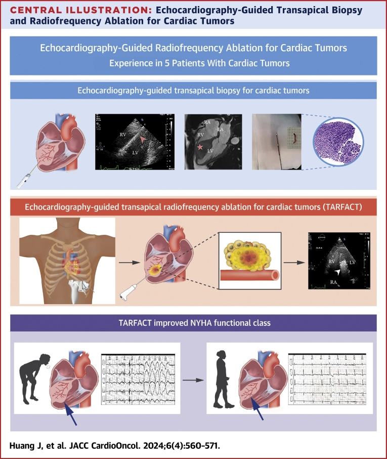
Background: Patients with cardiac tumors may present challenges for surgical resection due to poor clinical condition. Echocardiography-guided transapical radiofrequency ablation for cardiac tumors (TARFACT) potentially offers a less invasive palliative therapy option.
Objectives: This study aimed to evaluate the safety and efficacy of TARFACT.
Methods: Five patients with cardiac tumors (mucinous liposarcoma, myocardial hypertrophy with inflammatory cell infiltration mass, fibrous tissue tumor hyperplasia, myocardial clear cell sarcoma, and cardiac rhabdomyoma) were included. All patients underwent TARFACT and were assessed with electrocardiogram, echocardiographic imaging, biochemical analysis, and pathological confirmation.
Results: The median follow-up for all patients was 9 (range 4-12) months. Three surviving patients were alive at their last follow-up (9, 12, and 12 months, respectively), whereas 2 patients with late-stage tumors survived 6 months and 13 months after TARFACT, respectively. After TARFACT, all patients showed significant reductions in tumor size: the mean length decreased from 6.7 ± 2.0 cm to 4.7 ± 1.8 cm (P = 0.007); and the mean width decreased from 5.0 ± 2.1 cm to 2.5 ± 0.7 cm (P = 0.041). NYHA functional class also improved: median (IQR) decreased from 3.0 (1.5) to 2.0 (1.0) (P = 0.038), Peak E-wave on echocardiography showed a mean increase from 64.4 ± 15.7 cm/s to 76.6 ± 18.6 cm/s (P = 0.008), and NT-pro BNP levels had a median (IQR) reduction from 115.7 (252.1) pg/mL to 55.0 (121.6) pg/mL (P = 0.043).
Conclusions: TARFACT is a novel palliative treatment option for cardiac tumors, reducing accessible tumors and improving clinical symptoms in a preliminary group of patients. (Cardiac Tumors Interventional [Radio Frequency/Laser Ablation] Therapy [CTIH]; NCT02815553).
Keywords: cardiac tumors; transapical radiofrequency ablation for cardiac tumors TARFACT; tumor debulking ablation.
© 2024 The Authors.
Conflict of interest statement
This study was supported by National Natural Science Foundation of China grants No. 82230065, 82071932, and 82001831; Key Research and Development Program of Shaanxi Province grant No. 2022KWZ-19; Clinical Research Funding Project of Fourth Military Medical University grant No. 2021XD010; and Technology Upgrading Project of Fourth Military Medical University grant No. 2023XJSZ02. The authors have reported that they have no relationships relevant to the contents of this paper to disclose.
Figures


References
-
- Poterucha T.J., Kochav J., O'Connor D.S., Rosner G.F. Cardiac tumors: clinical presentation, diagnosis, and management. Curr Treat Options Oncol. 2019;20:66. - PubMed
-
- Maleszewski J.J., Basso C., Bois M.C., et al. The 2021 WHO Classification of Tumors of the Heart. J Thorac Oncol. 2022;17:510–518. - PubMed
-
- Rocca C., Pasqua T., Cerra M.C., Angelone T. Cardiac damage in anthracyclines therapy: focus on oxidative stress and inflammation. Antioxid Redox Signal. 2020;32:1081–1097. - PubMed
Associated data
LinkOut - more resources
Full Text Sources
Medical
Research Materials
