

Fabrication of Out-of-Plane High Channel Density Microelectrode Neural Array with 3D Recording and Stimulation Capabilities
- PMID: 39239464
- PMCID: PMC11376443
- DOI: 10.1109/jmems.2020.3004847
Fabrication of Out-of-Plane High Channel Density Microelectrode Neural Array with 3D Recording and Stimulation Capabilities
Abstract
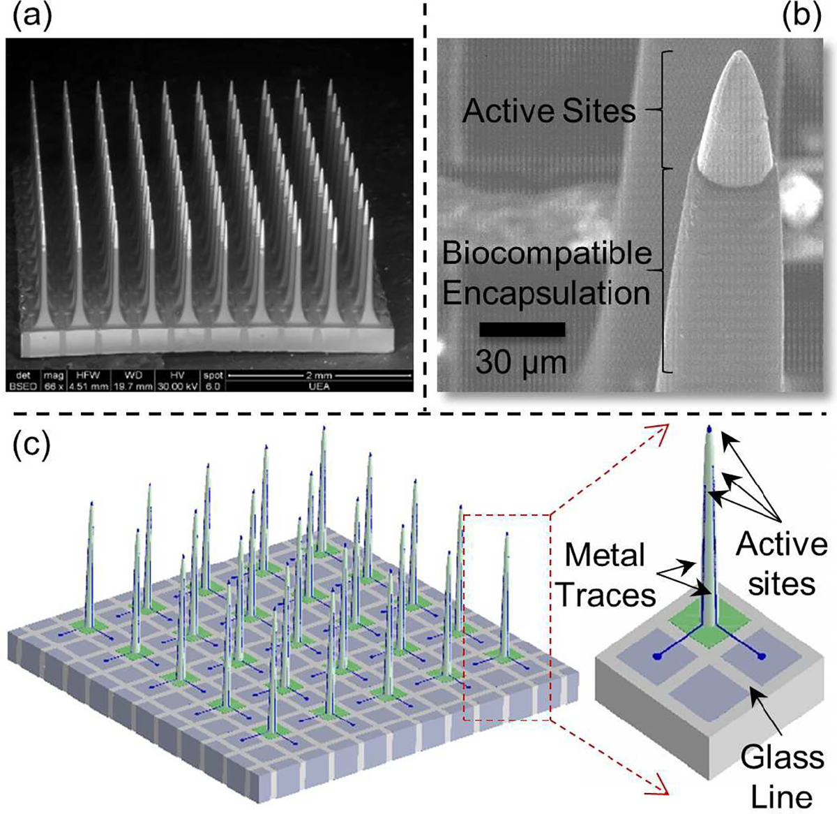
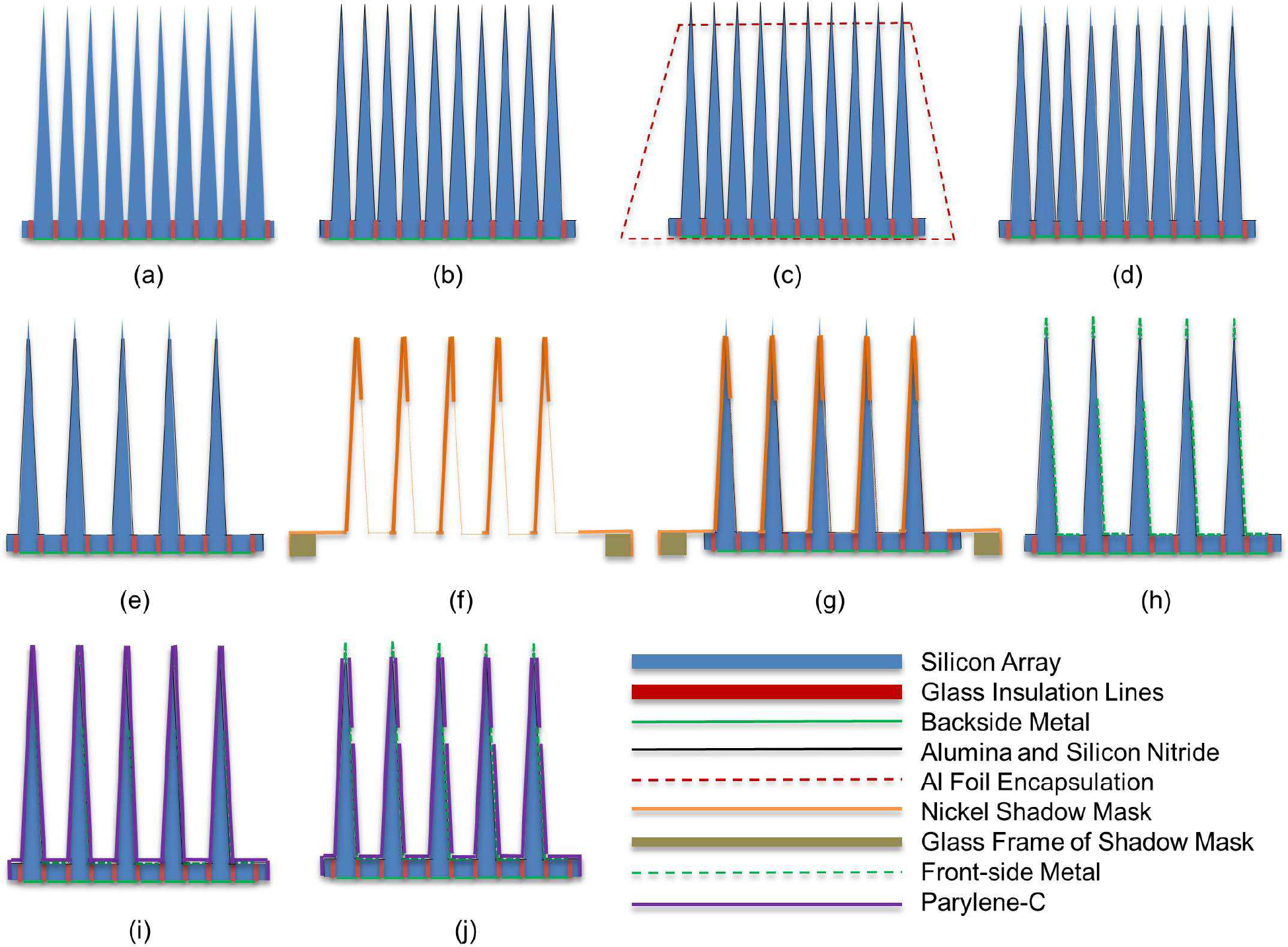
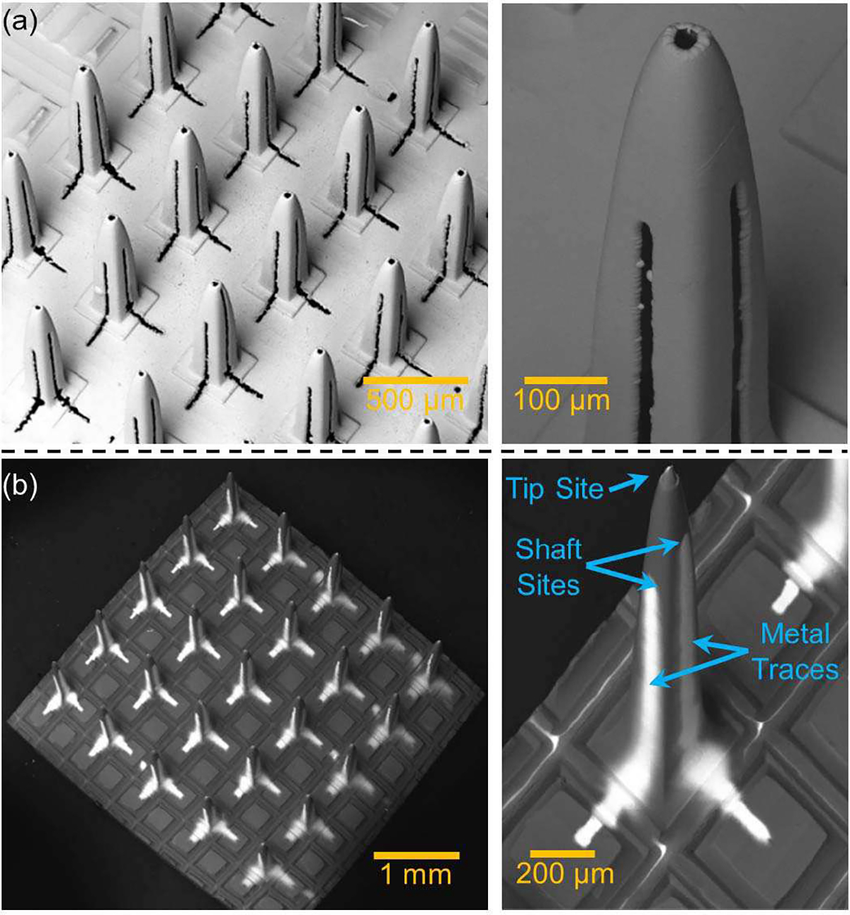
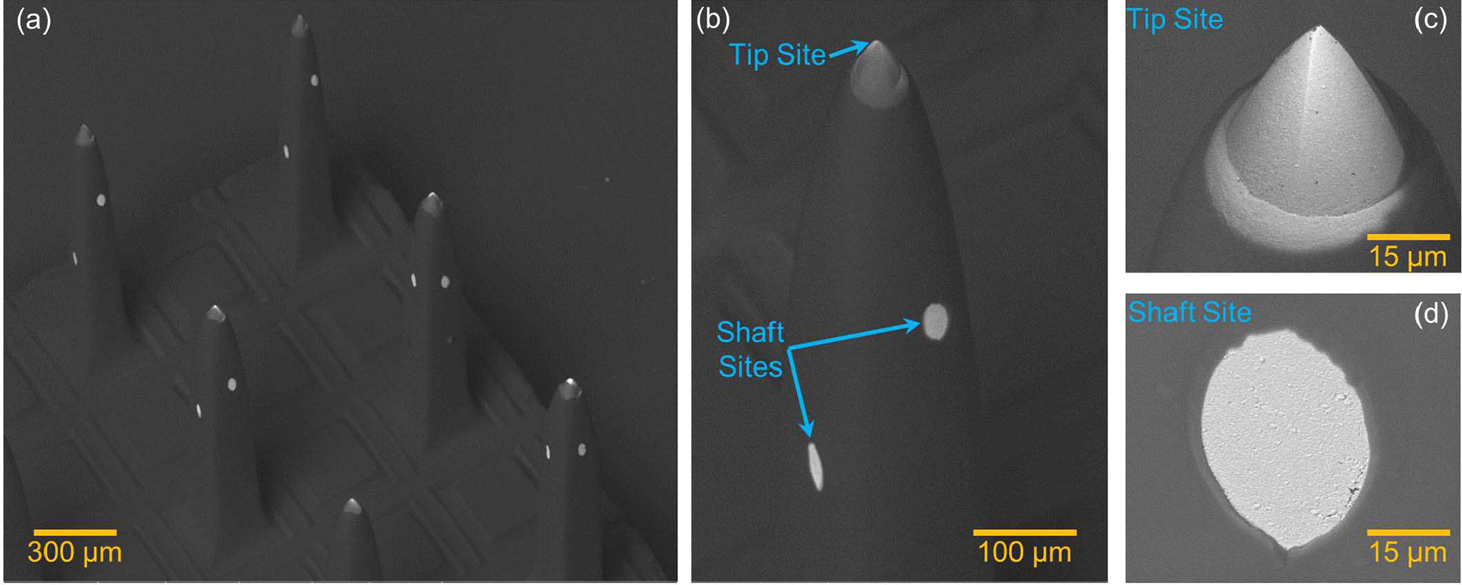
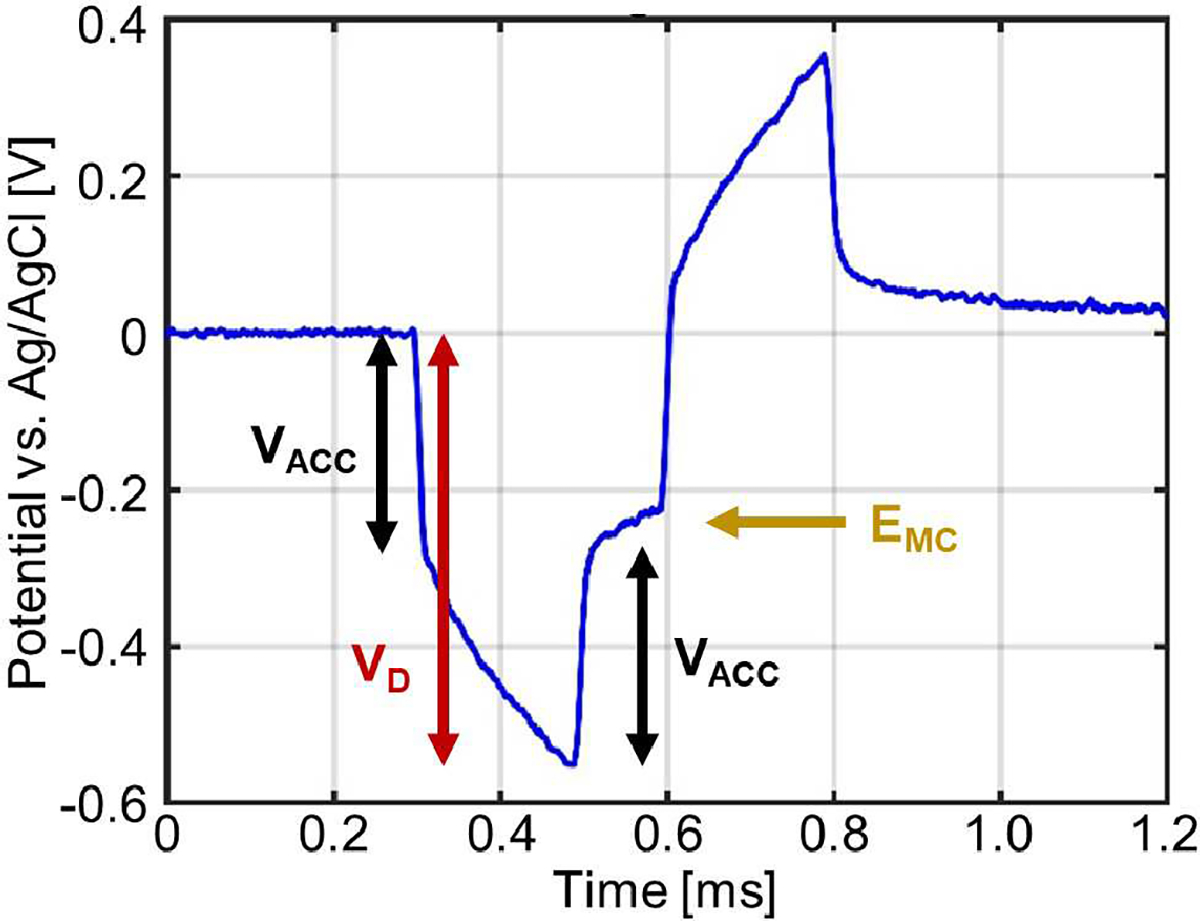
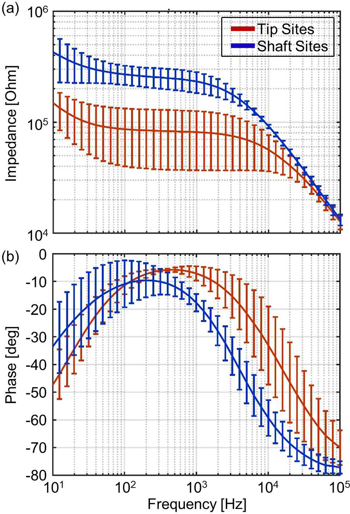
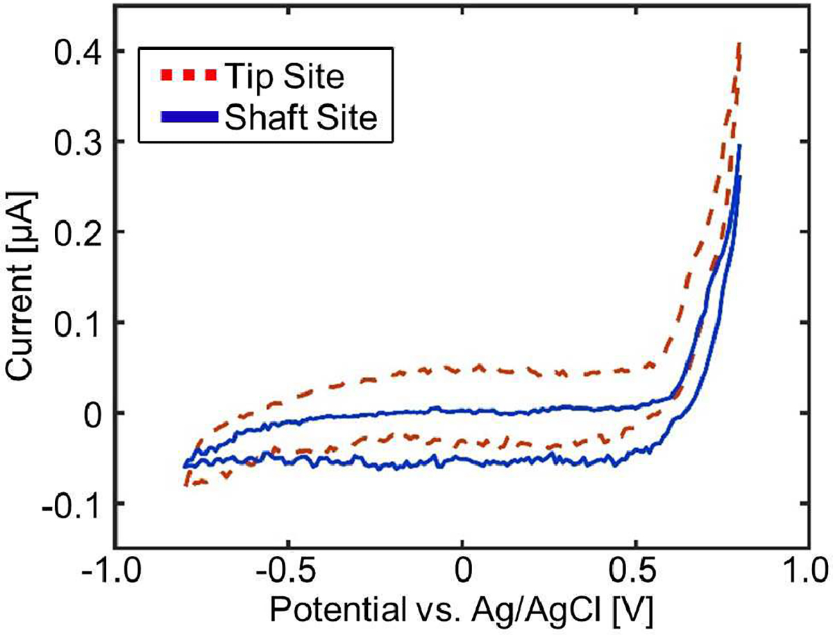
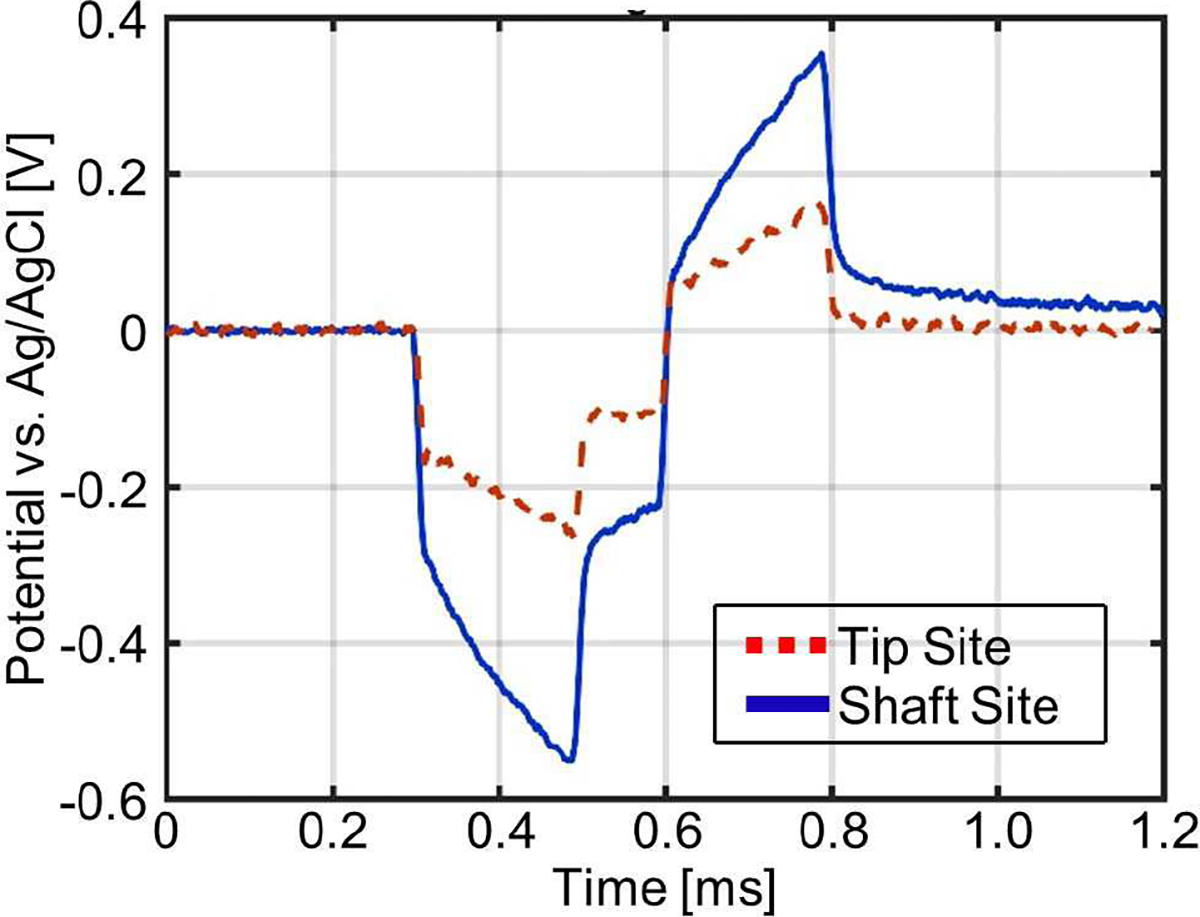
The Utah Electrode Array (UEA) and its different variants have become a gold standard in penetrating high channel count neural electrode for bi-directional neuroprostheses (simultaneous recording and stimulation). However, despite its usage in numerous applications, it has one major drawback of having only one active site per shaft, which is at the tip of the shaft. In this work, we are demonstrating a next-generation device, the Utah Multisite Electrode Array (UMEA), which is capable of having multiple sites around the shaft and also retaining the site at the tip. The UMEA can have up to 9 sites per shaft (hence can accommodate 900 active sites) while retaining the form factor of the conventional UEA with 100 sites. However, in this work and to show the proof of concept, the UMEA was fabricated with one active site at the tip and two around the shaft at different heights; thus, three active sites per shaft. The UMEA device is fabricated using a 3D shadow mask patterning technology, which is suitable for a batch fabrication process for these out-of-plane structures. The UMEA was characterized by in-vitro tests to showcase the electrochemical properties of the shaft sites for bi-directional neuroprostheses in contrast to the traditional tip sites of the standard UEA. The UMEA not only improves the channel density of conventional UEAs and hence can access a larger population of neurons, but also enhances the recording and stimulation capabilities from different layers of the human cortex without further increasing the risk of neuronal damage.
Keywords: Microelectrodes; Neural Electrodes; Shadow Mask; Utah Electrode Array; Utah Multisite Electrode Array.
Figures
References
-
- Benabid A-L, Pollak P, Louveau A, Henry S, and De Rougemont J, “Combined (thalamotomy and stimulation) stereotactic surgery of the VIM thalamic nucleus for bilateral Parkinson disease,” Stereotactic and functional neurosurgery, vol. 50, no. 1–6, pp. 344–346, 1987. - PubMed
-
- Odekerken VJ et al. , “GPi vs STN deep brain stimulation for Parkinson disease: three-year follow-up,” Neurology, vol. 86, no. 8, pp. 755–761, 2016. - PubMed
-
- Baizabal-Carvallo JF, Kagnoff MN, Jimenez-Shahed J, Fekete R, and Jankovic J, “The safety and efficacy of thalamic deep brain stimulation in essential tremor: 10 years and beyond,” J Neurol Neurosurg Psychiatry, vol. 85, no. 5, pp. 567–572, 2014. - PubMed
-
- Benabid AL et al. , “Long-term suppression of tremor by chronic stimulation of the ventral intermediate thalamic nucleus,” The Lancet, vol. 337, no. 8738, pp. 403–406, 1991. - PubMed
-
- Boccard SG et al. , “Targeting the affective component of chronic pain: a case series of deep brain stimulation of the anterior cingulate cortex,” Neurosurgery, vol. 74, no. 6, pp. 628–637, 2014. - PubMed
Grants and funding
LinkOut - more resources
Full Text Sources