. 2024 Aug 19;21(11):2233-2243.
doi: 10.7150/ijms.97051. eCollection 2024.

Magnetic nanomagnetic nanoparticles combining with Slit2 gene and bone marrow mononuclear cells to improve cognitive dysfunction in rats with chronic cerebral ischemia

Affiliations

Magnetic nanomagnetic nanoparticles combining with Slit2 gene and bone marrow mononuclear cells to improve cognitive dysfunction in rats with chronic cerebral ischemia

Nan Wang et al. Int J Med Sci. .

Abstract

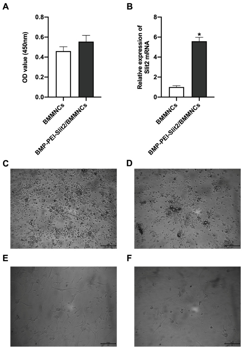
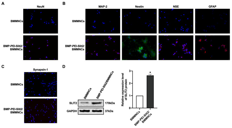
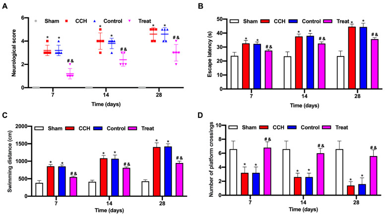
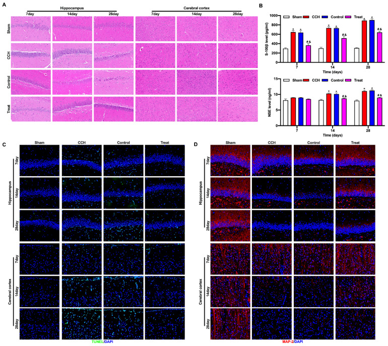
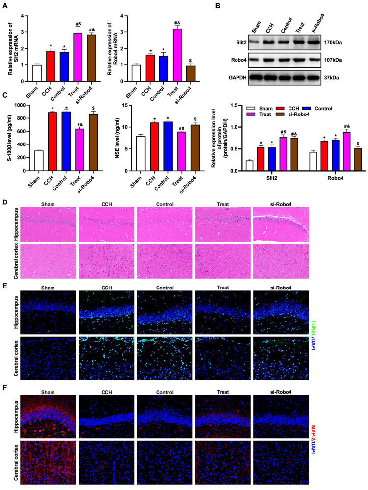
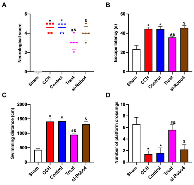
Purpose: Cognitive dysfunction caused by chronic cerebral hypoperfusion (CCH) is the leading cause of vascular dementia. Therefore, it is necessary to explore the mechanism that causes cerebral injury and find an effective therapy. Methods: Bone marrow mononuclear cells (BMMNCs) were extracted to detect the activity by CCK-8 kit and verify the transfection efficiency using reverse transcription-quantitative real-time polymerase chain reaction (RT-qPCR). A CCH rat model was established. Superparamagnetic iron oxide nanoparticles (BMPs)-PEI-Slit2/BMMNCs were injected into the tail vein and intervened with an external magnetic field. Hematoxylin and eosin staining was used to observe the pathological changes in brain tissue. The Slit/Robo pathway-related proteins Slit2 and Robo4 were detected by RT-qPCR and Western blotting. Results: The neurological score of the CCH group significantly increased compared with that of the sham group (P<0.05). The levels of brain injury markers S-100β and NSE were significantly higher in the CCH group than in the sham group (P<0.05). Neuronal apoptosis in the frontal cortex and hippocampus of CCH rats significantly increased compared with that of the sham group (P<0.05). The expression levels of Slit2 and Robo4 mRNAs and proteins in brain tissue of CCH rats significantly increased (P<0.05). The neurological function scores of CCH rats treated with BMP-PEI-Slit2/BMMNC significantly increased after Robo4 siRNA administration (P<0.05). Conclusion: BMP combination with the CCH-related gene Slit2 can effectively improve the efficiency of BMMNC transplantation in treatment.

Keywords: bone marrow mononuclear cells; chronic cerebral hypoperfusion; rat; superparamagnetic iron oxide nanoparticles.


Conflict of interest statement

Competing Interests: The authors have declared that no competing interest exists.

Figures

Figure 1
Figure 1
Preparation of BMP-PEI-Slit2 BMMNCs. BMMNC viability after BMP-PEI-Slit2 was co-cultured with BMMNCs for 24 hours detected by CCK-8 (A); and Slit2 mRNA expression detected by RT-qPCR (B). The primary cells (C), the cell after three days of induced culture (D), the cell after seven days of induced culture (E), and the cell after 14 days of induced culture (F) were observed by the inverted microscope. *, p < 0.05 compare with BMMNCs group. BMMNC: Bone marrow mononuclear cell; BMP: superparamagnetic iron oxide nanoparticle; PCR: polymerase chain reaction; PEI: Polyethylenimine.
Figure 2
Figure 2
BMP-PEI-Slit2 induced BMMNC differentiation into neuron-like cells. The expression of NeuN after three days of in vitro culture of BMP-PEI-Slit2/BMMNCs (A), and the expression of MAP-2, Nestin, NSE, GFAP (B), and synapsin-1 (C) after 14 days of culture were detected by immunofluorescence. The expression of Slit2 in BMP-PEI-Slit2/BMMNCs cultured in vitro for 24 hours was detected by Western blot (D). *, p < 0.05 compare with BMMNCs group. BMMNC: Bone marrow mononuclear cell; BMP: superparamagnetic iron oxide nanoparticle; PEI: Polyethylenimine.
Figure 3
Figure 3
BMP-PEI-Slit2/BMMNCs improved cognitive function in CCH rats. A CCH rat model was established. BMP-PEI-Slit2/BMMNCs were injected into the tail vein and intervened with an external magnetic field. The cognitive function was observed at 7, 14, and 28 days, respectively. The neuronal score of rats was assessed using the Bederson method (A). The learning and memory abilities of rats were detected by directional navigation (B) and spatial exploration tests (C and D) in the Morris water maze. *, p < 0.05 compare with Sham group; #, p < 0.05 compare with CCH group; &, p < 0.05 compare with Control group. BMMNC: Bone marrow mononuclear cell; BMP: superparamagnetic iron oxide nanoparticle; CCH: chronic cerebral hypoperfusion; PEI: Polyethylenimine.
Figure 4
Figure 4
BMP-PEI-Slit2/BMMNCs ameliorated brain tissue injury in CCH rats. The pathological changes in the rat brain tissue were observed by HE staining (A). The expression levels of brain injury markers S-100β and NSE were measured by ELISA (B). The neuronal apoptosis in the frontal cortex and hippocampus of rats was observed by TUNEL assay (C). The expression of MAP2 in the frontal cortex and hippocampus of rats was detected by immunofluorescence (D). *, p < 0.05 compare with Sham group; #, p < 0.05 compare with CCH group; &, p < 0.05 compare with Control group. BMMNC: Bone marrow mononuclear cell; BMP: superparamagnetic iron oxide nanoparticle; CCH: chronic cerebral hypoperfusion; ELISA: enzyme-linked immunosorbent assay; HE: hematoxylin and eosin; PEI: Polyethylenimine; TUNEL: terminal dUTP nick-end labeling.
Figure 5
Figure 5
BMP-PEI-Slit2/BMMNCs ameliorated brain injury in CCH rats by regulating the Slit2/Robo4 pathway. The Slit/Robo pathway-related proteins Slit2 and Robo4 were detected by RT-qPCR (A) and Western blot (B). The expression of brain injury markers S-100β and NSE levels were measured by ELISA (C). The pathological changes in the rat brain tissue were observed by HE staining (D). The neuronal apoptosis in the frontal cortex and hippocampus of rats was observed by TUNEL assay (E). The expression of MAP2 in the frontal cortex and hippocampus of rats was detected by immunofluorescence (F). *, p < 0.05 compare with Sham group; #, p < 0.05 compare with CCH group; &, p < 0.05 compare with Control group; $, p < 0.05 compare with Treat group. BMMNC: Bone marrow mononuclear cell; BMP: superparamagnetic iron oxide nanoparticle; CCH: chronic cerebral hypoperfusion; ELISA: enzyme-linked immunosorbent assay; HE: hematoxylin and eosin; PCR: polymerase chain reaction; PEI: Polyethylenimine; TUNEL: terminal dUTP nick-end labeling.
Figure 6
Figure 6
BMP-PEI-Slit2/BMMNCs improved cognitive function in CCH rats by regulating the Slit2/Robo4 pathway. The neuronal score of rats was assessed using the Bederson method (A). The learning and memory abilities of rats were detected by directional navigation (B) and spatial exploration tests (C and D) in the Morris water maze. *, p < 0.05 compare with Sham group; #, p < 0.05 compare with CCH group; &, p < 0.05 compare with Control group; $, p < 0.05 compare with Treat group. BMMNC: Bone marrow mononuclear cell; BMP: superparamagnetic iron oxide nanoparticle; CCH: chronic cerebral hypoperfusion; PEI: Polyethylenimine.

Similar articles

References

    1. Kalaria RN. The pathology and pathophysiology of vascular dementia. Neuropharmacol. 2018;134(Pt B):226–39. - PubMed
    1. Yu W, Li Y, Hu J, Wu J, Huang Y. A Study on the Pathogenesis of Vascular Cognitive Impairment and Dementia: The Chronic Cerebral Hypoperfusion Hypothesis. J Clin Med. 2022;11(16):4742. - PMC - PubMed
    1. Wang XX, Zhang B, Xia R, Jia QY. Inflammation, apoptosis and autophagy as critical players in vascular dementia. Eur Rev Med Pharmacol Sci. 2020;24(18):9601–14. - PubMed
    1. Wolters FJ, Ikram MA. Epidemiology of Vascular Dementia. Arterioscler Thromb Vasc Biol. 2019;39(8):1542–9. - PubMed
    1. Menarim BC, MacLeod JN, Dahlgren LA. Bone marrow mononuclear cells for joint therapy: The role of macrophages in inflammation resolution and tissue repair. World J Stem Cells. 2021;13(7):825–40. - PMC - PubMed

MeSH terms

LinkOut - more resources