. 2024 Jun 13;6(1):vdae098.
doi: 10.1093/noajnl/vdae098. eCollection 2024 Jan-Dec.

A systematic review of normal tissue neurovascular unit damage following brain irradiation-Factors affecting damage severity and timing of effects

Affiliations

A systematic review of normal tissue neurovascular unit damage following brain irradiation-Factors affecting damage severity and timing of effects

Annet Nakkazi et al. Neurooncol Adv. .

Abstract

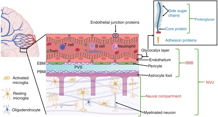
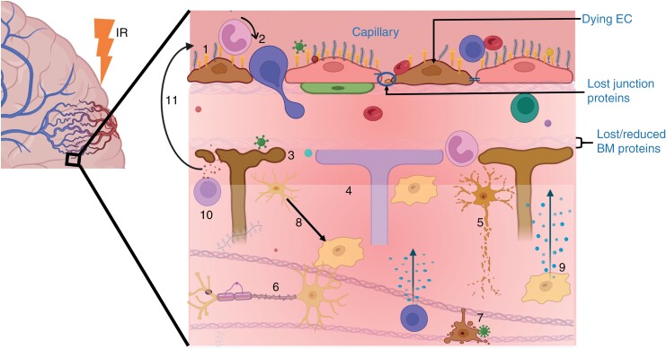
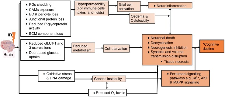
Background: Radiotherapy is key in the treatment of primary and secondary brain tumors. However, normal tissue is inevitably irradiated, causing toxicity and contributing to cognitive dysfunction. The relative importance of vascular damage to cognitive decline is poorly understood. Here, we systematically review the evidence for radiation-induced damage to the entire neurovascular unit (NVU), particularly focusing on establishing the factors that influence damage severity, and timing and duration of vascular effects relative to effects on neural tissue.

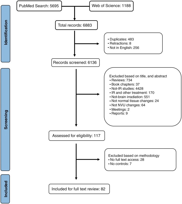
Methods: Using PubMed and Web of Science, we searched preclinical and clinical literature published between January 1, 1970 and December 1, 2022 and evaluated factors influencing NVU damage severity and timing of NVU effects resulting from ionizing radiation.

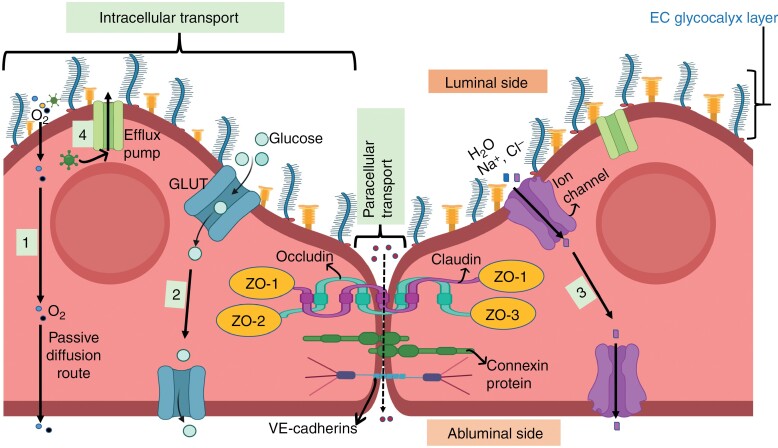
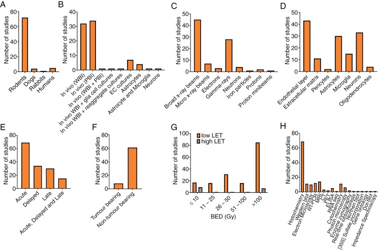
Results: Seventy-two rodents, 4 canines, 1 rabbit, and 5 human studies met inclusion criteria. Radiation increased blood-brain barrier (BBB) permeability, reduced endothelial cell number and extracellular matrix proteoglycans, reduced tight junction proteins, upregulated cellular adhesion molecule expression, reduced activity of glucose and BBB efflux transporters and activated glial cells. In the brain parenchyma, increased metalloproteinases 2 and 9 levels, demyelination, cell death, and inhibited differentiation were observed. Effects on the vasculature and neural compartment were observed across acute, delayed, and late timepoints, and damage extent was higher with low linear energy transfer radiation, higher doses, lower dose rates, broader beams, and in the presence of a tumor.

Conclusions: Irradiation of normal brain tissue leads to widespread and varied impacts on the NVU. Data indicate that vascular damage is in most cases an early effect that does not quickly resolve. More studies are needed to confirm sequence of damages, and mechanisms that lead to cognitive dysfunction.

Keywords: blood-brain barrier damage; brain irradiation; cognitive decline; neurotoxicity; neurovascular unit dysfunction.


Conflict of interest statement

None declared.

Figures

Figure 1.
Figure 1.
Components of the neurovascular unit in a healthy brain tissue. The BBB is the vessel component of the NVU, and is composed of the endothelial layer (endothelium and its glycocalyx), pericytes, endothelial basement membrane (EBM), perivascular space (PVS), parenchymal basement membrane (PBM), and astrocyte endfeet., Among these structures, the endothelial layer is considered to be the chief element of the BBB as it forms walls of the vessels, and regulates majority of the exchange/transport between blood and the brain tissue, (Figure 2). The neural compartment consists of the interstitial matrix and perineuronal network, neurons, interneurons, oligodendrocytes, and resident immune cells (mainly microglia), which work closely with the BBB to meet the structural, developmental, and functional demands of the brain.,, This synergistic interconnection of NVU components enables proper neuronal metabolic activity, effective waste removal, a sufficient and well-regulated cerebral blood flow, and a controlled neuroimmune response., If the NVU is impaired, for instance by ionising radiation (IR), its ability to meet the energy demands of the neuronal tissue may be hindered, which could ultimately result in loss of proper brain function. (Figure created with BioRender.)
Figure 2.
Figure 2.
Endothelial layer components, and their role in regulating transport across the BBB. The endothelial layer is polarized into luminal (blood-facing) and abluminal (brain-facing) plasma membrane domains, and is made up of ECs (or endothelium) with their outer glycocalyx layer. Cerebrovascular ECs lack fenestrations, thus nutrients and other molecules enter the brain tissue via transcellular (through ECs) and/or paracellular (in between ECs) pathways, depending on factors like their molecular weight, lipid solubility, and charge. The transcellular pathway acts as the major transport route, and it allows crossing of small hydrophilic and lipophilic molecules (molecular weight <500 Da) through ECs by diffusion or specific transport channels., Small lipophilic molecules, such as oxygen, carbon dioxide, and alcohol can passively cross the endothelial cell membranes unrestricted (1), but glucose, and water and small ion molecules require specific transporters (eg, GLUTs) (2) and ion channels (3), respectively. To control passive diffusion into the brain, ECs highly express efflux pumps/transporters (4), mainly P-glycoprotein (a glycosylated member of the ATP-binding cassette transporters) on the luminal side, which transport undesirable molecules like toxins back into the blood stream., Paracellular transport, on the other hand, is restricted by the 3 types of junctions that interconnect ECs; tight junctions (made of proteins, including zonula occludens—ZO, Occludin, and claudin), gap junctions (made of connexin proteins), and adherens junctions (made of proteins, such as VE-cadherins). These junction proteins only allow lipophilic, low molecular weight molecules to passively diffuse through the intercellular gaps, depending on hydrostatic, electrochemical, and osmotic gradient.,,, This restriction is achieved due to their complex and layered arrangement, which creates a trans-endothelial electrical resistance (TEER) of up to 5900 Ω cm2 (in vivo, rat), making the BBB the tightest barrier in the body when compared to other organs’ TEER values that are below 4000 Ω cm2,,,, However, the localization and expression of these junction proteins can be affected by stressors, such as upregulated Ca2+ signaling (due to increased intracellular Ca2+ levels) that has been reported to induce tight junction disassembly., Influx of molecules into the endothelium is also controlled by the glycocalyx (on the luminal side), a grass-like extracellular matrix (ECM) layer mainly composed of proteoglycans that mask cell adhesion molecules (CAMs). Proteoglycans consist of core proteins (mainly glypicans and syndecans) that are covalently bound to long unbranched glycosaminoglycan side chains (mostly chondroitin sulfate and heparan sulfate). CAMs include selectins (P and E-selectins, which are crucial in leukocyte adhesion) and immunoglobulin-like proteins (vascular cell adhesion molecule 1 [VCAM-1], intercellular adhesion molecules 1 and 2 [ICAM-1 and –2], and platelet/endothelial cell adhesion molecule 1 [PECAM-1, also known as CD31]). Immunoglobulin-like proteins are involved in cell-cell adhesion, EC migration and regulation of EC-matrix interactions, but their specific roles are not well established. If the endothelial layer is affected/damaged, toxic substances and peripheral immune cells can more easily enter the neural tissue, where they can induce unregulated immune responses and neuronal death. (Figure created with BioRender.)
Figure 3.
Figure 3.
PRISMA flow diagram showing inclusion and exclusion criteria of identified publications.
Figure 4.
Figure 4.
Summary of preclinical and clinical studies investigating radiation-induced effects on the NVU. A quantitative analysis of (A) subjects studied, (B) models, (C) ionizing radiation type, (D) NVU component studied, (E) timepoints studied, (F) studies with and without tumors present, (G) biologically effective doses (BED) used separated into high and low LET types, and (H) assays used.
Figure 5.
Figure 5.
A summary of radiation-induced NVU damage/changes, and how the neural tissue can be directly or indirectly affected. Indirect effects can start from BBB disruption: Loss of the glycocalyx proteoglycans results in exposure of CAMs, such as selectins, ICAM-1, and VCAM-1 (1), which enable circulating immune cells like leukocytes to infiltrate through the endothelium (2). Additionally, loss of ECs, junction proteins, pericytes, and basement membrane (BM) components allows easier influx of blood components (like toxins and immune cells) into the brain tissue where they can be harmful to astrocytes (3) and neural tissue cells (7). Direct effects to the neural tissue can involve loss of neuroblasts, interstitial ECM proteins (4), neurons (5), myelin sheath (6), oligodendrocytes and microglia (7), reduced stem cell proliferation and differentiation, and dysfunction of synaptic and volume transmission. In both pathways, infiltrating and resident immune cells become activated (8), which triggers signal transduction pathways, such as the nuclear factor kappa B, that mediate the production of proinflammatory cytokines, chemokines and inducible enzymes (9 and 10). These mediators in turn increase the BBB permeability (11), for instance, by further upregulating the expression of adhesion molecules on the endothelium, and the cycle repeats. (Figure created with BioRender.)
Figure 6.
Figure 6.
Radiation-induced NVU changes and possible biological processes that promote cognitive decline. Observations from the above studies provide clues on their possible linkage to some of the biological processes reported in neurodegenerative studies.

Similar articles

Cited by

References

    1. Armstrong CL, Shera DM, Lustig RA, Phillips PC.. Phase measurement of cognitive impairment specific to radiotherapy. Int J Radiat Oncol Biol Phys. 2012;83(3):e319–e324. - PMC - PubMed
    1. Galakhova AA, Hunt S, Wilbers R, et al.. Evolution of cortical neurons supporting human cognition. Trends Cogn Sci. 2022;26(11):909–922. - PMC - PubMed
    1. Zhu WM, Neuhaus A, Beard DJ, Sutherland BA, DeLuca GC.. Neurovascular coupling mechanisms in health and neurovascular uncoupling in Alzheimer’s disease. Brain. 2022;145(7):2276–2292. - PMC - PubMed
    1. Burnet NG, Thomas SJ, Burton KE, Jefferies SJ.. Defining the tumour and target volumes for radiotherapy. Cancer Imaging. 2004;4(2):153–161. - PMC - PubMed
    1. Baumann M, Krause M, Overgaard J, et al.. Radiation oncology in the era of precision medicine. Nat Rev Cancer. 2016;16(4):234–249. - PubMed

LinkOut - more resources