

Microstructural architecture of the bony scutes, spine, and rays of the bony fins in the common pleco (Hypostomus plecostomus)
- PMID: 39239634
- PMCID: PMC11376312
- DOI: 10.1080/23144599.2024.2374201
Microstructural architecture of the bony scutes, spine, and rays of the bony fins in the common pleco (Hypostomus plecostomus)
Abstract
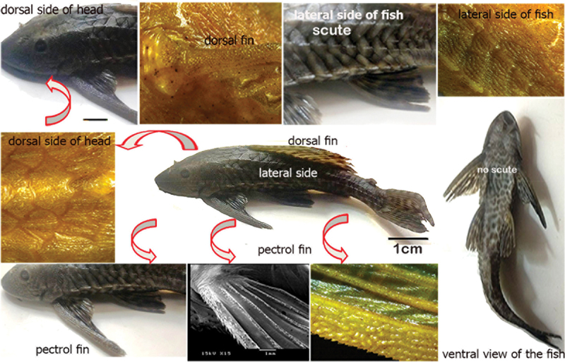
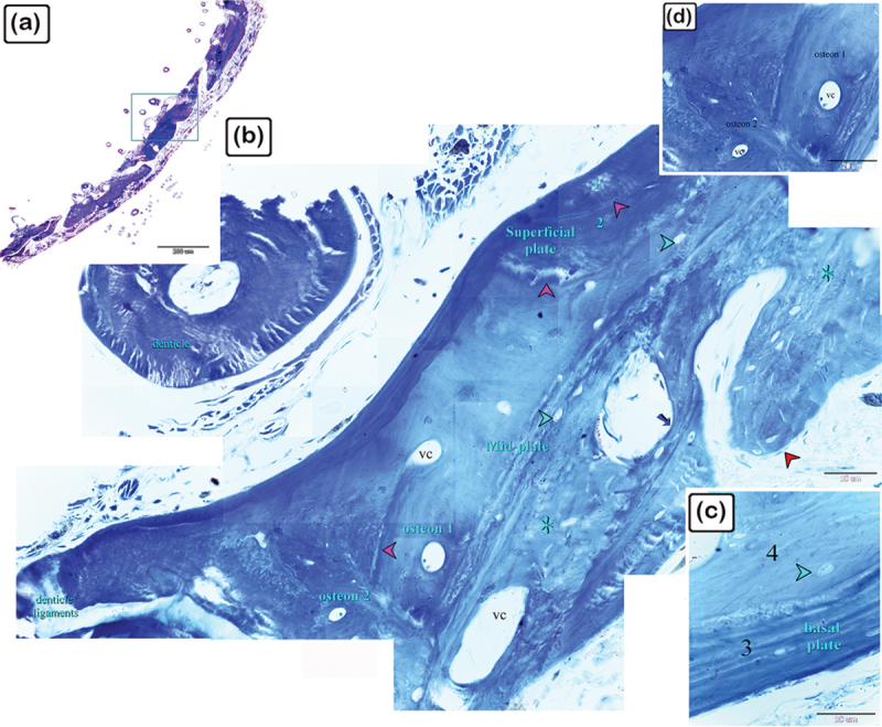
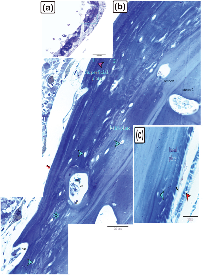
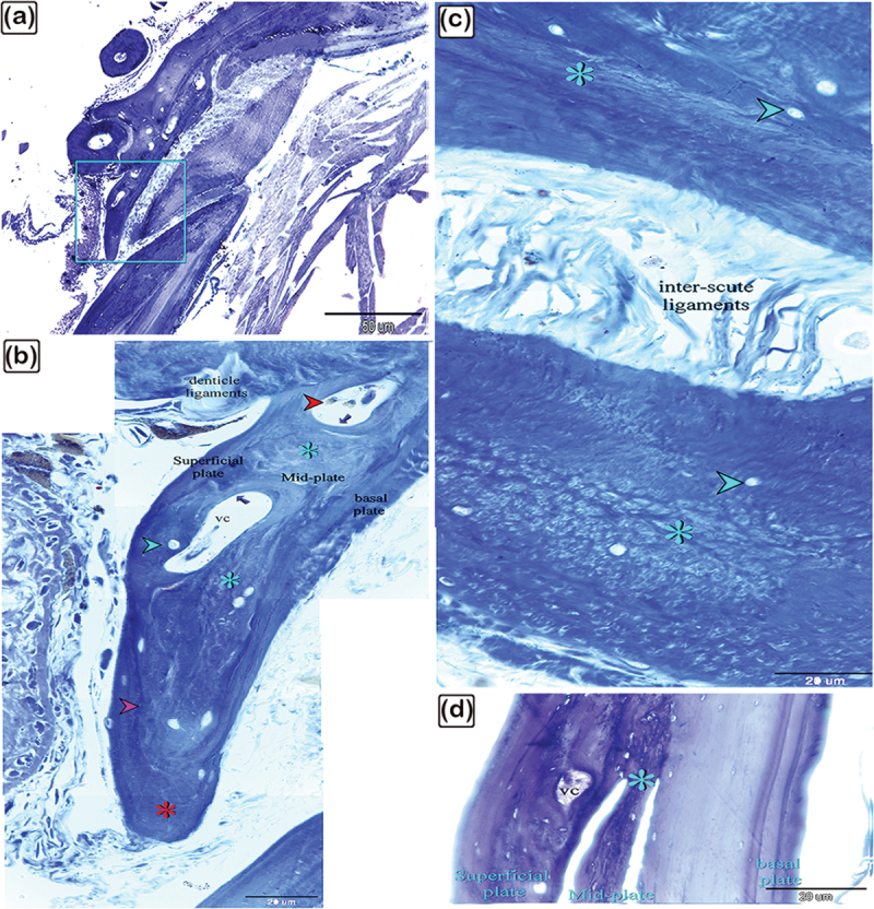
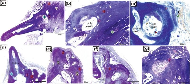
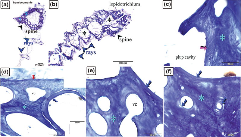
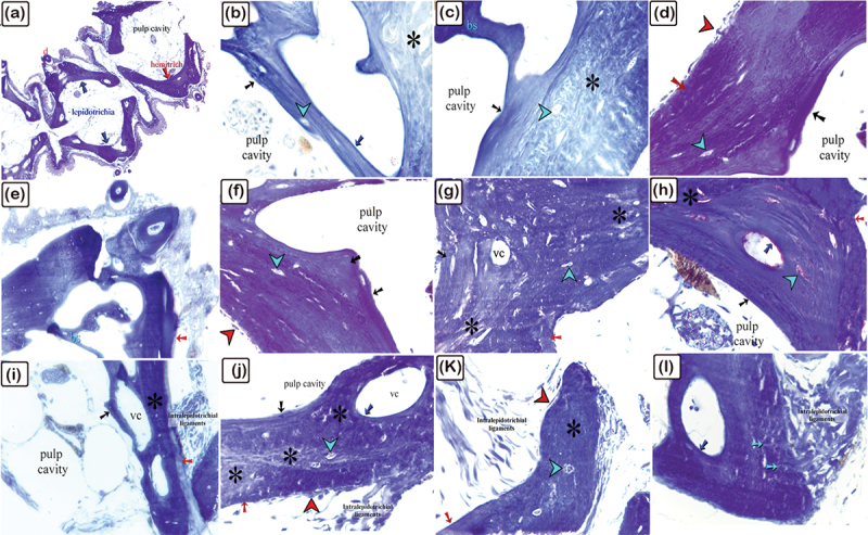
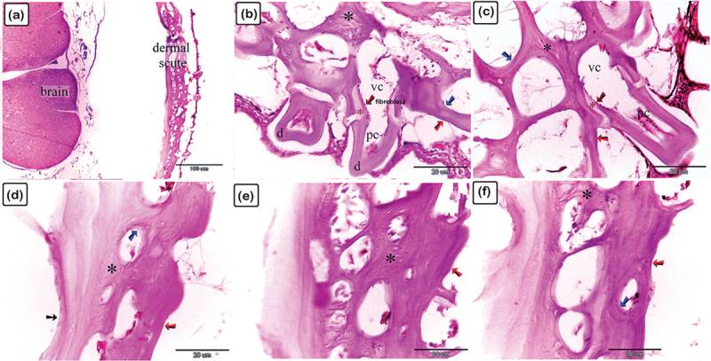
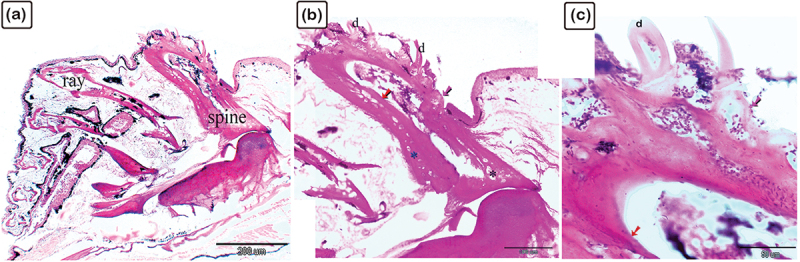
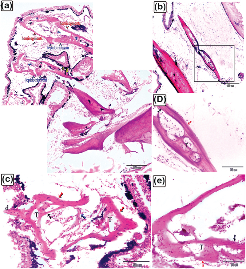
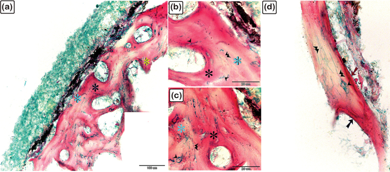
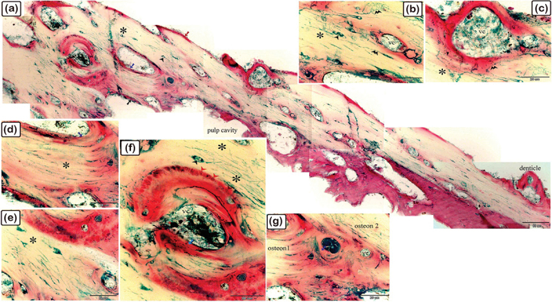
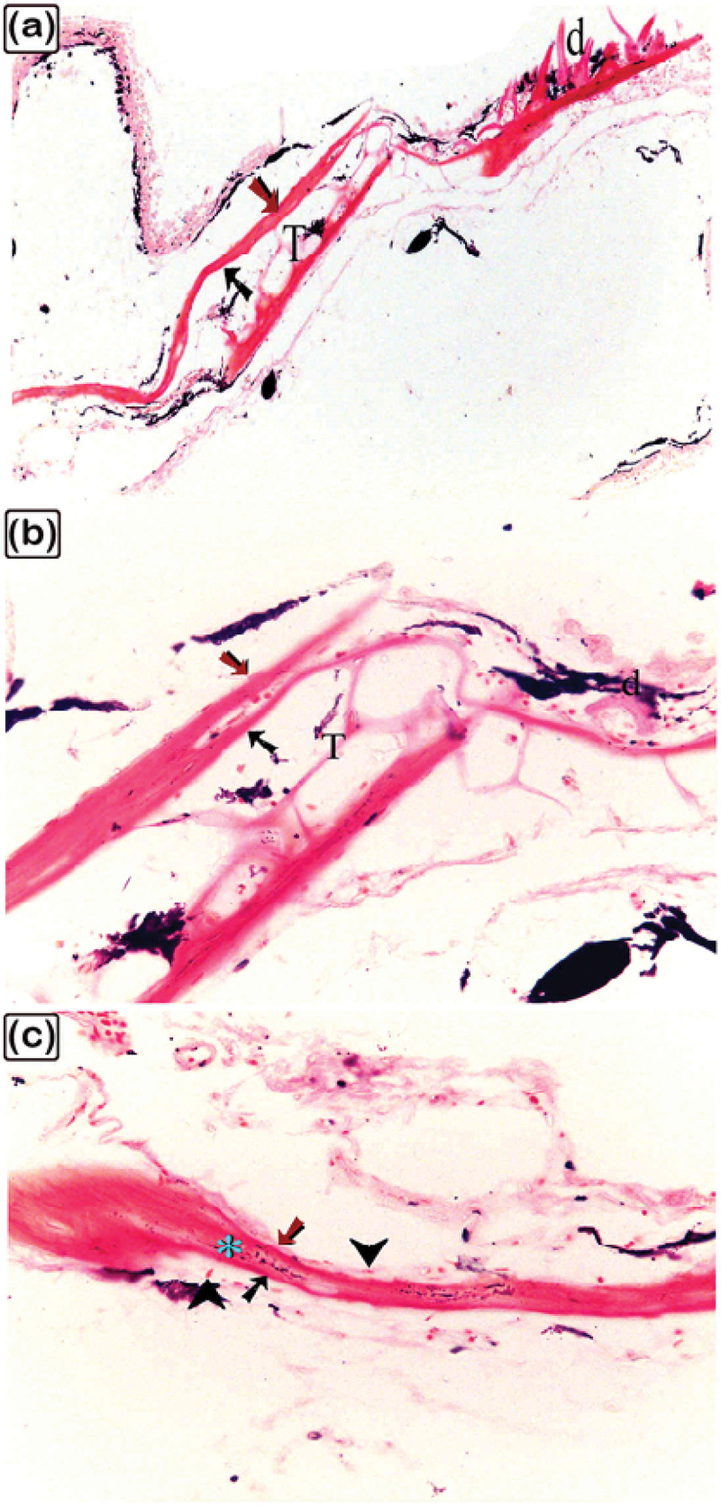
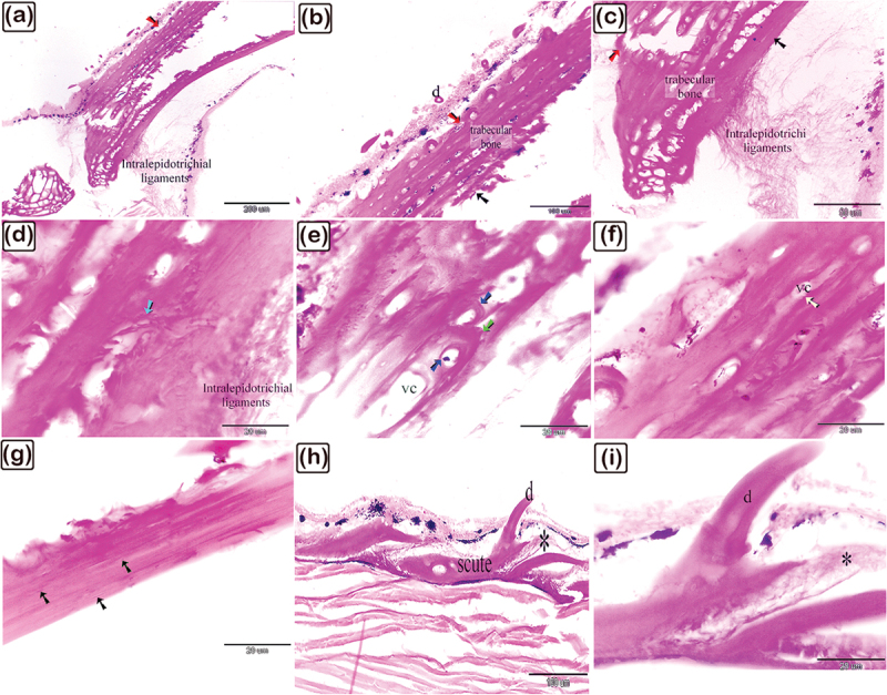
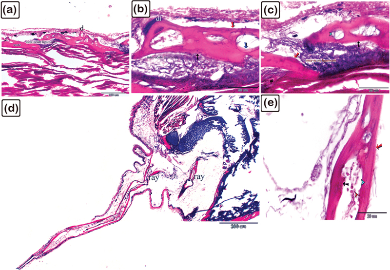
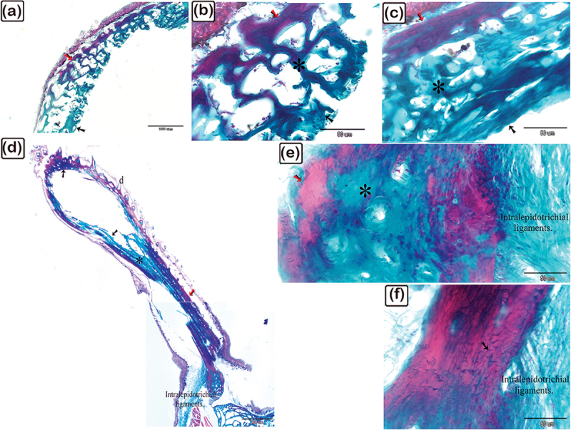
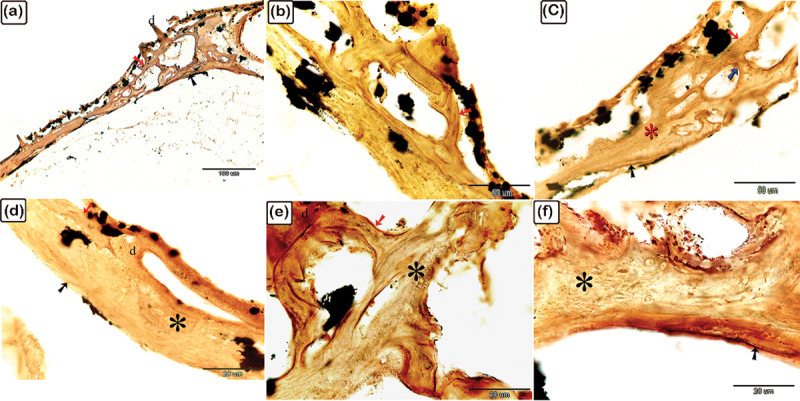
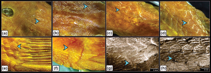
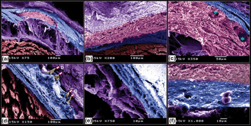
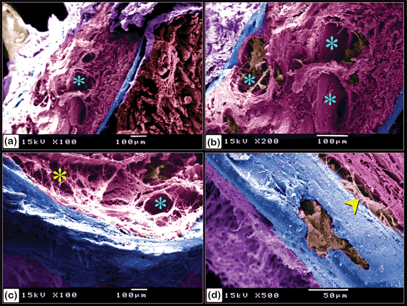
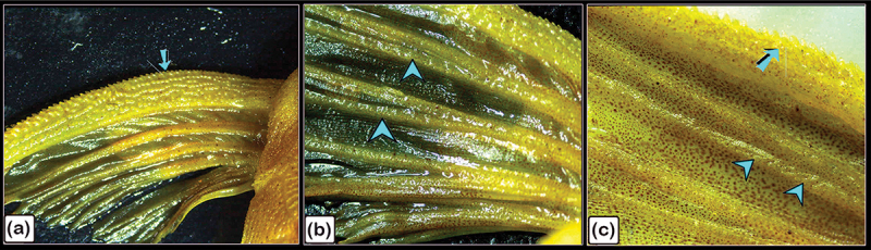
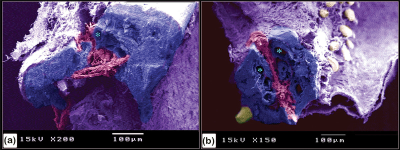
Studying scute and fin morphology are advantageous approaches for phylogenetic identification and provide information on biological linkages and evolutionary history that are essential for deciphering the fossil record. Despite this, no prior research has precisely characterized the histological structures of scutes in the common pleco. Therefore, this research investigated the microstructure and organization of bone tissue within the dermal skeleton, including the scutes and fins, in the common pleco, using light microscopy, stereomicroscopy, and scanning electron microscopy. The dermal scutes were organized in a pentagonal shape with denticular coverage and were obliquely aligned with the caudal portion pointing dorsally. The dermal scutes consisted of three distinct portions: the central, preterminal, and terminal portions. Each portion comprised three layers: a superficial bony plate, a basal bony plate, and a mid-plate. Both the superficial and basal bony plates were composed of lamellar bone and lamellar zonal bone, whilst the mid-plate consisted of secondary osteons and woven bone. In the terminal portion, the superficial and basal bony plates became thinner. The pectoral fin consists of spines and rays composed of lepidotrichium (two symmetrical hemi-rays). The spine contained centrifugal and centripetal lamellar and trabecular bones. A centripetal fibrous bone was implanted between the lamellar bones. Besides being oriented in a V shape, the hemi-rays were also composed of thin centrifugal and centripetal lamellar bones and trabecular bones. A fibrous bone was identified between the centrifugal and centripetal bones. The trabecular bone and lamellar bone were made up of bone spicules.
Keywords: Hypostomus plecostomus; bony fins; bony scutes; light microscope; scanning electron microscope; stereoscope.
© 2024 The Author(s). Published by Informa UK Limited, trading as Taylor & Francis Group.
Conflict of interest statement
No potential conflict of interest was reported by the author(s).
Figures
References
-
- Groombridge B. Global biodiversity: status of the earth’s living resources: a report. 1992.
-
- Phillips JB. Development of vertebrate anatomy [by] Joy B. Phillips. Missouri, USA: Mosby; 1975.
-
- Kent GC. Comparative anatomy of the vertebrates. Missouri, USA: Times Mirror/Mosby College Publishing; 1987.
-
- Elliott DG. Gross functional anatomy: integumentary System. In: Ostrander GK, editor. Handbook of experimental animals. The laboratory fish. London: Academic Press; 2000. p. 95–108.
LinkOut - more resources
Full Text Sources
Other Literature Sources