. 2024 Aug 22:14:1405689.
doi: 10.3389/fcimb.2024.1405689. eCollection 2024.

Analysis of miRNAs involved in mouse brain injury upon Coxsackievirus A6 infection

Affiliations

Analysis of miRNAs involved in mouse brain injury upon Coxsackievirus A6 infection

Yihao Sun et al. Front Cell Infect Microbiol. .

Abstract

Introduction: Coxsackievirus A6 (CV-A6) has emerged as the predominant epidemic strain responsible for hand, foot and mouth disease (HFMD). CV-A6 infection can result in severe clinical manifestations, including encephalitis, meningitis, and potentially life-threatening central nervous system disorders. Our previous research findings demonstrated that neonatal mice infected with CV-A6 exhibited limb weakness, paralysis, and ultimately succumbed to death. However, the underlying mechanism of CV-A6-induced nervous system injury remains elusive. Numerous reports have highlighted the pivotal role of miRNAs in various viral infections.

Methods: Separately established infection and control groups of mice were used to create miRNA profiles of the brain tissues before and after CV-A6 transfection, followed by experimental verification, prediction, and analysis of the results.

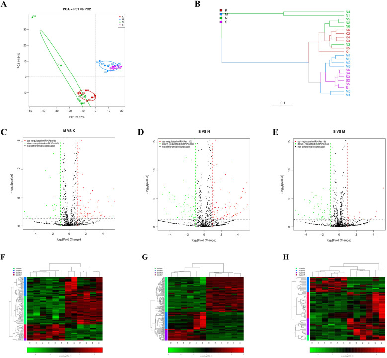
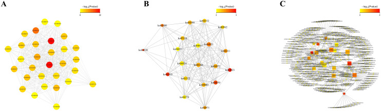
Results: At 2 days post-infection (dpi), 4 dpi, and 2dpi vs 4dpi, we identified 175, 198 and 78 significantly differentially expressed miRNAs respectively using qRT-PCR for validation purposes. Subsequently, we predicted target genes of these differentially expressed miRNAs and determined their potential targets through GO (Gene Ontology) enrichment analysis and KEGG (Kyoto Encyclopedia of Genes and Genomes) enrichment analysis. Finally, we verified the miRNA-mRNA pairing via double luciferase experiments while confirming functional enrichment of target genes through Western Blotting analyses.

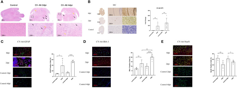
Discussion: The results from this study suggest that transcriptional regulation, neuronal necrosis, pro-inflammatory cytokine release, and antiviral immunity are all implicated in the pathogenesis of central nervous system injury in mice infected with CV-A6. Brain injury resulting from CV-A6 infection may involve multiple pathways, including glial cell activation, neuronal necrosis, synaptic destruction, degenerative diseases of the nervous system. It can even encompass destruction of the blood-brain barrier, leading to central nervous system injury. The dysregulated miRNAs and signaling pathways discovered in this study provide valuable insights for further investigations into the pathogenesis of CV-A6.

Keywords: Coxsackievirus A6 (CV-A6); brain; central nervous system; hand foot and mouth disease (HFMD); miRNA.


Conflict of interest statement

Authors YS, JW, SQ, and SS were employed by the company Wuhan Institute of Biological Products Co. Ltd. The remaining author declares that the research was conducted in the absence of any commercial or financial relationships that could be construed as a potential conflict of interest.

Figures

Figure 1
Figure 1
Brain tissues of mice were damaged after CV-A6 infection. (A) Histopathological changes in the brain tissue sections of mice were evaluated by H&E staining. The black arrow signifies the shrinkage of neurons, while the yellow arrow marks the site of bleeding. (B) Viral proteins in the brain tissue sections of mice were evaluated by immunohistochemical staining. The viral titer was expressed as the fold change of VP1 mRNA in the brain tissues of mice detected by qRT-PCR (n=6). The results were calculated as 2^-△Ct. (C) Colocalization of CV-A6 with GFAP in the mouse brain tissue, and the GFAP positive cell rate was determined. (D) Colocalization of CV-A6 with IBA-1 in the mouse brain tissue, and the IBA-1 positive cell rate was determined. (E) Colocalization of CV-A6 with NeuN in the mouse brain tissue, and the NeuN positive cell rate was determined. Original magnification, × 20, Scale bar = 500 μm; × 200, Scale bar = 50 μm; × 400, Scale bar = 20 μm. Red light represents CV-A6 staining, green light represents GFAP, IBA-1, and NeuN staining in different figures, and blue light represents DAPI staining. *p < 0.05; **p < 0.01; ***p < 0.001; **** p<0.00001.
Figure 2
Figure 2
The miRNA expression pattern in brain tissue of mice was analyzed after CV-A6 infection. (A) Principal component analysis (PCA) plot was used to visualize the sample groups under different environments or conditions, with dots of different colors or shapes representing each group. The scales of the horizontal and vertical coordinates are relative distances without practical significance. (B) Hierarchical clustering tree analysis was performed to assess the similarity between samples, with branch lengths indicating the distance between samples. Samples were distinguished by different colors based on grouping. (C–E) An expression difference volcano plot was generated to illustrate the fold-change (log (B/A)) value of gene expression differences between sample groups on the horizontal axis, and the statistical significance of gene expression changes (pValue) on the vertical axis. Smaller pValues corresponded to larger -log(pValue), indicating greater significance in differential gene expression. Each dot represented a gene, where red indicated up-regulated genes, green indicated down-regulated genes, and black indicated non-differential genes. (F–H) Heatmap of differentially expressed miRNAs. Each row represents a gene and each column represents a sample. The higher the color is, the higher the expression level is. S: mice in the 4dpi group, M: mice in the 2dpi group, N: control-4dpi group, K: control-2dpi group.
Figure 3
Figure 3
GO and KEGG enrichment analysis of miRNAs exhibiting significantly aberrant expression in mouse brain tissues following CV-A6 infection were performed. (A) GO enrichment analysis was conducted on miRNAs with significantly abnormal expression in the brain tissues of mice at 2dpi vs control-2dpi. (B) GO enrichment analysis was carried out on miRNAs with significantly abnormal expression in the brain tissues of mice at 4dpi vs control-2dpi. (C) GO enrichment analysis was carried out on miRNAs with significantly abnormal expression in the brain tissues of mice at 4dpi vs 2dpi. (D) KEGG enrichment analysis was performed on miRNAs with significantly abnormal expression in the brain tissues of mice at 2dpi vs control-2dpi. (E) KEGG enrichment analysis was executed on miRNAs with significantly abnormal expression in the brain tissues of mice at 4dpi vs control-4dpi. (F) KEGG enrichment analysis was executed on miRNAs with significantly abnormal expression in the brain tissues of mice at 4dpi vs 2dpi.
Figure 4
Figure 4
Functional enrichment correlation analysis was performed on the differentially expressed miRNA target genes in mouse brain tissues following CV-A6 infection. (A) The significantly enriched function-function interaction network diagram was presented, where circular nodes represented functional information and edges represented the correlation between functions. The color of nodes indicated the degree of function enrichment, namely P-value value. Nodes with higher degrees of enrichment had lower P-values and appeared red. (Only the top 50 functions with the highest degree of enrichment were included.) (B) Another significantly enriched function-function interaction network diagram was shown, using circular nodes to represent functional information and edges to represent correlations between functions. The color of nodes reflected the degree of function enrichment, namely P-value value. Nodes with higher degrees of enrichment had lower P-values and appeared red. (C) Additionally, a significantly enriched function-gene interaction network diagram was displayed, where square nodes represented functional information, circular nodes represented genes, and edges depicted correlations between genes and functions. The size of each node corresponded to its connectivity or degree; larger nodes indicated more connections.
Figure 5
Figure 5
Verification of RNA-seq using qPCR. (A) The 5 miRNAs changed at 2dpi vs control-2dpi. (B) The 9 miRNAs changed at 4dpi vs control-4dpi. (C) The 5 miRNAs changed at 4dpi vs 2dpi.The detection for each miRNA was repeated at least 3 times and the standard deviation was.
Figure 6
Figure 6
Validation of miRNA and target gene. (A) Schematic representation of the binding sites between mmu-miR-3473b and Dvl2. (B) Schematic illustration of the binding sites between mmu- novel-9 and Axin2. (C) Observed changes in reporter gene expression comparing Dvl2-WT and Dvl2-MUT groups. (D) Observed changes in reporter gene expression comparing Axin2-WT and Axin2-MUT groups. Luciferase reporter gene assay was conducted using 293T cells with firefly luciferase serving as an endogenous control. Data are presented as mean ± SD from three independent experiments (**p < 0.01).
Figure 7
Figure 7
Validation of functional enrichment. Brain tissues were obtained from 4dpi (n = 6) and control-4dpi group (n = 6) and subjected to Western blotting (n = 6). (A) Western blot analysis of Axin 2, GSK3-β, β-Catenin, LEF1, Cyclin D1 - C-terminal, PLCG1, IL-1, IL-6, and TNF-α. (B) Expression levels of Axin 2, GSK3-β, β-Catenin, LEF1, Cyclin D1 - C-terminal, PLCG1, IL-1, IL-6, and TNF-α. *p < 0.05, **p < 0.01, ***p < 0.001.

Similar articles

Cited by

References

    1. Alyu F., Miriş D. (2017). Inflammatory aspects of epileptogenesis: contribution of molecular inflammatory mechanisms. Acta neuropsychiatrica. 29, 1–16. doi: 10.1017/neu.2016.47 - DOI - PubMed
    1. Bernier A., Sagan S. M. (2018). The diverse roles of microRNAs at the host (-) virus interface. Viruses 10, 440. doi: 10.3390/v10080440 - DOI - PMC - PubMed
    1. Berry M., Ahmed Z., Morgan-Warren P., Fulton D., Logan A. (2016). Prospects for mTOR-mediated functional repair after central nervous system trauma. Neurobiol. diseas. 85, 99–110. doi: 10.1016/j.nbd.2015.10.002 - DOI - PubMed
    1. Bian L., Wang Y., Yao X., Mao Q., Xu M., Liang Z. (2015). Coxsackievirus A6: a new emerging pathogen causing hand, foot and mouth disease outbreaks worldwide. Expert Rev. anti-infective Ther. 13, 1061–1071. doi: 10.1586/14787210.2015.1058156 - DOI - PubMed
    1. Cai Y., Yu X., Hu S., Yu J. (2009). A brief review on the mechanisms of miRNA regulation. Genomics Proteomics Bioinf. 7, 147–154. doi: 10.1016/S1672-0229(08)60044-3 - DOI - PMC - PubMed

LinkOut - more resources