. 2024 Sep 6:12:RP92135.
doi: 10.7554/eLife.92135.

Early moderate prenatal alcohol exposure and maternal diet impact offspring DNA methylation across species

Affiliations

Early moderate prenatal alcohol exposure and maternal diet impact offspring DNA methylation across species

Mitchell Bestry et al. Elife. .

Abstract

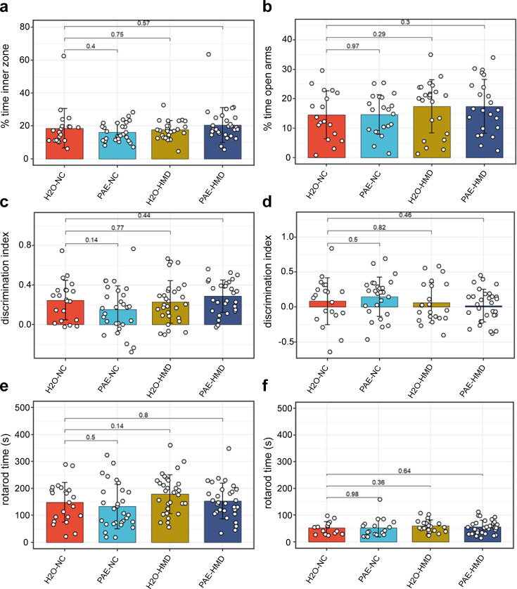
Alcohol consumption in pregnancy can affect genome regulation in the developing offspring but results have been contradictory. We employed a physiologically relevant murine model of short-term moderate prenatal alcohol exposure (PAE) resembling common patterns of alcohol consumption in pregnancy in humans. Early moderate PAE was sufficient to affect site-specific DNA methylation in newborn pups without altering behavioural outcomes in adult littermates. Whole-genome bisulfite sequencing of neonatal brain and liver revealed stochastic influence on DNA methylation that was mostly tissue-specific, with some perturbations likely originating as early as gastrulation. DNA methylation differences were enriched in non-coding genomic regions with regulatory potential indicative of broad effects of alcohol on genome regulation. Replication studies in human cohorts with fetal alcohol spectrum disorder suggested some effects were metastable at genes linked to disease-relevant traits including facial morphology, intelligence, educational attainment, autism, and schizophrenia. In our murine model, a maternal diet high in folate and choline protected against some of the damaging effects of early moderate PAE on DNA methylation. Our studies demonstrate that early moderate exposure is sufficient to affect fetal genome regulation even in the absence of overt phenotypic changes and highlight a role for preventative maternal dietary interventions.

Keywords: DNA methylation; alcohol; developmental biology; epigeneome; genetics; genomics; human; mouse.

Plain language summary

Drinking excessive amounts of alcohol during pregnancy can cause foetal alcohol spectrum disorder and other conditions in children that affect their physical and mental development. Many countries advise women who are pregnant or trying to conceive to avoid drinking alcohol entirely. However, surveys of large groups of women in Western countries indicate that most women continue drinking low to moderate amounts of alcohol until they discover they are pregnant and then stop consuming alcohol for the rest of their pregnancy. It remains unclear how this common drinking pattern affects the foetus. The instructions needed to build and maintain a human body are stored within molecules of DNA. Some regions of DNA called genes contain the instructions to make proteins, which perform many tasks in the body. Other so-called ‘non-coding’ regions do not code for any proteins but instead have roles in regulating gene activity. One way cells control which genes are switched on or off is adding or removing tags known as methyl groups to certain locations on DNA. Previous studies indicate that alcohol may affect how children develop by changing the patterns of methyl tags on DNA. To investigate the effect of moderate drinking during the early stages of pregnancy, Bestry et al. exposed pregnant mice to alcohol and examined how this affected the patterns of methyl tags on DNA in their offspring. The experiments found moderate levels of alcohol were sufficient to alter the patterns of methyl tags in the brains and livers of the newborn mice. Most of the changes were observed in non-coding regions of DNA, suggesting alcohol may affect how large groups of genes are regulated. Fewer changes in the patterns of methyl tags were found in mice whose mothers had diets rich in two essential nutrients known as folate and choline. Further experiments found that some of the affected mouse genes were similar to genes linked to foetal alcohol spectrum disorder and other related conditions in humans. These findings highlight the potential risks of consuming even moderate levels of alcohol during pregnancy and suggest that a maternal diet rich in folate and choline may help mitigate some of the harmful effects on the developing foetus.


Conflict of interest statement

MB, AL, NK, EC, CB, JF, EE, JC, EM, JH, DH, SB, RL, MS, DM No competing interests declared

Figures

Figure 1.
Figure 1.. Overview of prenatal alcohol exposure (PAE) model.
A schematic representation of the experiment design is shown in the figure. Fifteen dams were allocated to each treatment group. PAE mice were exposed to ethanol (10% vol/vol in non-acidified reverse osmosis drinking water ad libitum) from 1 week before pregnancy to gestational days 8–10 and the remaining mice received water (H2O). The PAE and H2O groups received either normal chow (NC) or a high methyl donor (HMD) diet (NC containing 20 mg/kg folate and 4970 mg/kg choline) from 1 week before pregnancy until birth.
Figure 2.
Figure 2.. PAE and HMD effects on dam characteristics.
(a) Dam weight progression was significantly affected by HMD but not PAE by quadratic mixed effects model without interaction. (b) Trajectory of liquid consumption across pregnancy was affected by PAE and HMD by quadratic mixed effects model. PAE and HMD significantly interacted with trimester of pregnancy. (c) Litter size (N=40) and (d) pup sex ratios (N=36) were not significantly associated with PAE or HMD by unpaired t-test or ANOVA. All line and bar plots show mean and standard deviation. NC = normal chow, HMD = high methyl diet, PAE = prenatal alcohol exposure. Comparisons show p-value by unpaired t-test compared to the H2O-NC baseline treatment group.
Figure 3.
Figure 3.. Prenatal alcohol exposure (PAE) and high methyl donor (HMD) effects on dam characteristics.
There was no significant difference in the average gain of weight in dams between (a) days 1–17 and (b) days 1–19 by treatment group. Both timepoints were included due to some pregnancies ending by day 19. (c) Dams given supplemented chow consumed significantly lower total quantity of liquid across pregnancy. Bar plots show mean and standard deviation for each treatment group. Each point represents one dam. (d) The trajectory of chow consumed by dams across pregnancy significantly varied with the addition of treatments. Points show mean and standard deviation for each treatment group. Statistical analysis involved linear mixed effects regression comparing trajectories of treatment groups to H2O-NC baseline control group. N=36.
Figure 4.
Figure 4.. No evidence for global disruption of methylation by prenatal alcohol exposure (PAE).
The figure shows methylation levels averaged across cytosine-guanines (CpGs) in different regulatory genomic contexts. Neither brain tissue (a and b) nor liver tissue (c and d) were grossly affected by PAE exposure (blue bars). Bars represent means and standard deviation.
Figure 5.
Figure 5.. Prenatal alcohol exposure (PAE) was associated with site-specific differences in offspring DNA methylation.
The majority of differentially methylated regions (DMRs) lost methylation with PAE in (a) brain and (b) liver of mice given normal chow (NC). Each point represents one DMR. Point colour indicates change in DNA methylation with PAE. PAE was also associated with lower methylation in the DMRs identified in the promoter of the Impact gene in (c) brain and (d) liver, within NC mice. Each plot represents a separate treatment group. Each blue vertical line indicates a cytosine-guanine (CpG) site, with the height and corresponding left y-axis indicating the methylation ratio. The grey line and corresponding right y-axis indicate coverage at each CpG site. The black horizontal dotted line indicates 40% methylation for comparison purposes. The x-axis indicates the base position on chromosome 18, with the pink shaded area highlighting the DMR. DMR plots include 200 base pair flanking regions on each side of the DMR. DMRs identified in (e) brain and (f) liver were enriched in intergenic and inter-CpG regions, whilst being underrepresented in CpG and gene regions. The bar plot compares the number of whole-genome bisulfite sequencing (WGBS) DMRs in red to a set of equivalent randomly generated regions in blue. (g) Gene ontology analysis of liver DMRs shows enrichment within neuronal cellular components and biological processes. BP/red point = biological process, CC/blue point = cellular component. X-axis of point indicates FDR of ontology. Size of point indicates number of overlapping genes with ontology. There were insufficient number of DMRs identified in the brain for a gene ontology analysis.
Figure 6.
Figure 6.. High methyl donor (HMD) partially mitigated effects of prenatal alcohol exposure (PAE) on offspring DNA methylation.
Average DNA methylation effect sizes above 30% with PAE were observed in some (a) brain and (b) liver differentially methylated regions (DMRs) in normal chow (NC) mice. Mean absolute difference in methylation with PAE is reduced within the HMD mice in (c) brain and (d) liver. Each point represents one DMR. Point colour indicates change in DNA methylation with PAE. Points with a high number of cytosine-guanines (CpGs) and methylation difference are annotated with associated gene if located within a genic region. HMD was associated with (e) higher methylation in the DMR identified proximal to Lamb1 on chromosome 12 in brain and (f) lower methylation in the DMR identified proximal to Socs5 on chromosome 17 in liver. Each plot represents a separate treatment group. Each blue vertical line indicates a CpG site, with the height and corresponding left y-axis indicating the methylation ratio. The grey line and corresponding right y-axis indicate coverage at each CpG site. The black horizontal line indicates (e) 40% and (f) 80% methylation for comparison purposes. The x-axis indicates the base position on the chromosome, with the pink shaded area highlighting the DMR. DMR plots include 200 base pair flanking regions on each side of the DMR.
Figure 7.
Figure 7.. PAE had no significant effect on other assessed behavioural outcomes.
PAE and HMD had no significant effect on anxiety as evident by no significant difference by unpaired t-test in the (a) percent time in the inner zone in the open field test (N=104) and (b) percent time open arms in the elevated plus maze test (N=85). PAE and HMD had no significant effect on spatial recognition as evident by no significant difference by unpaired t-test in the discrimination index in (c) object recognition (N=108) and (d) object in place test (N=98). PAE and HMD had no significant effect on motor coordination and balance as evident by no significant difference by unpaired t-test in times in (e) first rotarod test (N=112) and (f) second rotarod test (N=87). Bars show mean and standard deviation. Each point represents one mouse. NC = normal chow, HMD = high methyl diet, PAE = prenatal alcohol exposure. Time interval for each mouse was (a–c) 300 s and (d) 180 s.
Figure 8.
Figure 8.. HMD was associated with increased locomotor activity.
HMD was associated with increased locomotor activity compared to NC, indicated by significantly greater total distance travelled in the (a) open field test (N=104), (b) object recognition test (N=108), (c) elevated plus maze test (N=88), and (d) object in place test (N=98) by unpaired t-test. Bars show mean and standard deviation. Each point represents one mouse. NC = normal chow, HMD = high methyl diet, PAE = prenatal alcohol exposure. Time interval for each mouse was (a–c) 300 s and (d) 180 s.
Figure 9.
Figure 9.. Seven prenatal alcohol exposure (PAE) differentially methylated regions (DMRs) identified in the murine model were successfully replicated in the Lussier et al., 2018 human fetal alcohol spectrum disorder (FASD) cohort.
Examples of two PAE DMRs that had significantly lower DNA methylation with a clinical diagnosis of FASD in the Lussier et al., 2018 cohort (a and c), while their mouse liftover DMR also had significantly lower DNA methylation with PAE in the murine model experiment (b and d).

Update of

  • doi: 10.1101/2023.06.15.544516
  • doi: 10.7554/eLife.92135.1
  • doi: 10.7554/eLife.92135.2

References

    1. Abbott CW, Rohac DJ, Bottom RT, Patadia S, Huffman KJ. Prenatal ethanol exposure and neocortical development: A transgenerational model of FASD. Cerebral Cortex. 2018;28:2908–2921. doi: 10.1093/cercor/bhx168. - DOI - PMC - PubMed
    1. Aryee MJ, Jaffe AE, Corrada-Bravo H, Ladd-Acosta C, Feinberg AP, Hansen KD, Irizarry RA. Minfi: a flexible and comprehensive Bioconductor package for the analysis of Infinium DNA methylation microarrays. Bioinformatics. 2014;30:1363–1369. doi: 10.1093/bioinformatics/btu049. - DOI - PMC - PubMed
    1. Autism Spectrum Disorders Working Group of The Psychiatric Genomics Consortium Meta-analysis of GWAS of over 16,000 individuals with autism spectrum disorder highlights a novel locus at 10q24.32 and a significant overlap with schizophrenia. Molecular Autism. 2017;8:21. doi: 10.1186/s13229-017-0137-9. - DOI - PMC - PubMed
    1. Bestry M, Symons M, Larcombe A, Muggli E, Craig JM, Hutchinson D, Halliday J, Martino D. Association of prenatal alcohol exposure with offspring DNA methylation in mammals: a systematic review of the evidence. Clinical Epigenetics. 2022;14:12. doi: 10.1186/s13148-022-01231-9. - DOI - PMC - PubMed
    1. Chen Y-L, Yang S-S, Peng H-C, Hsieh Y-C, Chen J-R, Yang S-C. Folate and vitamin B12 improved alcohol-induced hyperhomocysteinemia inrats. Nutrition. 2011;27:1034–1039. doi: 10.1016/j.nut.2010.10.019. - DOI - PubMed

Associated data