. 2024 Oct 16;15(10):e0131724.
doi: 10.1128/mbio.01317-24. Epub 2024 Sep 6.

FabR, a regulator of membrane lipid homeostasis, is involved in Klebsiella pneumoniae biofilm robustness

Affiliations

FabR, a regulator of membrane lipid homeostasis, is involved in Klebsiella pneumoniae biofilm robustness

Ibrahima Dramé et al. mBio. .

Abstract

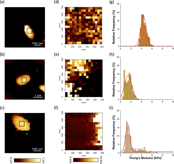
Biofilm is a dynamic structure from which individual bacteria and micro-aggregates are released to subsequently colonize new niches by either detachment or dispersal. Screening of a transposon mutant library identified genes associated with the alteration of Klebsiella pneumoniae biofilm including fabR, which encodes a transcriptional regulator involved in membrane lipid homeostasis. An isogenic ∆fabR mutant formed more biofilm than the wild-type (WT) strain and its trans-complemented strain. The thick and round aggregates observed with ∆fabR were resistant to extensive washes, unlike those of the WT strain. Confocal microscopy and BioFlux microfluidic observations showed that fabR deletion was associated with biofilm robustness and impaired erosion over time. The genes fabB and yqfA associated with fatty acid metabolism were significantly overexpressed in the ∆fabR strain, in both planktonic and biofilm conditions. Two monounsaturated fatty acids, palmitoleic acid (C16:1) and oleic acid (C18:1), were found in higher proportion in biofilm cells than in planktonic forms, whereas heptadecenoic acid (C17:1) and octadecanoic acid, 11-methoxy (C18:0-OCH3) were found in higher proportion in the planktonic lifestyle. The fabR mutation induced variations in the fatty acid composition, with no clear differences in the amounts of saturated fatty acids (SFA) and unsaturated fatty acids for the planktonic lifestyle but lower SFA in the biofilm form. Atomic force microscopy showed that deletion of fabR is associated with decreased K. pneumoniae cell rigidity in the biofilm lifestyle, as well as a softer, more elastic biofilm with increased cell cohesion compared to the wild-type strain.IMPORTANCEKlebsiella pneumoniae is an opportunistic pathogen responsible for a wide range of nosocomial infections. The success of this pathogen is due to its high resistance to antibiotics and its ability to form biofilms. The molecular mechanisms involved in biofilm formation have been largely described but the dispersal process that releases individual and aggregate cells from mature biofilm is less well documented while it is associated with the colonization of new environments and thus new threats. Using a multidisciplinary approach, we show that modifications of bacterial membrane fatty acid composition lead to variations in the biofilm robustness, and subsequent bacterial detachment and biofilm erosion over time. These results enhance our understanding of the genetic requirements for biofilm formation in K. pneumoniae that affect the time course of biofilm development and the embrittlement step preceding its dispersal that will make it possible to control K. pneumoniae infections.

Keywords: Klebsiella pneumoniae; atomic force microscopy; biofilm robustness; membrane fatty acid.


Conflict of interest statement

The authors declare no conflict of interest.

Figures

Fig 1
Fig 1
The transcription repressor FabR is related to biofilm formation in K. pneumoniae. Biofilms were formed in M63B1-0.4% Glc in glass tubes for 18 hours (a), in a 96-well microtiter plate for 5 hours (b) and 24-well microtiter plate for 5 hours (c), and biomass was then quantified by crystal violet staining or CFU determination as described in Materials and Methods. Biofilms formed by the GFP-expressing WT strains (d), ∆fabR (e), and ∆fabR(pSTAB-fabR) (f) in 96-well microtiter plates with coverslip bottom were observed using confocal microscopy after 5 hours of incubation at ×10 magnification. Biovolume (µm3) (g) and thickness (h) of the biofilm formed by each strain were determined using the IMARIS 9.8.2 software package. Data are expressed as means  ±  SEM (N  =  3). Statistical analysis: one-way ANOVA with post hoc Dunn’s test (****P ≤ 0.0001; ***P ≤ 0.001; **P ≤ 0.01; *P ≤ 0.05).
Fig 2
Fig 2
The absence of FabR alters the structure of mature biofilm. The architecture of 24-hour-old biofilms formed by GFP-expressing WT (a), ∆fabR (b), and ∆fabR(pSTAB-fabR) (c) strains in 96-well microplates with glass bottom was observed by confocal microscopy. Upper images represent the 3D reconstructions of biofilms, and lower images represent one optical section of the z-stack, with the position indicated by a red line in the upper images. Scale bars correspond to 100 µm. Each image is representative of three independent biological replicates.
Fig 3
Fig 3
The absence of FabR leads to a stable biofilm without aggregate detachment and shear-erosion over time. The kinetics of biofilm formation by the GFP-expressing WT (a), ∆fabR (b), and ∆fabR(pSTAB-fabR) (c) strains was followed in the BioFlux microfluidic system at 37°C under a shear force of 0.5 dyn/cm² using epifluorescence microscope (Axio observer 7, Zeiss) at a magnification of 20×. Images were acquired in real time at T = 0 hour and then every hour, and each image represents one time point. The yellow arrows indicate the comet-like structures formed in the WT strain. The white arrows indicate the flow direction. Each image sequence is representative of three independent biological replicates.
Fig 4
Fig 4
The absence of FabR leads to the formation of a robust biofilm more resistant to shear forces than the WT strain. The behavior of K. pneumoniae biofilm under increasing shear forces was analyzed in the BioFlux microfluidic system using an epifluorescence microscope (Axio observer 7, Zeiss) at a magnification of 20×. Twenty-four-hour-old biofilms formed by the WT (a), ∆fabR (b), and ∆fabR(pSTAB-fabR) (c) strains under shear forces of 0.5 dyn/cm² were subjected to a gradually increasing shear force from 0.5 to 10 dyn/cm2. Images were acquired after each increase in shear force. Each image sequence is representative of three independent biological replicates.
Fig 5
Fig 5
FabR regulates the expression of fabB and yqfA genes in planktonic and biofilm cells. Differential expressions of the genes fabA, fabB, yqfA, and desA involved in fatty acid metabolism in K. pneumoniae WT, ∆fabR, and ∆fabR(pSTAB-fabR) strains were determined by RT-qPCR. RNA isolated from (a) planktonic culture and (b) 24-hour-old biofilm formed by the indicated strains were used, and the fold change in transcript abundance was determined relative to the transcript abundance values of the WT strain. (c) Analysis of fabR gene expression by RT-qPCR in planktonic bacteria compared to that in bacteria grown in biofilm. The fabR expression in biofilm is used as the reference condition. The expression data were normalized using both rpoD and proC reference genes. Data are expressed as means  ±  SEM (N  =  3). Statistical analysis: two-way ANOVA with post hoc Tukey test (***P ≤ 0.001; **P ≤ 0.01; *P ≤ 0.05).
Fig 6
Fig 6
Identification and quantification of the total fatty acid composition in planktonic and biofilm cultures were conducted using GC-MS and GC-flame ionization detector. Fatty acid results are presented in mol% for both biofilm and planktonic cultures in the WT strain (a and d). The fatty acid content was also analyzed in WT, ∆fabR, and ∆fabR(pSTAB-fabR) planktonic cultures (b and e) and biofilm cultures (c and f). The amount of SFA and UFA in mol% was determined for each corresponding condition (d, e, and f). The results correspond to at least three or four biologically independent samples. Statistical significances were determined by post hoc Tukey test after a two-way analysis of variance (****P ≤ 0.0001; ***P ≤ 0.001; **P ≤ 0.01; *P ≤ 0.05).
Fig 7
Fig 7
Imaging and nanomechanical properties of K. pneumoniae single cell. Height image of WT (a), ∆fabR (b), and ∆fabR(pSTAB-fabR) (c) individual cell interactions between the AFM colloidal probe (silica microsphere) and single planktonic cells were probed on areas of 500 × 500 nm on top of each strain. Elasticity maps (d–f) and statistical distribution of the Young’s modulus (g–i) were determined for each strain (respectively WT (d, g), ∆fabR (e, h), and ∆fabR(pSTAB-fabR) (f, i). Force curves were recorded on four cells from three independent cultures (a total of 256 force curves per cell corresponding to 16 × 16 pixels).
Fig 8
Fig 8
Imaging and nanomechanical properties of K. pneumoniae biofilms. Height images of 5-hour-old biofilms of WT (a), ∆fabR (b), and ∆fabR(pSTAB-fabR) (c) were recorded in QI mode. Measurement of the interaction between AFM colloidal probe and area of 50 µm × 50 µm on formed biofilms. (d–f) Elasticity maps and representative force-indentation curves were obtained on each biofilm (black line), fitted with the Hertz model (red line). (g–i) Statistical distribution of Young’s modulus determined for each strain. Force curves were recorded on three areas of biofilm and from three independent cultures (a total of 256 force curves per measure corresponding to 16 × 16 pixels).
Fig 9
Fig 9
Homotypic interactions between individual cells and 5-hour-old biofilms of K. pneumoniae. Adhesion force maps (a–c), adhesion force histograms, and typical force-distance curves (d–f) were obtained by recording force curves on 50 µm × 50 µm biofilm surface in WT (d), ∆fabR (e), and ∆fabR(pSTAB-fabR) (f) strains. All force curves were recorded on biofilm from three independent cultures and with 16 × 16 pixels per condition corresponding to 256 force curves.

References

    1. Stoodley P, Sauer K, Davies DG, Costerton JW. 2002. Biofilms as complex differentiated communities. Annu Rev Microbiol 56:187–209. doi:10.1146/annurev.micro.56.012302.160705 - DOI - PubMed
    1. Flemming H-C, Wingender J. 2010. The biofilm matrix. Nat Rev Microbiol 8:623–633. doi:10.1038/nrmicro2415 - DOI - PubMed
    1. Hou J, Veeregowda DH, van de Belt-Gritter B, Busscher HJ, van der Mei HC. 2018. Extracellular polymeric matrix production and relaxation under fluid shear and mechanical pressure in Staphylococcus aureus biofilms. Appl Environ Microbiol 84:e01516-17. doi:10.1128/AEM.01516-17 - DOI - PMC - PubMed
    1. Guilhen C, Forestier C, Balestrino D. 2017. Biofilm dispersal: multiple elaborate strategies for dissemination of bacteria with unique properties. Mol Microbiol 105:188–210. doi:10.1111/mmi.13698 - DOI - PubMed
    1. Tierra G, Pavissich JP, Nerenberg R, Xu Z, Alber MS. 2015. Multicomponent model of deformation and detachment of a biofilm under fluid flow. J R Soc Interface 12:20150045. doi:10.1098/rsif.2015.0045 - DOI - PMC - PubMed

MeSH terms

LinkOut - more resources