. 2024 Oct 22;98(10):e0091124.
doi: 10.1128/jvi.00911-24. Epub 2024 Sep 6.

A cardioviral 2C-ATP complex structure reveals the essential role of a conserved arginine in regulation of cardioviral 2C activity

Affiliations

A cardioviral 2C-ATP complex structure reveals the essential role of a conserved arginine in regulation of cardioviral 2C activity

Qing-Yi He et al. J Virol. .

Abstract

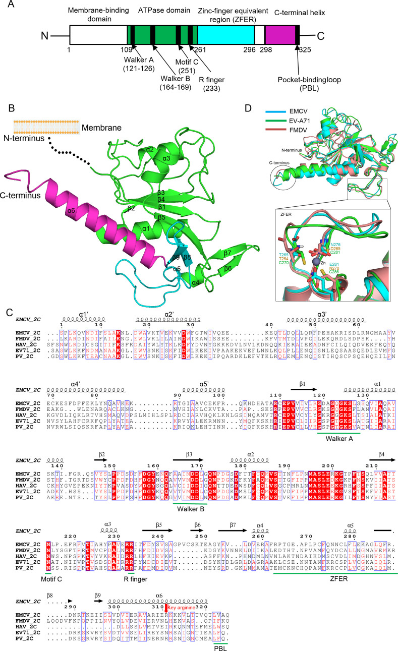
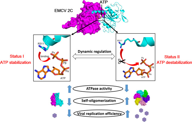
2C is a highly conserved picornaviral non-structural protein with ATPase activity and plays a multifunctional role in the viral life cycle as a promising target for anti-picornavirus drug development. While the structure-function of enteroviral 2Cs have been well studied, cardioviral 2Cs remain largely uncharacterized. Here, an endogenous ATP molecule was identified in the crystal structure of 2C from encephalomyocarditis virus (EMCV, Cardiovirus A). The ATP is bound into the ATPase active site with a unique compact conformation. Notably, the γ-phosphate of ATP directly interacts with Arg311 (conserved in cardioviral 2Cs), and its mutation significantly inhibits the ATPase activity. Unexpectedly, this mutation remarkably promotes 2C self-oligomerization and viral replication efficiency. Molecular dynamic simulations showed that the Arg311 side chain is highly dynamic, indicating it may function as a switch between the activation state and the inhibition state of ATPase activity. A hexameric ring model of EMCV 2C full length indicated that the C-terminal helix may get close to the N-terminal amphipathic helices to form a continuous positive region for RNA binding. The RNA-binding studies of EMCV 2C revealed that the RNA length is closely associated with the RNA-binding affinities and indicated that the substrate may wrap around the outer surface of the hexamer. Our studies provide a biochemical framework to guide the characterization of EMCV 2C and the essential role of arginine in cardioviral 2C functions.

Importance: Encephalomyocarditis virus (Cardiovirus A) is the causative agent of the homonymous disease, which may induce myocarditis, encephalitis, and reproductive disorders in various mammals. 2C protein is functionally indispensable and a promising target for drug development involving broad-spectrum picornaviral inhibitors. Here, an endogenous ATP molecule with a unique conformation was discovered by a combination of protein crystallography and high-performance liquid chromatography in the encephalomyocarditis virus (EMCV) 2C structure. Biochemical and structural characterization analysis of EMCV 2C revealed the critical role of conserved Arg311 in ATPase activity and self-oligomerization of EMCV 2C. The viral replication kinetics and infectivity study suggested that the residue negatively regulated the infectivity titer and virus encapsulation efficiency of EMCV and is, therefore, crucial for 2C protein to promote viral replication. Our systemic structure-function analysis provides unique insights into the function and regulation mechanism of cardioviral 2C protein.

Keywords: 2C; ATPase activity; crystal structure; encephalomyocarditis virus; endogenous ATP; molecular dynamic simulations.


Conflict of interest statement

The authors declare no conflict of interest.

Figures

Fig 1
Fig 1
Biochemical characterization of soluble full-length EMCV 2C (2C-FL). (A) Expression and purification of EMCV MBP-2C-FL in E. coli. Lanes 1–2: elution of MBP-EMCV 2C from Ni-NTA resin. Lanes 3–4: flow-through of the sample from lanes 1–2 after TEV protease digestion (remove MBP tag) that was reloaded on Ni-NTA. M, protein marker; Mw, molecular weight standards. The standard molecular weight was indicated on the top in kilodalton. (B) ATP hydrolysis by EMCV MBP-2C-FL (0.25 µM) is plotted as the function of ATP concentration. The data were fitted with non-linear regression using the Michaelis–Menten equation to calculate kinetic parameters (Km, Vmax, kcat, and R2 of the fitting are indicated). Data are presented as the average (±standard error of the mean) from three independent experiments. (C) Oligomeric state of recombinant EMCV MBP-2C-FL in solution analyzed by analytical ultracentrifugation (AUC).
Fig 2
Fig 2
Overview of EMCV 2C-∆N structure. (A) Domain/motif organization of EMCV 2C full-length protein with annotated conserved motifs. The ATPase domain, ZFER, and pocket-binding domain (PBD, including C-terminal helix) are colored in green, cyan, and magenta, respectively. (B) Overall crystal structure of EMCV 2C-∆N colored the same as above. (C) Structure-based sequence alignment of EMCV 2C (Cardiovirus genus) with its representative homologs from various picornaviruses performed using Clustal X (version 1.81) and ESPript 3. They include FMDV (Aphthovirus genus), HAV (Hepatovirus genus), PV (Enterovirus genus), and EV71 (Enterovirus genus). The conserved residues are boxed in blue. Identical conserved and low conserved residues are highlighted in red background and red letters, respectively. The major elements are labeled using green bars. The key arginine (Arg311) in EMCV 2C is highlighted using a red arrow. It is noted that the helices are labeled with a superscript (α1′-α5′) in the EMCV 2C N-terminal domain structure (1–108 aa) predicted by AlphaFold2. (D) Upper panel: structural comparison of EMCV 2C (cyan) with that of EV-A71 (green) and FMDV (pink). The PBL and ZFER are highlighted using an ellipse and a rectangle, respectively. Lower panel: close-up view of the ZFER among the three 2C proteins.
Fig 3
Fig 3
The C-terminal helix mediated self-oligomerization of EMCV 2C. (A) Crystal packing analysis of EMCV 2C-∆N crystals reveals 10 molecules in the lattice that are connected by the C-terminal helix. (B) Close-up view of the C-terminal helix of one molecule binding into a hydrophobic pocket (referred to as pocket) in the adjacent 2C molecule. (C) The detailed analysis of PBD–pocket interaction between the two 2C molecules. (D) Oligomeric state of recombinant EMCV 2C-∆N variants (wild type and mutants) in solution analyzed by gel filtration chromatography (Superdex 75 10/300 GL). The elution volumes of marker proteins (~158, ~44, and ~17 kDa) are marked using arrows.
Fig 4
Fig 4
Identification of an endogenous ATP molecule in EMCV 2C-∆N. (A) Crystallographic analysis of the ATP-like molecule and its neighboring region in EMCV 2C-∆N structure. Electron density map (2Fo-Fc) of the ATP-like molecule is shown at a 1.5σ level. (B) HPLC analysis of the ATP-like molecule from EMCV 2C-∆N extract. The major peak of EMCV 2C extract has an identical retention time (~5.5 min) with that of ATP in a mixture of standard ATP/ADP/AMP samples. (C) The detailed contact analysis between the ATP molecule and the protein. The H-bonds are shown using red dash lines. (D) Structural comparison of EMCV 2C-∆N with EV71 2C-∆N (PDB ID: 5GRB). Subunits A and B of EMCV 2C-∆N dimer are shown in green and magenta, respectively, while the two subunits of EV71 2C-∆N dimer are shown in purple and cyan, respectively. Subunit A in the two 2Cs are structurally superimposed. The ATP in EMCV 2C-∆N and ATP-γ-S in EV71 2C-∆N are shown as yellow and gray sticks, respectively. (E) Close-up view of the ATP-binding regions of EMCV/EV71 2C-∆N. The critical residue Arg311 in EMCV 2C-∆N and corresponding residue Asn316 in EV71 2C-∆N are shown as magenta and cyan sticks, respectively.
Fig 5
Fig 5
Impact of Arg311 mutation on EMCV 2C ATPase activity and viral replication. (A) ATP activity of MBP-tagged EMCV MBP-2C-FL WT and R311S mutant in the presence or absence of RNA. The ATP hydrolysis rate was measured in the reaction mixture [1,000 µM ATP, 50 nM MBP-2C-FL, and 0 or 5 µM 30-nt RNA as used in the surface plasmon resonance (SPR) assay], and the ATPase activity is defined as “micromole of ATP hydrolyzed per micromole of enzyme per minute.” Data represent the mean ± SEM (n = 3 technically independent samples). The MBP-tagged pMAL vector was used as a negative control. (B) BHK-21 cells were infected with mutant or WT virus at a multiplicity of infection (MOI) of 5 or 0.01, respectively. The resulting virus was harvested at different times, titered, and expressed as a 50% tissue culture infectious dose (TCID50). The mean values ± SD (repeated measures ANOVA, n = 3, no significant differences identified) are shown. (C) The replication abilities of EMCV-WT and EMCV mutant virus (R311S) were analyzed by the plaque assay on BHK-21 cells. (D) Structure-based sequence alignment of C-terminal helix of 2C from several cardioviral species. The key arginine is highlighted using a red arrow.
Fig 6
Fig 6
Microsecond-long MD simulations of ATP-binding EMCV 2C-∆N. (A) RMSD calculated for ATP and Arg311 (Cα atom) along the MD trajectories. In each case, the system has been superimposed onto the initial minimized structure that was built from the crystal structure as explained in Materials and Methods. (B) Evolution of Arg311 side chain nitrogen (NH1) to ATP (γ-phosphate) distances in the MD trajectory (1,000 ns). (C) Distance distribution analysis of the Arg311-ATP distances during the MD trajectories. (D) Snapshots of the distances between Arg311 (NH1) and ATP (γ-phosphate) at the initial state (0 ns), the minimal distance (465.6 ns), and the maximal distance (558.9 ns), respectively. (E) Structural superposition of the three above statuses describing the shift (red arrows) of Arg311 side chain nitrogen during the MD.
Fig 7
Fig 7
Hexameric ring model of EMCV 2C-FL. (A) Overall structure of EMCV 2C-FL hexameric model. The six subunits are shown as different colors, and each subunit is composed of NTD and CTD. (B–C) A molecular surface representation of hexameric model shown in front view (B) and back view (C), colored by its local electrostatic potential (blue, +7KT; red, −7KT). (D) Close-up view of the C-terminal helix and its neighboring two helices in NTD. The three helices constitute a continuous region with predominantly electropositive charges for potential substrate RNA binding. (E) Structural comparison of EMCV 2C-FL dimer derived from the above model with EMCV 2C-∆N dimer. Subunits A and B of EMCV 2C-FL dimer are shown in cyan and magenta, respectively, while the two subunits of EMCV 2C-∆N dimer are shown in pink and orange, respectively. Subunit A in the two dimers is structurally superimposed. (F) Close-up view of the ATPase active site of the two dimers. The critical residues are shown as sticks.
Fig 8
Fig 8
RNA-binding characteristics of EMCV 2C-FL by SPR. (A–C) Kinetic-binding analysis of EMCV MBP-2C-FL to different types of RNA. The colored curves are the experimental traces obtained from SPR experiments, and black curves are the best global fits to the data using 1:1 ligand binding model. The values of association rate (ka), dissociation rate (kd), and subsequent dissociation constant (KD) were calculated. The maximum response unit (Rmax) represents the theoretical maximum EMCV MBP-2C-FL binding capacity of the surface (RU). Each biotinylated RNA fragment (5 nM) was immobilized on Series S Sensor Chip SA, and the EMCV MBP-2C-FL proteins were set up in the concentration range of 31.25–1,000 nM. (D) The affinity constants for RNA binding by EMCV MBP-2C-FL.
Fig 9
Fig 9
A supposed mechanism on the regulation of Arg311 on EMCV 2C function. An endogenous ATP is bound by Arg311 in EMCV 2C. The Arg311–ATP interaction in 2C is highly dynamic, and it may function as a switch to keep a balance between ATPase activation and inhibition. In status I (ATP stabilization), the Arg311 in the C-terminal helix of adjacent subunits can stabilize ATP (γ-phosphate) to stimulate its ATPase activity (ATP hydrolysis). However, the self-oligomerization, infectivity titer, and replication efficiency are inhibited. In status II (ATP destabilization), Arg311 moves far away from ATP with their interactions being disrupted. At this stage, self-oligomerization and replication efficiency are promoted.

Similar articles

Cited by

References

    1. Zell R, Delwart E, Gorbalenya AE, Hovi T, King AMQ, Knowles NJ, Lindberg AM, Pallansch MA, Palmenberg AC, Reuter G, Simmonds P, Skern T, Stanway G, Yamashita T, Ictv Report Consortium . 2017. ICTV virus taxonomy profile: picornaviridae. J Gen Virol 98:2421–2422. doi:10.1099/jgv.0.000911 - DOI - PMC - PubMed
    1. Liang Z, Kumar ASM, Jones MS, Knowles NJ, Lipton HL. 2008. Phylogenetic analysis of the species Theilovirus: emerging murine and human pathogens. J Virol 82:11545–11554. doi:10.1128/JVI.01160-08 - DOI - PMC - PubMed
    1. Reddacliff LA, Kirkland PD, Hartley WJ, Reece RL. 1997. Encephalomyocarditis virus infections in an Australian zoo. J Zoo Wildl Med 28:153–157. - PubMed
    1. LaRue R, Myers S, Brewer L, Shaw DP, Brown C, Seal BS, Njenga MK. 2003. A wild-type porcine encephalomyocarditis virus containing A short poly(C) tract is pathogenic to mice, pigs, and cynomolgus macaques. J Virol 77:9136–9146. doi:10.1128/jvi.77.17.9136-9146.2003 - DOI - PMC - PubMed
    1. Jones P, Cordonnier N, Mahamba C, Burt FJ, Rakotovao F, Swanepoel R, André C, Dauger S, Bakkali Kassimi L. 2011. Encephalomyocarditis virus mortality in semi-wild bonobos (Pan panicus). J Med Primatol 40:157–163. doi:10.1111/j.1600-0684.2010.00464.x - DOI - PubMed

MeSH terms

LinkOut - more resources