. 2024;41(2):115-132.
doi: 10.3233/CBM-230523.

RANK in cancer-associated fibroblasts: A valuable prognostic determinant for metastasis in early-stage breast cancer patients

Affiliations

RANK in cancer-associated fibroblasts: A valuable prognostic determinant for metastasis in early-stage breast cancer patients

María Belén Giorello et al. Cancer Biomark. 2024.

Abstract

Background: The molecular system of receptor activator of nuclear factor kappa-β (RANK) and its ligand (RANKL) plays a role in a variety of physiological and pathological processes. These encompass the regulation of bone metabolism, mammary gland development, immune function, as well as their involvement and tumorigenesis. Nevertheless, limited knowledge exists regarding their function within the tumor microenvironment.

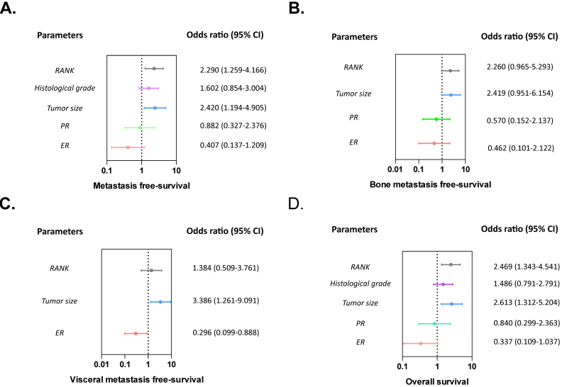
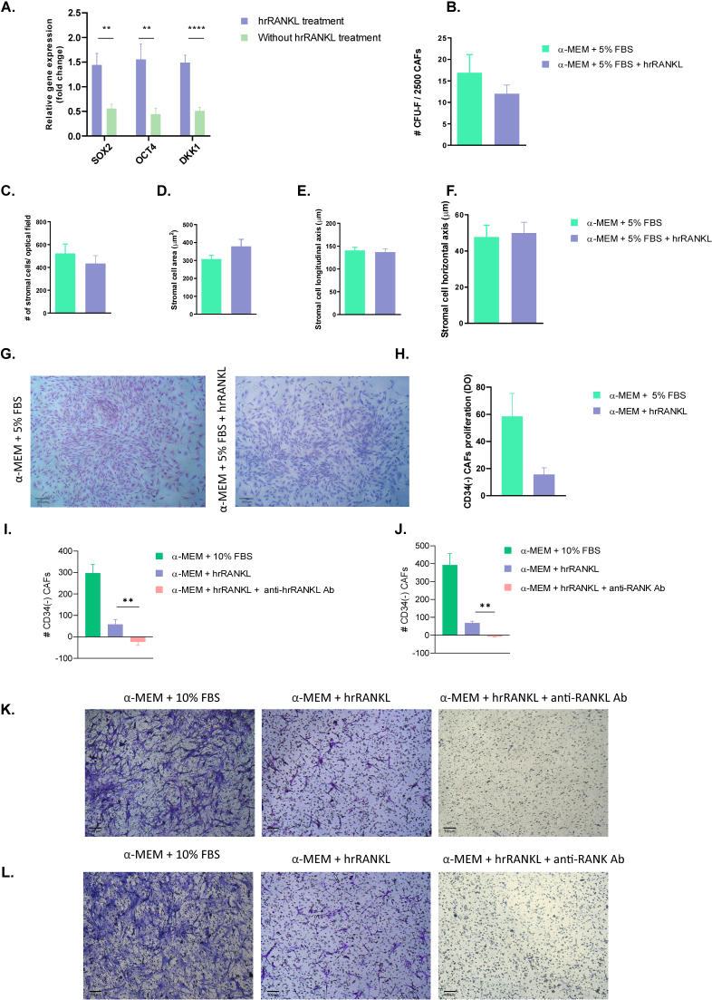
Methods and results: We explored the significance of RANK expression in cancer-associated fibroblasts (CAFs) as a prognostic biomarker in early breast cancer patients (BCPs) by immunohistochemistry. Results reveal a significant correlation between high RANK expression in CAFs and an increased risk of metastasis (p= 0.006), shorter metastasis-free survival (MFS) [p= 0.007, OR (95%CI) = 2.290 (1.259-4.156)], and lower overall survival (OS) [p= 0.004, OR (95%CI) = 2.469 (1.343-4.541)]. Upon analyzing the phenotype of CD34(-) CAFs isolated from primary tumors in BCPs, we observed co-expression of RANK with CD105 marker by immunofluorescence and flow cytometry, characteristic of mesenchymal stem/stromal cells (MSCs), suggesting the possible cellular origin. Also RANKL-RANK system increase the OCT-4, SOX-2 and DKK-1 (dickkopf 1) gene expression in CD34(-) CAFs by RT-PCR. Moreover, this system plays a crucial role in the migration of these CD34(-) CAFs.

Conclusions: These results support the clinical relevance of RANK in CAFs and propose its potential as a future therapeutic target in the treatment of early BCPs.

Keywords: Cancer-associated fibroblasts; RANK/RANKL; breast cancer; metastatic occurrence; prognostic factor.


Conflict of interest statement

The authors declare there are no potential conflicts of interest with respect to the research, authorship and/or publication of this article. Consent to participate Informed consent was obtained from all individual participants included in the study.

Figures

Figure 1.
Figure 1.
Expression of RANK in CAFs from the primary tumor of BCPs. Left panel: representative example of RANK immunostaining (brown chromogen) in stromal cells assessed in the primary tumor tissue of a BCP. Right panel: isotype control. Nuclei were stained with hematoxylin (purple color). Original magnification: × 400. Scale bars represent 25 μm. B. Association of RANK expression with local relapse-free survival (RFS), metastasis-free survival (MFS), bone metastasis-free survival (BMFS), visceral metastasis-free survival (VMFS), mix metastasis-free survival (MMFS), and overall survival (OS) in early invasive ductal BCPs. Kaplan-Meier curves (univariate analysis) marked in green represent data from samples with high RANK expression, while blue curves represent samples with negative/low RANK expression. The Log Rank (Mantel-Cox) test was used to assess the Kaplan-Meier curves. *p-value < 0.050. C. Details of RFS, MFS, BMFS, VMFS, MMFS and OS for the negative/low and high RANK expression groups.
Figure 2.
Figure 2.
Forest plot showed odds ratios for the multivariate association between classical prognosis factors and RANK, and metastasis-free survival (A), bone metastasis-free survival (B), visceral metastasis-free survival (C), and overall survival (D) in early invasive ductal BCPs.
Figure 3.
Figure 3.
Phenotypic characterization of CD34(-) CAFs. A. Representative flow cytometry surface antigen histograms of CAFs from a representative BCP. Isotype control (). B. Co-expression of RANK and CD105 in CAFs of breast tumors. A representative dot plot of RANK-CD105 co-expression, CD34-CD105, and CD34-RANK in fibroblasts isolated from the primary tumors of BCPs (I/II). C. Dual staining of RANK (green) and CD105 (red) by immunofluorescence in CD34(-) fibroblasts isolated from the primary tumors of BCPs. Counterstained with DAPI. Magnification 400X. The scale corresponds to 50 μm. D. Dual staining of RANK (green) and CD105 (red) by immunofluorescence in paraffin-embedded breast tissue from BCPs. Magnification 200X. The scale corresponds to 200 μm.
Figure 4.
Figure 4.
Effect of RANKL-RANK system on the self-renewal, proliferation abilities, expression of pluripotency factors and migration of CD34(-) CAFs in BCPs. A. Gene expression of self–renewal and pluripotency factors in CAFs from BCPs treatment with and without RANKL. Expression of OCT-4, SOX-2 and DKK-1 by quantitative real-time polymerase chain reaction (RT-PCR). All the results were normalized against a set of reference genes. B. CFU-F Assay: The self-renewal capacity of CD34(-) CAFs from BCPs was assessed in αMEM basal medium supplemented with 5% fetal bovine serum (FBS) and in the presence of hrRANKL (25 ng/ml). The CFU-F assay was also conducted in the presence of 20% FBS as a standard positive control (#CFU-F/2500 CAFs =29.22 ± 5.11). C. Stromal cell density per optical microscope field in each CFU-F. D. Area of stromal cells in typical regions of each CFU-F culture. E. Length of stromal cells in typical regions of CFU-F cultures. F. Width of stromal cells in typical regions of CFU-F cultures. G. CFU-F size observed for a representative BCP in supplemented αMEM added with 5% FBS and in supplemented α MEM added with 5% SBF + 25 ng/ml hrRANKL. Giemsa staining (40X). H. Proliferation of CD34(-) CAFs in BCPs was evaluated in supplemented αMEM added with 5% and 10% SBF without hrRANKL, and in the presence of 25 ng/ml hrRANKL. Percentage increase relative to baseline (negative control) is plotted. All values are expressed as mean ± SE. Unpaired t-test with Welch’s correction was used for statistical analysis. Asterisks indicate a significant difference (*p < 0.0500). I and K. Migration of CD34(-) CAFs in BCPs was assessed in supplemented basal medium (α MEM) with I) 10% FBS (positive control), II) 50 ng/ml of hrRANKL and III) 50 ng/ml of hrRANKL and 3.3 μg/ml of anti-RANKL Ab. Values are expressed as mean ± standard error (SE). Unpaired t-test with Welch’s correction was used for statistical analysis, (**p= 0.0089). J and L. Migration of CD34(-) CAFs in BCP was assessed in supplemented basal medium (αMEM) with I) 10% FBS (positive control), II) 50 ng/ml of hrRANKL and III) 50 ng/ml of hrRANKL and 5 μg/ml of anti-RANK Ab. Values are expressed as mean ± standard error (SE). Unpaired t-test with Welch’s correction was used for statistical analysis, (**p= 0.0049).

References

    1. Lorusso G. and Rüegg C., The tumor microenvironment and its contribution to tumor evolution toward metastasis, Histochem. Cell Biol. 130 (2008), 1091–1103. doi: 10.1007/s00418-008-0530-8. - DOI - PubMed
    1. Bissell M.J., Radisky D.C., Rizki A., Weaver V.M. and Petersen O.W., The organizing principle: Microenvironmental influences in the normal and malignant breast, Differentiation 70 (2002), 537–546. doi: 10.1046/j.1432-0436.2002.700907.x. - DOI - PMC - PubMed
    1. Kalluri R. and Zeisberg M., Fibroblasts in cancer, Nat. Rev. Cancer 6 (2006), 392–401. doi: 10.1038/nrc1877. - DOI - PubMed
    1. Giorello M.B., Borzone F.R., Labovsky V., Piccioni F.V. and Chasseing N.A., Cancer-Associated Fibroblasts in the Breast Tumor Microenvironment, J. Mammary Gland Biol. Neoplasia 26 (2021), 135–155. doi: 10.1007/s10911-020-09475-y. - DOI - PubMed
    1. Sarkar M., Nguyen T., Gundre E., Ogunlusi O., El-Sobky M., Giri B. and Sarkar T.R., Cancer-associated fibroblasts: The chief architect in the tumor microenvironment, Front. Cell Dev. Biol. 11 (2023), 1–24. doi: 10.3389/fcell.2023.1089068. - DOI - PMC - PubMed

MeSH terms

LinkOut - more resources