. 2024 Dec 2;84(23):4031-4048.
doi: 10.1158/0008-5472.CAN-24-1102.

MYC Drives mRNA Pseudouridylation to Mitigate Proliferation-Induced Cellular Stress during Cancer Development

Affiliations

MYC Drives mRNA Pseudouridylation to Mitigate Proliferation-Induced Cellular Stress during Cancer Development

Jane Ding et al. Cancer Res. .

Abstract

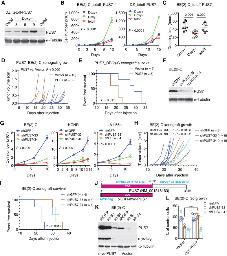
Pseudouridylation is a common RNA modification that is catalyzed by the family of pseudouridine synthases (PUS). Pseudouridylation can increase RNA stability and rigidity, thereby impacting RNA splicing, processing, and translation. Given that RNA metabolism is frequently altered in cancer, pseudouridylation may be a functionally important process in tumor biology. Here, we show that the MYC family of oncoproteins transcriptionally upregulates PUS7 expression during cancer development. PUS7 is essential for the growth and survival of MYC-driven cancer cells and xenografts by promoting adaptive stress responses and amino acid biosynthesis and import. ATF4, a master regulator of stress responses and cellular metabolism, was identified as a key downstream mediator of PUS7 functional activity. Induction of ATF4 by MYC oncoproteins and cellular stress required PUS7, and ATF4 overexpression overcame the growth inhibition caused by PUS7 deficiency. Mechanistically, PUS7 induced pseudouridylation of MCTS1 mRNA, which enhanced its translation. MCTS1, a noncanonical translation initiation factor, drove stress-induced ATF4 protein expression. A PUS7 consensus pseudouridylation site in the 3' untranslated region of ATF4 mRNA was crucial for the induction of ATF4 by cellular stress. These findings unveil an MYC-activated mRNA pseudouridylation program that mitigates cellular stress induced by MYC stimulation of proliferation and biomass production, suggesting that targeting PUS7 could be a therapeutic strategy selectively against MYC-driven cancers. Significance: Oncogene activation of mRNA pseudouridylation is a mechanism that facilitates metabolic reprogramming and adaptive responses to overcome cellular stress during cancer development.


Conflict of interest statement

S. Sudarshan reports grants from NIH and the Department of Veterans Affairs during the conduct of the study. H.-F. Ding reports grants from NIH during the conduct of the study. No disclosures were reported by the other authors.

Figures

Figure 1.
Figure 1.
PUS7 is a direct transcriptional target of MYC oncoproteins. A, Microarray data show mRNA expression of PUS family genes in non–MYCN-amplified neuroblastoma SK-N-AS cells with MYCN overexpression compared with vector control cells (dashed line). B and C, qRT-PCR (B) and immunoblotting (C) show MYCN-mediated upregulation of PUS7 mRNA and protein in non–MYCN-amplified neuroblastoma SK-N-AS cells with inducible MYCN overexpression in the absence of Doxy and SHEP1 cells with constitutive MYCN overexpression. PUS7 protein levels were quantified against α-tubulin (α-tub). D and E, qRT-PCR (D) and immunoblotting (E) show downregulation of PUS7 mRNA and protein expression by shRNA-mediated MYCN knockdown in MYCN-amplified neuroblastoma cell lines. PUS7 protein levels were quantified against GAPDH. F, Immunoblotting show MYC upregulation of PUS7 in P493-6 cells with inducible MYC expression in the absence of Doxy (tetoff-MYC). PUS7 protein levels were quantified against β-actin. G, ChIP-qPCR show endogenous MYCN binding to the promoter and first intron of PUS7 in MYCN-amplified neuroblastoma BE(2)-C cells. The MDM2 and DDIT3 promoters were used as positive control for MYCN and ATF4 binding, respectively. The number associated with each primer set indicates the position of the forward primer relative to its target gene transcription start site (+1). H, Violin plot shows higher PUS7 expression in MYCN-amplified neuroblastoma tumors [the Sequencing Quality Control (SEQC) cohort, n = 498]. P values were calculated using one-way ANOVA. I, RNA-seq data show increased Pus7 mRNA expression in B cells during lymphoma development in Eµ-myc mice. PCR data (B, D, and G) are presented as mean ± SD (B and D, n = 4; G, n = 3) and analyzed using two-way ANOVA. **, P < 0.01; ***, P < 0.001.
Figure 2.
Figure 2.
High PUS7 expression promotes neuroblastoma cell proliferation and tumorigenicity. A, Immunoblotting of PUS7 in neuroblastoma SK-N-DZ cells, with inducible PUS7 expression in the absence of Doxy (Doxy−). B, PUS7 overexpression (Doxy−) promoted the proliferation of BE(2)-C and SK-N-DZ neuroblastoma cells. The same cell lines cultured in the presence of Doxy (Doxy+, no PUS7 induction) and their parental tetoff cell lines were used as control. C, PUS7 overexpression reduced the cell population doubling time based on the data from B. D and E, PUS7 overexpression accelerated BE(2)-C xenograft growth (D) and decreased event-free survival (E) of xenograft-bearing NOD/SCID mice. Log-rank test P value is indicated. F, Immunoblotting of PUS7 in BE(2)-C expressing shGFP or shPUS7. G, PUS7 knockdown inhibited the proliferation of neuroblastoma cell lines. H and I, PUS7 knockdown impeded BE(2)-C xenograft growth (H) and prolonged event-free survival (I) of xenograft-bearing NOD/SCID mice. Log-rank test P value is indicated. J, Diagram illustrates shPUS7-33 and shPUS7-34 targeting the PUS7 3′-UTR and CDS, respectively. The myc-PUS7 construct lacks the PUS7 3′-UTR, making it resistant to downregulation by shPUS7-33. K, Immunoblotting of PUS7 in BE(2)-C cells expressing shGFP, shPUS7-33, or shPUS7-34 that were then infected with vector or myc-PUS7 lentiviruses. L, Cell growth assay shows PUS7 expression abrogated the growth inhibitory effect of shPUS7-33 but not that of shPUS7-34. All cell growth data (B, G, and L) are presented as mean ± SD (n = 4) and quantitative data (B, D, G, H, and L) were analyzed using two-way ANOVA.  ***, P < 0.001.
Figure 3.
Figure 3.
PUS7 sustains the expression of genes involved in stress responses and amino acid metabolism. A, GO analysis of microarray data shows the top biological processes for genes downregulated by PUS7 knockdown. B, Volcano plot shows the representative genes involved in stress responses and amino acid metabolism. C, Gene set enrichment analysis of microarray data shows the downregulation of genes involved in AAR by PUS7 knockdown. D, Volcano plot shows the co-upregulation of PUS7 and representative genes involved in amino acid biosynthesis and transport. E and F, qRT-PCR (E) and immunoblotting (F) confirm the downregulation of mRNA and protein expression for representative genes involved in stress responses and amino acid metabolism. qRT-PCR data (E) are presented as mean ± SD (n = 4) and analyzed using two-way ANOVA. ***, P < 0.001.
Figure 4.
Figure 4.
PUS7 is required for sustaining amino acid biosynthesis and transport and stress responses. A, Diagram for incorporation of 13C from glucose into serine, glycine, and alanine. Fold changes (numbers in parentheses) in mRNA expression of relevant enzymes were from microarray profiling. B, PUS7 knockdown reduced 13C6-glucose flux into serine and glycine production but had no effect on alanine production from 13C6-glucose. C, Labeling patterns of metabolites derived from 13C5-15N2-glutamine via anaplerosis. Fold changes (numbers in parentheses) in mRNA expression of relevant enzymes were from microarray profiling. D, PUS7 knockdown reduced incorporation of 13C and 15N from glutamine into asparagine. Data (B and D) are presented as mean ± SD (n = 6) and analyzed using two-tailed Student t test. ***, P < 0.001. E,3H-serine uptake assays show PUS7 regulation of serine transport. Data are presented as mean ± SD of three biological replicates and analyzed using two-way ANOVA. **, P < 0.01; ***, P < 0.001; ****, P < 0.0001. F and G, Immunoblotting shows that PUS7 knockdown diminished stress-induced upregulation of ATP4 protein and its downstream targets involved in stress responses and amino acid synthesis and transport. HisOH, AAR inducer; tunicamycin, ER stress inducer. H, qRT-PCR shows PUS7 knockdown diminished ER stress–induced upregulation of ATP4 and its target genes. Data are presented as mean ± SD (n = 4) and analyzed using two-way ANOVA. ***, P < 0.001.
Figure 5.
Figure 5.
PUS7 protects cells from MYC-induced cellular stress by enabling ATF4-mediated adaptive responses. A–C, Immunoblot analysis shows PUS7 knockdown (A) or inhibition (B and C) abrogated MYCN- or MYC-induced ATF4 and proteins for amino acid synthesis and transport. Protein levels were quantified against α-tubulin (A and B) or β-actin (C). D and E, MYCN activation by 4-hydroxytamoxifen (4OHT) promoted cell proliferation but sensitized cells to PUS7 knockdown (D) or inhibition (E). Data are presented as mean ± SD (n = 4) and analyzed using two-way ANOVA. F, Immunoblot analysis of autophagy (LC3B-II) in SHEP MYCN-ER cells without or with MYCN activation (4OHT) and PUS7 knockdown. G, ATF4 overexpression alleviated the growth inhibitory effect of PUS7 knockdown on neuroblastoma cell lines. H, qRT-PCR shows ATF4 overexpression abrogated PUS7 knockdown–mediated repression of genes for amino acid synthesis and transport. Data are presented as mean ± SD (n = 4) and analyzed using two-way ANOVA. ***, P < 0.001; ****, P < 0.0001.
Figure 6.
Figure 6.
PUS7 targets MCTS1 to sustain ATF4 mRNA translation. A, Diagram of MCTS1 mRNA with indicated PUS7-dependent PSI (Ψ) sites. B, Quantification of Ψ fractions at MCTS1 mRNA Ψ sites shows significant reduction in Ψ levels after PUS7 knockdown. Data represent two biological replicates with 100 or more mRNA reads. C, PUS7 knockdown eliminated the subset of MCTS1 mRNA with two or more Ψ modifications. Nanopore data (B and C) were analyzed using Fisher exact test. D, PUS7 knockdown reduced the Ψ-strength of MCTS1 mRNA, defined as the sum of Ψ fractions at all Ψ sites within each MCTS1 mRNA molecule. E, Volcano plot shows proteomic profiling of PUS7 knockdown vs. control BE(2)-C cells. MCTS1 and the ATF4 target ASNS are highlighted along with several translation initiation factors. F and G, Immunoblotting (F) and quantification (G) of MCTS1 protein expression in HeLa and BE(2)-C cells without (shGFP) or with PUS7 knockdown (shPUS7). Quantitative data (G) are from two or more independent experiments, with samples from each experiment analyzed by immunoblotting in two or more technical replicates. Data were analyzed by one-way ANOVA. H, Polysome profiling show PUS7 knockdown reduced polysome-associated MCTS1 mRNA. Data are representative of two independent experiments and are presented as mean ± SD of three technical replicates. I, Immunoblotting shows MCTS1 knockdown abrogated serine deprivation (AAR)–induced and tunicamycin (Tm)–induced ATF4 protein expression in BE(2)-C cells. ATF4 protein levels were quantified against α-tubulin. *, P < 0.05; **, P < 0.01; ***, P < 0.001; ****, P < 0.0001.
Figure 7.
Figure 7.
The one amino acid–stop codon uORF1 in the 5′-UTR of ATF4 mRNA is critical for stress-induced ATF4 protein expression. A, Diagram of the ATF4 V2 mRNA with indicated uORFs and the main ORF. The uORF1 nucleotide sequence is shown. B–E, Immunoblotting (B and D) and quantification (C and E) of ATF4 protein expression after the AAR (HisOH; B and C) or ER stress (tunicamycin). D and E, 293T-ATF4 KO cells expressing WT or mutant ATF4 V2 in the presence of Doxy (teton). ATF4 protein levels were quantified against β-actin. Data are representative of three independent experiments. F and G, Immunoblotting (F) and quantification (G) of ATF4 protein expression 24 hours after cotransfection of 293T-ATF4 KO cells with vector (pCDH) or pCDH-MYCN in combination with either pGenLenti-ATF4 V1 or pCW57.1-ATF4 V2, including WT or uORF1 mutants. Data represent results from two independent experiments.
Figure 8.
Figure 8.
PUS7 consensus pseudouridylation site in the ATF4 mRNA 3′-UTR regulates the timing of stress-induced ATF4 protein expression. A, Diagram of ATF4-201 transcript (ATF4 V1 mRNA) with indicated PUS7-dependent Ψ sites following ER stress. B, Quantification of Ψ fractions at ATF4-201 transcript Ψ sites shows >50% reduction in Ψ levels following PUS7 knockdown. Data represent two biological replicates with 32–35 mRNA reads. C, PUS7 knockdown largely eliminated Ψ modifications in ATF4-201 transcripts under ER stress. Data were analyzed using Fisher exact test. D, PUS7 knockdown reduced the Ψ strength of ATF4-201 transcripts. E,In vitro pseudouridylation of ATF4-201 transcript U1984 by immunopurified PUS7. F–I, Immunoblotting (F and H) and quantification (G and I) of ATF4 protein expression at various time points following AAR (HisOH) or ER stress (tunicamycin, Tm)). Quantitative data (G and I) are presented as mean ± SD (n = 3). J, Model for MYC/MYCN activation of the PUS7-MCTS1-ATF4 axis to mitigate cellular stress induced by oncogenic activation. *, P < 0.05; **, P < 0.01; ***, P < 0.001; ****, P < 0.0001. (J, Created with BioRender.com.)

References

    1. Roundtree IA, Evans ME, Pan T, He C. Dynamic RNA modifications in gene expression regulation. Cell 2017;169:1187–200. - PMC - PubMed
    1. Delaunay S, Helm M, Frye M. RNA modifications in physiology and disease: towards clinical applications. Nat Rev Genet 2024;25:104–22. - PubMed
    1. Adachi H, De Zoysa MD, Yu Y-T. Post-transcriptional pseudouridylation in mRNA as well as in some major types of noncoding RNAs. Biochim Biophys Acta Gene Regul Mech 2019;1862:230–9. - PMC - PubMed
    1. Borchardt EK, Martinez NM, Gilbert WV. Regulation and function of RNA pseudouridylation in human cells. Annu Rev Genet 2020;54:309–36. - PMC - PubMed
    1. Rodell R, Robalin N, Martinez NM. Why U matters: detection and functions of pseudouridine modifications in mRNAs. Trends Biochem Sci 2024;49:12–27. - PMC - PubMed

MeSH terms