Efficient data labeling strategies for automated muscle segmentation in lower leg MRIs of Charcot-Marie-Tooth disease patients
- PMID: 39241036
- PMCID: PMC11379393
- DOI: 10.1371/journal.pone.0310203
Efficient data labeling strategies for automated muscle segmentation in lower leg MRIs of Charcot-Marie-Tooth disease patients
Abstract
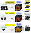
We aimed to develop efficient data labeling strategies for ground truth segmentation in lower-leg magnetic resonance imaging (MRI) of patients with Charcot-Marie-Tooth disease (CMT) and to develop an automated muscle segmentation model using different labeling approaches. The impact of using unlabeled data on model performance was further examined. Using axial T1-weighted MRIs of 120 patients with CMT (60 each with mild and severe intramuscular fat infiltration), we compared the performance of segmentation models obtained using several different labeling strategies. The effect of leveraging unlabeled data on segmentation performance was evaluated by comparing the performances of few-supervised, semi-supervised (mean teacher model), and fully-supervised learning models. We employed a 2D U-Net architecture and assessed its performance by comparing the average Dice coefficients (ADC) using paired t-tests with Bonferroni correction. Among few-supervised models utilizing 10% labeled data, labeling three slices (the uppermost, central, and lowermost slices) per subject exhibited a significantly higher ADC (90.84±3.46%) compared with other strategies using a single image slice per subject (uppermost, 87.79±4.41%; central, 89.42±4.07%; lowermost, 89.29±4.71%, p < 0.0001) or all slices per subject (85.97±9.82%, p < 0.0001). Moreover, semi-supervised learning significantly enhanced the segmentation performance. The semi-supervised model using the three-slices strategy showed the highest segmentation performance (91.03±3.67%) among 10% labeled set models. Fully-supervised model showed an ADC of 91.39±3.76. A three-slice-based labeling strategy for ground truth segmentation is the most efficient method for developing automated muscle segmentation models of CMT lower leg MRI. Additionally, semi-supervised learning with unlabeled data significantly enhances segmentation performance.
Copyright: © 2024 Lee et al. This is an open access article distributed under the terms of the Creative Commons Attribution License, which permits unrestricted use, distribution, and reproduction in any medium, provided the original author and source are credited.
Conflict of interest statement
The authors have declared that no competing interests exist.
Figures


Similar articles
-
Collaborative Learning for Annotation-Efficient Volumetric MR Image Segmentation.J Magn Reson Imaging. 2024 Oct;60(4):1604-1614. doi: 10.1002/jmri.29194. Epub 2023 Dec 29. J Magn Reson Imaging. 2024. PMID: 38156427
-
Charcot-Marie-Tooth disease type 1A duplication: spectrum of clinical and magnetic resonance imaging features in leg and foot muscles.Brain. 2006 Feb;129(Pt 2):426-37. doi: 10.1093/brain/awh693. Epub 2005 Nov 29. Brain. 2006. PMID: 16317020
-
Quantitative MRI outcome measures in CMT1A using automated lower limb muscle segmentation.J Neurol Neurosurg Psychiatry. 2024 May 14;95(6):500-503. doi: 10.1136/jnnp-2023-332454. J Neurol Neurosurg Psychiatry. 2024. PMID: 37979968
-
Muscle fat quantification using magnetic resonance imaging: case-control study of Charcot-Marie-Tooth disease patients and volunteers.J Cachexia Sarcopenia Muscle. 2019 Jun;10(3):574-585. doi: 10.1002/jcsm.12415. Epub 2019 Mar 15. J Cachexia Sarcopenia Muscle. 2019. PMID: 30873759 Free PMC article.
-
A review of self-supervised, generative, and few-shot deep learning methods for data-limited magnetic resonance imaging segmentation.NMR Biomed. 2024 Aug;37(8):e5143. doi: 10.1002/nbm.5143. Epub 2024 Mar 24. NMR Biomed. 2024. PMID: 38523402 Review.
References
MeSH terms
LinkOut - more resources
Full Text Sources
Medical

