. 2024 Sep 6;10(36):eadl6162.
doi: 10.1126/sciadv.adl6162. Epub 2024 Sep 6.

Lung-innervating nociceptor sensory neurons promote pneumonic sepsis during carbapenem-resistant Klebsiella pneumoniae lung infection

Affiliations

Lung-innervating nociceptor sensory neurons promote pneumonic sepsis during carbapenem-resistant Klebsiella pneumoniae lung infection

Prabhu Raj Joshi et al. Sci Adv. .

Abstract

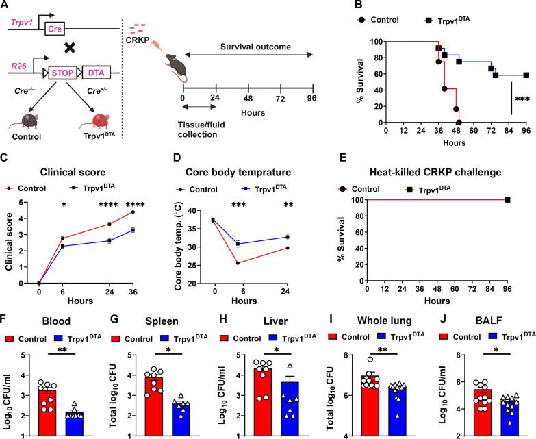
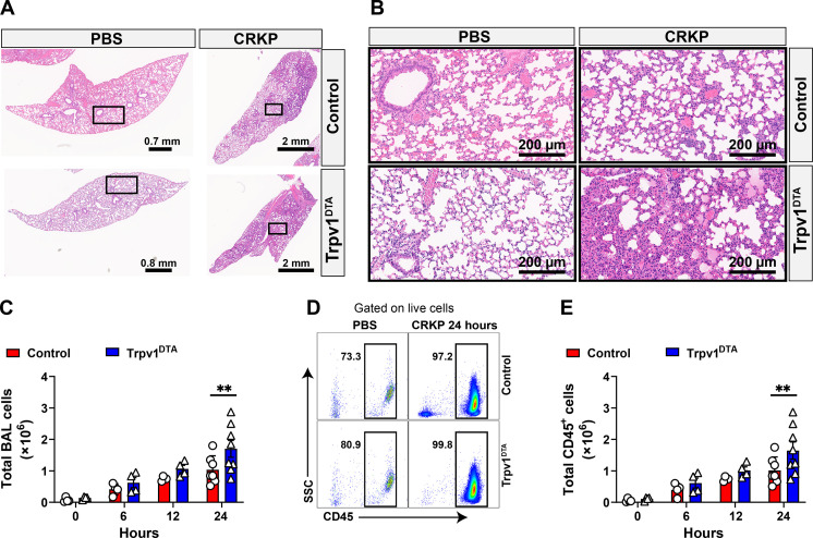
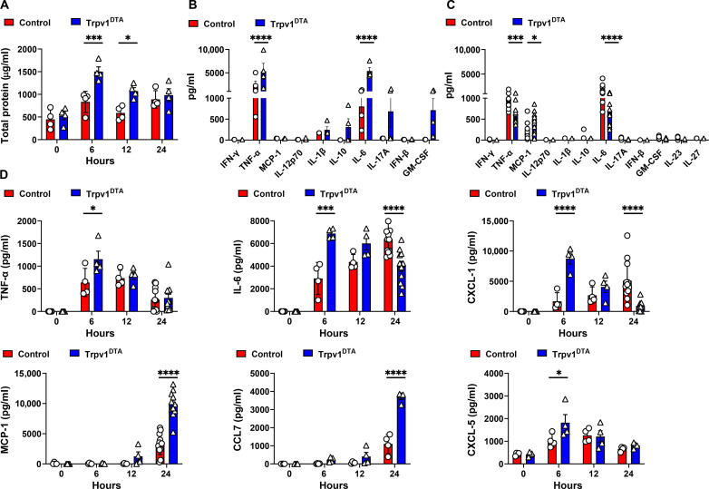
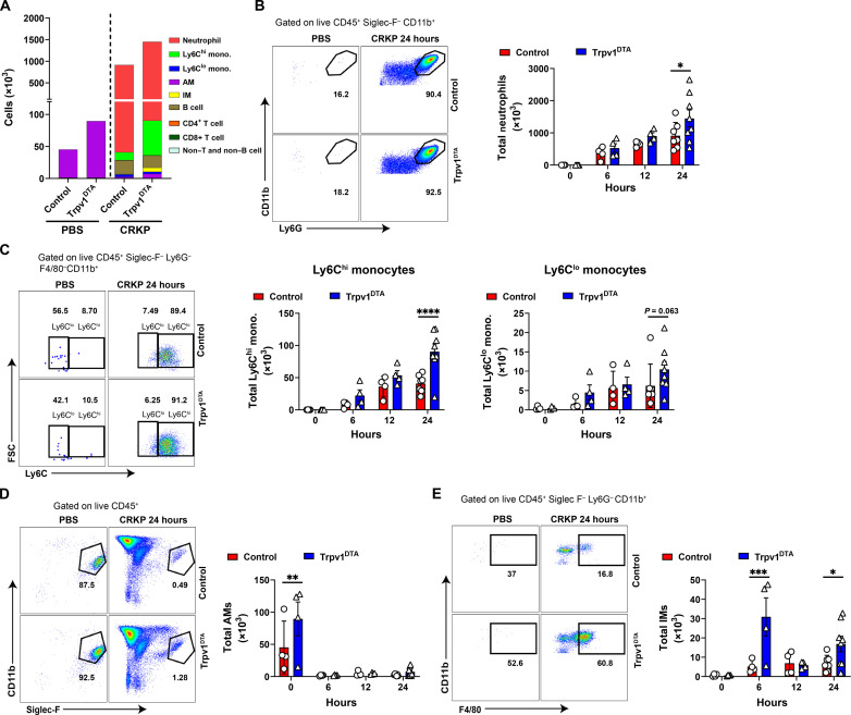
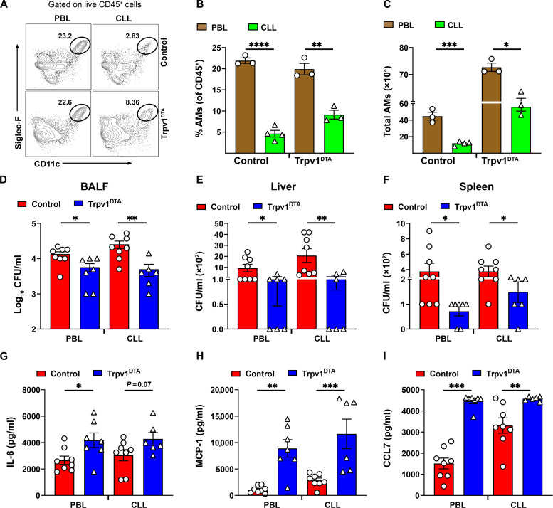
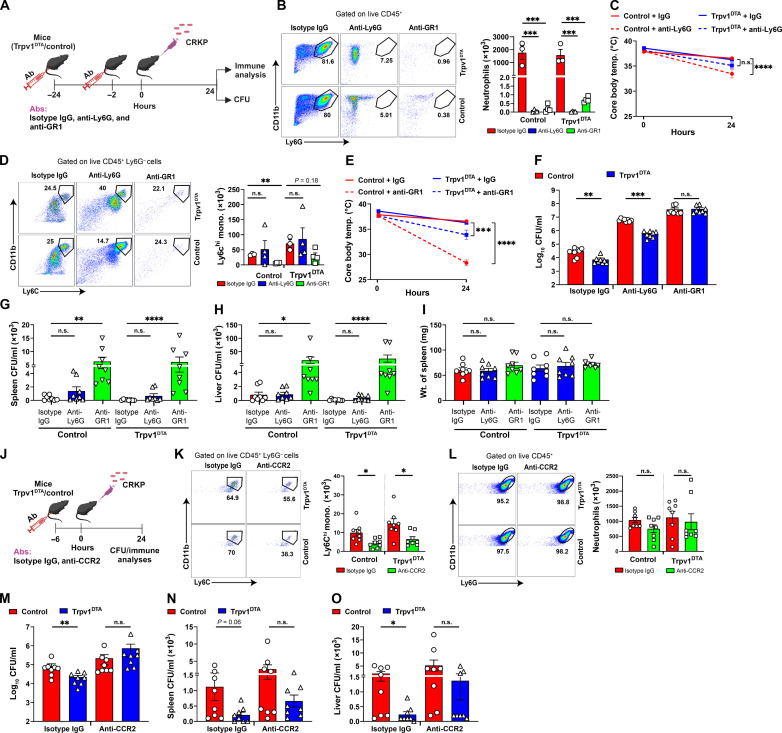
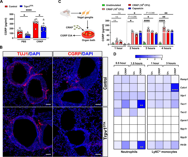
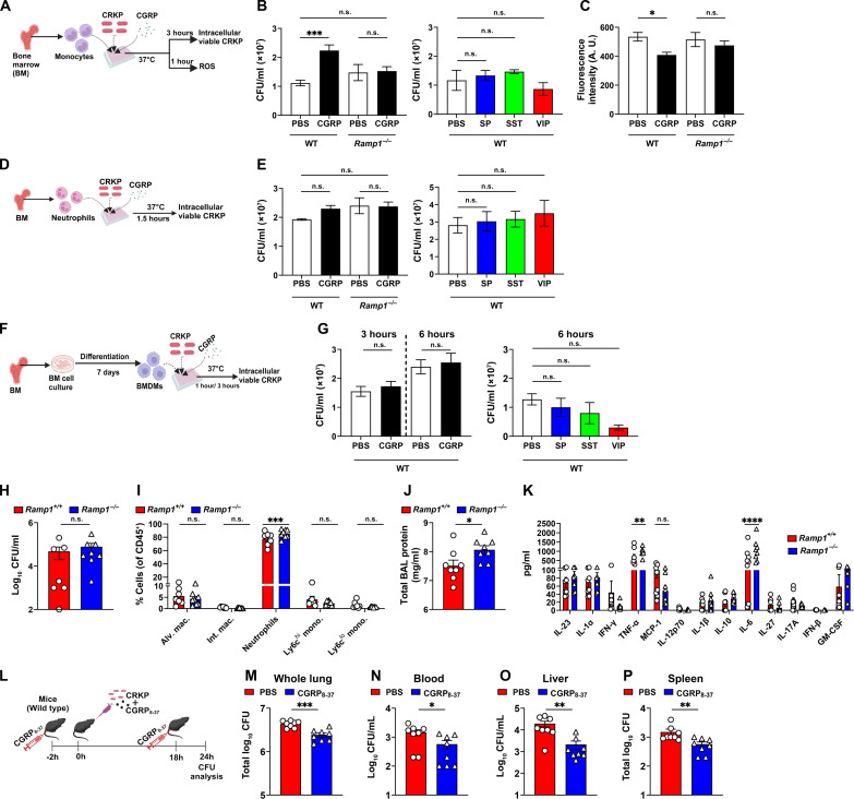
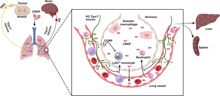
Carbapenem-resistant Klebsiella pneumoniae (CRKP) causes Gram-negative lung infections and fatal pneumonic sepsis for which limited therapeutic options are available. The lungs are densely innervated by nociceptor sensory neurons that mediate breathing, cough, and bronchoconstriction. The role of nociceptors in defense against Gram-negative lung pathogens is unknown. Here, we found that lung-innervating nociceptors promote CRKP pneumonia and pneumonic sepsis. Ablation of nociceptors in mice increased lung CRKP clearance, suppressed trans-alveolar dissemination of CRKP, and protected mice from hypothermia and death. Furthermore, ablation of nociceptors enhanced the recruitment of neutrophils and Ly6Chi monocytes and cytokine induction. Depletion of Ly6Chi monocytes, but not of neutrophils, abrogated lung and extrapulmonary CRKP clearance in ablated mice, suggesting that Ly6Chi monocytes are a critical cellular population to regulate pneumonic sepsis. Further, neuropeptide calcitonin gene-related peptide suppressed the induction of reactive oxygen species in Ly6Chi monocytes and their CRKP-killing abilities. Targeting nociceptor signaling could be a therapeutic approach for treating multidrug-resistant Gram-negative infection and pneumonic sepsis.


Figures

Fig. 1.
Fig. 1.. Nociceptor neurons suppress host protection against CRKP pneumonia and pneumonic sepsis.
(A) Genetic strategy for ablating TRPV1+ neurons in mice (left) and mouse model of CRKP lung infection (right). Control and Trpv1DTA mice were infected with the lethal dose (109 CFU per mouse) of CRKP intranasally. (B to D) Survival curve (B), clinical score (C), and core body temperature (D) of control and Trpv1DTA mice over time. Data in (B) include Trpv1DTA mice (n = 12) and littermate control mice (n = 12) from three independent experiments. Data in (C) and (D) are the means ± SEM and involve Trpv1DTA mice (n = 8 to 12 mice per group) and control littermates (n = 8 to 12 mice per group). (E) Survival curve of control (n = 8) and Trpv1DTA mice (n = 8) after intranasal challenge with heat-killed CRKP bacteria (109 CFU per mouse). (F to J) CFUs recovered in blood (F), spleen (G), liver (H), whole lung (I), and BALF (J) from control and Trpv1DTA mice at 24 hpi. Each symbol denotes a mouse [(F) to (J)]. Data in (F) to (J) are the means ± SEM and at least two independent experiments with n = 7 to 12 mice per group. Statistical analysis was done by log-rank (Mantel-Cox) test [(B) and (E)], two-way analysis of variance (ANOVA) with Sidak’s multiple comparison’s test (C), repeated measures two-way ANOVA with Sidak’s multiple comparison’s test (D), Mann-Whitney test [(F), (H), and (I)], and two-tailed unpaired t test [(G) and (J)]. Levels of significance for all statistical analyses: *P < 0.05, **P < 0.01, ***P < 0.001, ****P < 0.0001. BioRender.com was used to create schematic in (A).
Fig. 2.
Fig. 2.. Nociceptors suppress leukocyte influx during CRKP lung infection.
(A and B) H&E-stained sections of lungs: ×0.9 to ×1.1 magnification (A) and ×20 magnification of selected area (in rectangles) (B) of control and Trpv1DTA mice with PBS inoculation and after CRKP infection (24 hpi). Representative images were selected from at least 12 lobes of n = 3 to 4 mice in each group. Scale bars are shown in each figure. (C to E) Total live cells (C), representative flow cytometry plots of total leukocytes (CD45+ cells) (D), and total CD45+ cells (E), in BALF of Trpv1DTA and control mice at 6, 12, and 24 hpi with CRKP. Data in (C) and (E) are the means ± SEM and involve n = 4 mice per group for 0-, 6-, and 12-hour time course analyses and n = 7 to 8 mice per group for 24-hour time course analyses. Statistical analysis was done by two-way ANOVA of two-stage liner step-up procedure of Benjamini, Krieger, and Yekutieli posttests with the following significance levels: **P < 0.01. BAL, bronchoalveolar lavage.
Fig. 3.
Fig. 3.. Ablation of nociceptors alter the kinetics of cytokine and chemokine induction after CRKP infection.
(A) Total proteins in BALF over time of CRKP infection. (B and C) Panels of inflammatory cytokines measured by LEGENDplex assay in BALF at 6 (B) and 24 hpi (C). (D) Measurement of selected cytokines and chemokines (TNF-α, IL-6, CXCL-1, MCP-1, CCL7, and CXCL-5) by ELISA in BALF over time of CRKP infection. Data in (A) to (D) are the means ± SEM and involve Trpv1DTA mice (n = 4 mice per group for 0, 6, and 12 hours and n = 4 to 11 mice per group for 24 hours) and control littermates (n = 4 mice per group for 0, 6, and 12 hours and n = 4 to 12 mice per group for 24 hours). Data were analyzed by two-way ANOVA with Sidak’s multiple comparison’s posttests [(A) and (D)] and two-way ANOVA of two-stage liner step-up procedure of Benjamini, Krieger, and Yekutieli posttests [(B) and (C)] with the following significance levels: *P < 0.05, ***P < 0.001, ****P < 0.0001.
Fig. 4.
Fig. 4.. Nociceptors dampen recruitment of neutrophils, monocytes, and interstitial macrophages into airways during CRKP infection.
(A) Numbers of leukocyte subsets quantified in BALF of Trpv1DTA and control littermates at 24 hpi or of PBS treatment. (B to E) Representative flow cytometry plots and absolute total numbers of neutrophils (CD11b+ Ly6G+) (B), Ly6Chi and Ly6Clo monocytes (C), AMs (Siglec-F+CD11bCD11c+ F4/80+) (D), and interstitial macrophages (IMs; CD11b+ F4/80+) (E) in BALF of Trpv1DTA and control mice over time after CRKP infection or PBS treatment. Data in (A) to (E) are the means ± SEM and involve Trpv1DTA mice (n = 4 per group for 0, 6, and 12 hours and n = 8 per group for 24 hours) and control littermates (n = 4 per group for 0, 6, 12 hours and n = 7 per group for 24 hours). Data were analyzed by two-way ANOVA of two-stage liner step-up procedure of Benjamini, Krieger, and Yekutieli posttests with the following significance levels: *P < 0.05, **P < 0.01, ***P < 0.001, ****P < 0.0001.
Fig. 5.
Fig. 5.. Depletion of AMs does not affect host protection against CRKP in nociceptor-ablated mice.
(A) Representative flow cytometry plots showing CLL-mediated depletion of AMs in control and Trpv1DTA mice. PBL was used for control group of mice. CLL or PBL (70 μl per mouse) was inoculated via intratracheal route at 48 hours before the CRKP infection. (B and C) CLL-mediated depletion of AMs and their quantitative data at baseline after 48 hours of inoculation. (D to F) The number of CFUs recovered in BALF (D), liver (E), and spleen (F) from control and Trpv1DTA mice at 24 hours CRKP after infection with and without AM depletion. (G to I) Measurement of IL-6 (G), MCP-1 (H), and CCL7 (I) in BALF of AM-depleted control and Trpv1DTA mice at 24 hpi. Each symbol represents a mouse. Data in (B) to (I) are the means ± SEM. Statistical analysis was done by unpaired t test [(B), (C), and (G)] and Mann-Whitney test [(D) to (F) and (H) and (I)] with the following significance levels: *P < 0.05, **P < 0.01, ***P < 0.001, ****P < 0.0001.
Fig. 6.
Fig. 6.. Monocytes are critical for the control of CRKP dissemination and pneumonic sepsis.
(A) Schematic showing depletion of neutrophils and monocytes and CRKP infection. (B and D) Flow cytometry plots, total neutrophils (B), and total monocytes (D) in isotype immunoglobulin G (IgG)–, anti-Ly6G–, and anti-GR1–treated control and Trpv1DTA mice (n = 3 to 4 per group) at 24 hpi. (C and E) Core body temperature of isotype IgG–treated versus anti-Ly6G–treated mice [(C), n = 8 per group] and isotype IgG–treated versus anti-GR1–treated mice [(E), n = 8 per group] over time. n.s., not significant. (F to I) CFUs in BALF (F), spleen (G), and liver (H) and weight of spleen (I) in neutrophil/monocyte-depleted Trpv1DTA and control mice (n = 8 per group) at 24 hpi. (J) Schematic showing anti-CCR2–mediated depletion of Ly6Chi monocytes and CRKP infection. (K and L) Flow cytometry plots, total Ly6Chi monocytes (K), and total neutrophils (L) in isotype IgG– and anti-CCR2–treated Trpv1DTA and control mice (n = 8 per group) at 24 hpi. (M to O) CFUs in BALF (M), spleen (N), and liver (O) in monocyte-depleted Trpv1DTA and control mice (n = 8 per group) after 24 hpi. Data in (B) to (I) and (K) to (O) are the means ± SEM. Statistical analysis: one-way ANOVA with Holm-Sidak’s comparisons posttests [(B) and (I)], Brown-Forsythe and Welch ANOVA with Dunnett’s T3 multiple comparisons posttests (D), repeated measures two-way ANOVA with Sidak’s comparisons posttests [(C) and (E)], Mann-Whitney test [(F) and (M) to (O)], Kruskal-Wallis test with Dunn’s multiple comparisons test [(G) and (H)], and unpaired t test [(K) and (L)]. Level of significance: *P < 0.05, **P < 0.01, ***P < 0.001, ****P < 0.0001. BioRender.com was used to create schematics in (A) and (J).
Fig. 7.
Fig. 7.. CRKP infection induces the release of CGRP in the airways and the neuropeptide receptor expression by monocytes.
(A) Levels of CGRP in BALF of PBS-treated and CRKP-infected control and Trpv1DTA mice (n = 8 per group). (B) Stained sections of lungs showing β-tubulin III (TUJ1) and CGRP fibers of control and Trpv1DTA mice. Representative images were selected from a total of nine images of three mice in each group (three images per mouse). Scale bars, 50 μm. DAPI, 4′,6-diamidino-2-phenylindole. (C) Left: Schematic showing organotypic culture of VG (n = 4 VGs per well) and their stimulation with CRKP to measure CGRP release. Right: Time course of CGRP release from VG culture after stimulation with CRKP bacteria (106 CFU per well and 108 CFU per well). Unstimulated wells are VG culture with media only. Capsaicin (1.5 μM) was used as positive control. (D) Heatmap showing relative mRNA levels of neuropeptide receptors in CRKP-infected monocytes and neutrophils at indicated time points of infection (n ≥ 3 per group). Un., unstimulated cells. Data in (A), (C), and (D) are the means ± SEM. Statistical analysis: Brown-Forsythe and Welch ANOVA of two-stage liner step-up procedure of Benjamini, Krieger, and Yekutieli posttests (A), one-way ANOVA with Sidak’s multiple comparison test (C), and two-way ANOVA with Sidak’s multiple comparisons posttests (D). Level of significance: *P < 0.05, **P < 0.01, ****P < 0.0001. BioRender.com was used to create schematic in (C).
Fig. 8.
Fig. 8.. CGRP-RAMP1 signaling dampens anti-CRKP response in monocytes.
(A) Schematic showing monocyte and CRKP coculture in the presence and absence of neuropeptide. (B and C) Intracellular viable CFUs (B) and levels of ROS (C), in CRKP-infected wild-type (WT) and Ramp/ monocytes. (D) Schematic of neutrophil and CRKP coculture with and without neuropeptide. (E) Intracellular viable CFUs in CRKP-infected WT and Ramp/ neutrophils. (F) Schematic showing BMDM isolation and coculture with CRKP in the presence and absence of neuropeptides. (G) Intracellular viable CFUs in WT BMDMs. Data in (B), (C), (E), and (G) involve three to eight samples per group. (H to K) CRKP CFUs (H), proportions of myeloid subsets (I), total BAL protein (J), and panel of 13 mouse inflammatory cytokines/chemokines (K), measured in BALF of Ramp1+/+ and Ramp1/ mice (n = 7 to 8 per group) at 24 hpi. (L) Schematic showing administration of CGRP8–37 (5 μg per mouse) in C57BL/6 J mice and CRKP infection. (M to P) CFUs in whole lung (M), blood (N), liver (O), and spleen (P) of PBS-treated and CGRP8–37-treated mice (n = 8 per group) after 24 hpi. Data in (B), (C), (E), (G), (H) to (K) and (M) to (P) are the means ± SEM. Statistical analysis: one-way ANOVA with Sidak’s multiple comparisons posttests [(B), (C), (E), and (G)], Mann-Whitney test [(H), (O), and (P)], unpaired t test [(J), (M), and (N)], and two-way ANOVA of two-stage liner step-up procedure of Benjamini, Krieger, and Yekutieli posttests [(I) and (K)]. Levels of significance: *P < 0.05, **P < 0.01, ***P < 0.001, ****P < 0.0001. BioRender.com was used to create schematics in [(A), (D), (F), and (L)].
Fig. 9.
Fig. 9.. Model of nociceptor regulation of CRKP-induced pneumonic sepsis.
Lung-innervating nociceptor neurons suppress the recruitment of neutrophils and Ly6Chi monocytes to the airspaces of lungs during CRKP lung infection. CRKP infection induces vagal TRPV1+ neurons for releasing CGRP, which in turn acts on its receptor on Ly6Chi monocytes to dampen the ROS production. This leads to an increased CRKP survival in the lungs and enhanced dissemination of bacteria to vital organs. BioRender.com was used to create model.

References

    1. Bengoechea J. A., Pessoa J. S., Klebsiella pneumoniae infection biology: Living to counteract host defences. FEMS Microbiol. Rev. 43, 123–144 (2019). - PMC - PubMed
    1. Mizgerd J. P., Acute lower respiratory tract infection. N. Engl. J. Med. 358, 716–727 (2008). - PMC - PubMed
    1. Kang C. I., Song J. H., Kim S. H., Chung D. R., Peck K. R., Thamlikitkul V., Wang H., So T. M., Hsueh P. R., Yasin R. M., Carlos C. C., Van P. H., Perera J., Risk factors and pathogenic significance of bacteremic pneumonia in adult patients with community-acquired pneumococcal pneumonia. J. Infect. 66, 34–40 (2013). - PubMed
    1. Nair H., Simoes E. A., Rudan I., Gessner B. D., Azziz-Baumgartner E., Zhang J. S. F., Feikin D. R., Mackenzie G. A., Moiisi J. C., Roca A., Baggett H. C., Zaman S. M., Singleton R. J., Lucero M. G., Chandran A., Gentile A., Cohen C., Krishnan A., Bhutta Z. A., Arguedas A., Clara A. W., Andrade A. L., Ope M., Ruvinsky R. O., Hortal M., McCracken J. P., Madhi S. A., Bruce N., Qazi S. A., Morris S. S., El Arifeen S., Weber M. W., Scott J. A. G., Brooks W. A., Breiman R. F., Campbell H.; Severe Acute Lower Respiratory Infections Working Group , Global and regional burden of hospital admissions for severe acute lower respiratory infections in young children in 2010: A systematic analysis. Lancet 381, 1380–1390 (2013). - PMC - PubMed
    1. Zhang H., Wang J., Zhou W., Yang M., Wang R., Yan X., Cai Y., Risk factors and prognosis of carbapenem-resistant Klebsiella pneumoniae infections in Respiratory Intensive Care Unit: A retrospective study. Infect Drug Resist. 14, 3297–3305 (2021). - PMC - PubMed

Publication types

MeSH terms

LinkOut - more resources