. 2024 Sep 17;5(9):101715.
doi: 10.1016/j.xcrm.2024.101715. Epub 2024 Sep 5.

Acutely blocking excessive mitochondrial fission prevents chronic neurodegeneration after traumatic brain injury

Affiliations

Acutely blocking excessive mitochondrial fission prevents chronic neurodegeneration after traumatic brain injury

Preethy S Sridharan et al. Cell Rep Med. .

Abstract

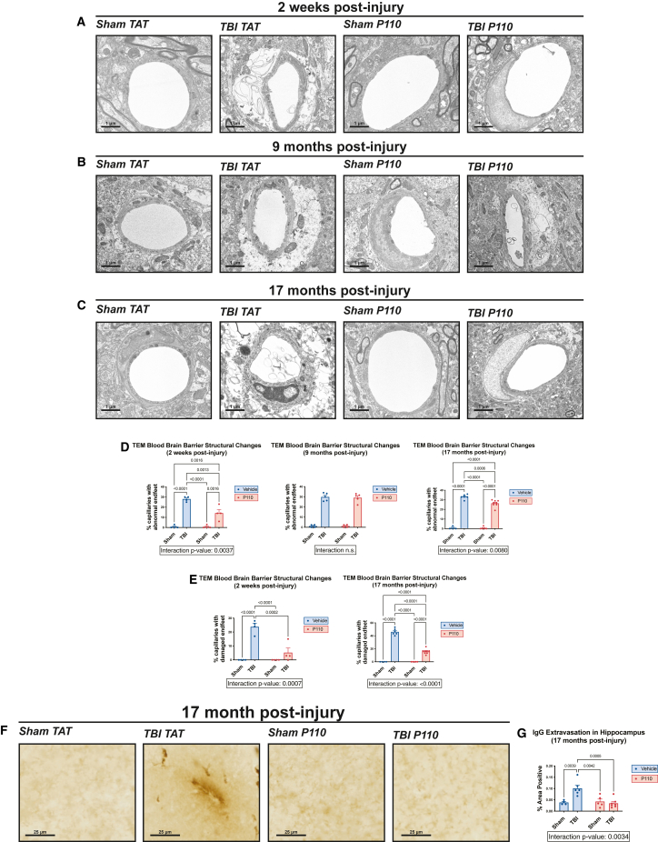
Progression of acute traumatic brain injury (TBI) into chronic neurodegeneration is a major health problem with no protective treatments. Here, we report that acutely elevated mitochondrial fission after TBI in mice triggers chronic neurodegeneration persisting 17 months later, equivalent to many human decades. We show that increased mitochondrial fission after mouse TBI is related to increased brain levels of mitochondrial fission 1 protein (Fis1) and that brain Fis1 is also elevated in human TBI. Pharmacologically preventing Fis1 from binding its mitochondrial partner, dynamin-related protein 1 (Drp1), for 2 weeks after TBI normalizes the balance of mitochondrial fission/fusion and prevents chronically impaired mitochondrial bioenergetics, oxidative damage, microglial activation and lipid droplet formation, blood-brain barrier deterioration, neurodegeneration, and cognitive impairment. Delaying treatment until 8 months after TBI offers no protection. Thus, time-sensitive inhibition of acutely elevated mitochondrial fission may represent a strategy to protect human TBI patients from chronic neurodegeneration.

Keywords: Drp1; Fis1; blood-brain barrier; mitochondria; mitochondrial fission; mitochondrial fusion; neurodegeneration; neuroprotection; oxidative stress; traumatic brain injury.


Conflict of interest statement

Declaration of interests X.Q. is an inventor of P110 and holds patents related to P110.

Figures

None
Graphical abstract
Figure 1
Figure 1
Prevention of chronic cognitive impairment after TBI by acute inhibition of pathologically excessive mitochondrial fission (A) Western blot and quantification of Drp1, phospho-Drp1 (pDrp1) S616, and Fis1 in mouse hippocampus at 24 h and 2 weeks post-injury show early elevation in Fis1 expression, but not Drp1 or pDrp1 S616 (n = 5 mice/group, two-tailed Student’s t test). (B) Western blot and quantification show that AD subjects with TBI history have elevated Fis1, but Fis1 is not elevated in subjects with AD alone (n = 3 patients/group, one-way ANOVA and Tukey’s post hoc analysis). (C) Co-immunoprecipitation of Drp1 with Fis1 shows increased interaction in the cerebellum of TBI TAT animals and reduced interaction in P110-treated animals (n = 3 mice/group). (D) Experimental schematic of injury and treatment strategy for early transient P110 treatment and experimental schematic of injury and treatment strategy for delayed transient P110 treatment. (E) Novel object recognition task at 2 weeks, 9 months, and 17 months post-injury shows significant cognitive impairment that is prevented by early transient P110 treatment (n = 35–40 mice/group for 2 weeks cohort, 36–40 mice/group for 9 months cohort, and n = 4–8 mice/group for 17 months cohort; two-way ANOVA and Tukey’s post hoc analysis). (F) Novel object recognition task at 9 months post-injury shows that cognitive impairment cannot be rescued with delayed transient P110 treatment (n = 6–7 mice/group; two-way ANOVA and Tukey’s post hoc analysis).
Figure 2
Figure 2
Prevention of chronic mitochondrial fragmentation and bioenergetic impairment after TBI by acute inhibition of pathologically excessive mitochondrial fission (A–C) Transmission electron microscopy (TEM) of hippocampal mitochondria collected 2 weeks, 9 months, and 17 months post-injury shows excessive mitochondrial fragmentation after TBI. Fragmentation is prevented with early transient P110 treatment. Structural changes are quantified by aspect ratio measurements. (D–F) Quantification of TEM images (n = 4–7 mice/group, with 700–1,000 mitochondria quantified and averaged per animal; two-way ANOVA and Tukey’s post hoc analysis). (G) Hippocampal synaptosome mitochondrial bioenergetics shows respiratory suppression 2 weeks after TBI, and P110 improves basal respiration, maximal respiration, and spare respiratory capacity (n = 16 samples (4 animals)/group; two-way ANOVA and Tukey’s post hoc analysis). Data are the means ± SEM. Two-way ANOVA tests for interaction between injury effect and treatment effect.
Figure 3
Figure 3
Prevention of chronic hippocampal lipid peroxidation after TBI by acute inhibition of excessive mitochondrial fission (A–C) Representative 4-HNE staining of the hippocampal CA3 region shows increased lipid peroxidation at 2 weeks, 9 months, and 17 months post-injury. Early transient P110 treatment prevents 4-HNE elevation. (D–F) Quantification of lipid peroxidation from CA3 region of hippocampus (n = 3 mice/group; two-way ANOVA and Tukey’s post hoc analysis).
Figure 4
Figure 4
Prevention of chronic microglial hyperactivation and lipid droplet accumulation after TBI by acute inhibition of excessive mitochondrial fission (A) Representative Iba1 staining of hippocampus with skeletonized microglia. (B and C) Sholl analysis of hippocampal microglia 9 months post-injury shows a decrease in microglial ramification after TBI, which is prevented by early transient P110 treatment. (D) Representative BODIPY and Iba1 staining of hippocampus shows an increase in lipid droplet-positive microglia at 9 months post-injury, which is prevented by early transient P110 treatment. (E) Quantification of microglial lipid droplets in hippocampus (n = 3 mice/group; two-way ANOVA and Tukey’s post hoc analysis). Data are the means ± SEM. Two-way ANOVA tests for interaction between injury effect and treatment effect.
Figure 5
Figure 5
Prevention of chronic axonal neurodegeneration after TBI by acute inhibition of pathologically excessive mitochondrial fission (A and B) Representative images of neurodegeneration measured by silver staining of the hippocampus at 2 weeks and 9 months post-injury show significant protection with early transient P110 treatment. (C) Quantification of silver stain images (n = 5 mice/group, with 6 sections/animal counted and normalized to area; two-way ANOVA and Tukey’s post hoc analysis). Data are the means ± SEM. Two-way ANOVA tests for interaction between injury effect and treatment effect.
Figure 6
Figure 6
Prevention of chronically excessive death of young hippocampal neurons after TBI by acute inhibition of pathologically excessive mitochondrial fission (A and B) Representative images of BrdU-positive cell survival, which is impaired 2 weeks and 9 months post-injury and prevented by early transient P110 treatment. (C) BrdU-positive cell survival is impaired 9 months post-injury and prevented by early transient P110 treatment (n = 4–5 mice/group, with 6 sections/animal counted and normalized to area; two-way ANOVA and Tukey’s post hoc analysis). Data are the means ± SEM. Two-way ANOVA tests for interaction between injury effect and treatment effect.
Figure 7
Figure 7
Prevention of chronic BBB deterioration after TBI is prevented by acute inhibition of excessive mitochondrial fission (A–C) Transmission electron microscopy shows significant deterioration of hippocampal astrocytic endfeet 2 weeks and 17 months post-injury, with minor swelling present at 9 months post-injury. Early transient P110 treatment mitigates deterioration of astrocytic endfeet. (D) Quantification of abnormal astrocytic endfeet, including both minor swelling and severe damage (n = 5–7 mice/group, with 30 capillaries counted per animal; two-way ANOVA and Tukey’s post hoc analysis). (E) Quantification of severely damaged astrocytic endfeet (n = 5–7 mice/group, with 30 capillaries counted per animal; two-way ANOVA and Tukey’s post hoc analysis). Data are the means ± SEM. Two-way ANOVA tests for interaction between injury effect and treatment effect. (F) Representative images of IgG staining show extravasation of IgG into brain parenchyma, indicating BBB deterioration. (G) Quantification of % hippocampal area positive for IgG extravasation (n = 3–7/group; two-way ANOVA and Tukey’s post hoc analysis).

References

    1. Ma V.Y., Chan L., Carruthers K.J. Incidence, Prevalence, Costs, and Impact on Disability of Common Conditions Requiring Rehabilitation in the United States: Stroke, Spinal Cord Injury, Traumatic Brain Injury, Multiple Sclerosis, Osteoarthritis, Rheumatoid Arthritis, Limb Loss, and Back Pain. Arch. Phys. Med. Rehabil. 2014;95:986–995.e1. doi: 10.1016/j.apmr.2013.10.032. - DOI - PMC - PubMed
    1. McKee A.C., Cairns N.J., Dickson D.W., Folkerth R.D., Keene C.D., Litvan I., Perl D.P., Stein T.D., Vonsattel J.-P., Stewart W., et al. The first NINDS/NIBIB consensus meeting to define neuropathological criteria for the diagnosis of chronic traumatic encephalopathy. Acta Neuropathol. 2016;131:75–86. doi: 10.1007/s00401-015-1515-z. - DOI - PMC - PubMed
    1. Gardner R.C., Burke J.F., Nettiksimmons J., Kaup A., Barnes D.E., Yaffe K. Dementia Risk After Traumatic Brain Injury vs Nonbrain Trauma: The Role of Age and Severity. JAMA Neurol. 2014;71:1490–1497. doi: 10.1001/jamaneurol.2014.2668. - DOI - PMC - PubMed
    1. Barker S., Paul B.D., Pieper A.A. Increased Risk of Aging-Related Neurodegenerative Disease after Traumatic Brain Injury. Biomedicines. 2023;11:1154. doi: 10.3390/biomedicines11041154. - DOI - PMC - PubMed
    1. Edwards G., Moreno-Gonzalez I., Soto C. Amyloid-beta and tau pathology following repetitive mild traumatic brain injury. Biochem. Biophys. Res. Commun. 2017;483:1137–1142. doi: 10.1016/j.bbrc.2016.07.123. - DOI - PubMed

MeSH terms