. 2024 Oct 3;111(10):2253-2264.
doi: 10.1016/j.ajhg.2024.08.008. Epub 2024 Sep 5.

Hearing restoration by gene replacement therapy for a multisite-expressed gene in a mouse model of human DFNB111 deafness

Affiliations

Hearing restoration by gene replacement therapy for a multisite-expressed gene in a mouse model of human DFNB111 deafness

Luoying Jiang et al. Am J Hum Genet. .

Erratum in

Abstract

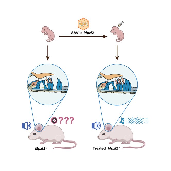
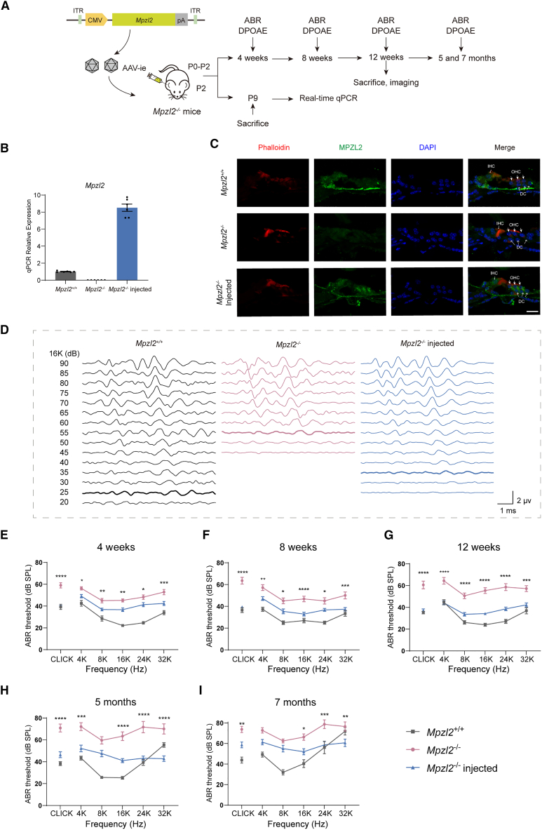
Gene therapy has made significant progress in the treatment of hereditary hearing loss. However, most research has focused on deafness-related genes that are primarily expressed in hair cells with less attention given to multisite-expressed deafness genes. MPZL2, the second leading cause of mild-to-moderate hereditary deafness, is widely expressed in different inner ear cells. We generated a mouse model with a deletion in the Mpzl2 gene, which displayed moderate and slowly progressive hearing loss, mimicking the phenotype of individuals with DFNB111. We developed a gene replacement therapy system mediated by AAV-ie for efficient transduction in various types of cochlear cells. AAV-ie-Mpzl2 administration significantly lowered the auditory brainstem response and distortion product otoacoustic emission thresholds of Mpzl2-/- mice for at least seven months. AAV-ie-Mpzl2 delivery restored the structural integrity in both outer hair cells and Deiters cells. This study suggests the potential of gene therapy for MPZL2-related deafness and provides a proof of concept for gene therapy targeting other deafness-related genes that are expressed in different cell populations in the cochlea.

Keywords: DFNB111; MPZL2; gene therapy; hereditary hearing loss.


Conflict of interest statement

Declaration of interests The authors declare no competing interests.

Figures

None
Graphical abstract
Figure 1
Figure 1
Generation and characterization of Mpzl2–/– mice (A) The CRISPR-Cas9 strategy for disrupting WT Mpzl2. The gRNAs were designed to target exon 2 to yield a 343-bp deletion. (B) Sequencing results of Mpzl2−/− and Mpzl2+/+ mice. (C and D) Representative data of ABR thresholds (C) and DPOAE thresholds (D) were obtained from Mpzl2−/− mice (n = 5; pink) and Mpzl2+/+ mice (n = 5; gray) at 4 weeks, 8 weeks, 12 weeks, 5 months, and 7 months of age. Data are presented as the mean ± SEM. p < 0.05, ∗∗p < 0.01, ∗∗∗p < 0.001, ∗∗∗∗p < 0.0001 by two-way ANOVA with Bonferroni correction. (E and F) Representative confocal images in the middle turn of whole-mount cochleae from Mpzl2−/− and Mpzl2+/+ mice stained with an anti-Myosin VIIa (green) and anti-Sox2 (red) antibody to label HCs (E) and SCs (F) at 12 weeks of age. Degeneration and disorganization of HCs and SCs were seen in Mpzl2−/− mice compared with Mpzl2+/+ controls. Scale bar, 20 μm. IPhC, inner phalangeal cells; IBC, inner border cells; HeC, Hensen’s cells.
Figure 2
Figure 2
Administration of the AAV-ie-CMV-Mpzl2 system restored ABR thresholds of Mpzl2−/− mice (A) Experimental workflow. (B) Quantification of Mpzl2 mRNA expression in P9 Mpzl2+/+ mice, untreated Mpzl2−/− mice, and the treated ears of Mpzl2−/− mice by real-time qPCR (n = 6 for each group). ∗∗∗∗p < 0.0001 vs. Mpzl2+/+; one-way ANOVA with Bonferroni correction. (C) Confocal images of cochleae from 4-week-old Mpzl2+/+, Mpzl2−/−, and injected Mpzl2−/− mice stained with an anti-MPZL2 (green) antibody and Alexa Fluor 555–phalloidin (red). Scale bar, 20 μm. (D) Representative ABR traces from the three groups of mice (Mpzl2+/+, Mpzl2−/−, and Mpzl2−/− injected) in response to 16 kHz were recorded 12 weeks after injection. The bold line in each group indicates the threshold. (E–I) Comparison of ABR thresholds in Mpzl2+/+ mice (gray), untreated Mpzl2−/− mice (pink), and the injected ears of treated Mpzl2−/− mice (blue) at 4 weeks (E), 8 weeks (F), 12 weeks (G), 5 months (H), and 7 months (I). Asterisks indicate the statistical differences between the injected Mpzl2−/− mice and untreated Mpzl2−/− mice. The numbers of mice in the Mpzl2+/+, Mpzl2−/−, and Mpzl2−/− injected ear groups at 4 weeks were n = 14, 25, and 27, respectively, at 8 weeks n = 13, 14, and 26, respectively, at 12 weeks, n = 21, 25, and 22, respectively, at 5 months n = 20, 12, and 14, respectively, and 7 months n = 13, 16, and 15, respectively. Data are shown as the mean ± SEM. p < 0.05, ∗∗p < 0.01, ∗∗∗p < 0.001, ∗∗∗∗p < 0.0001 by two-way ANOVA with Bonferroni correction.
Figure 3
Figure 3
AAV-ie-CMV-Mpzl2 delivery lowered DPOAE thresholds of Mpzl2−/− mice Comparison of DPOAE thresholds in Mpzl2+/+ mice (gray), untreated Mpzl2−/− mice (pink), and the injected ears of treated Mpzl2−/− mice (blue) at 4 weeks (A), 8 weeks (B), 12 weeks (C), 5 months (D), and 7 months (E). Asterisks indicate the statistical differences between the injected Mpzl2−/− mice and untreated Mpzl2−/− mice. The numbers of mice in the Mpzl2+/+, Mpzl2−/−, and Mpzl2−/− injected ear groups at 4 weeks were n = 14, 25, and 27, respectively, at 8 weeks n = 13, 14, and 26, respectively, at 12 weeks, n = 21, 25, and 22, respectively, at 5 months n = 20, 12, and 14, respectively, and 7 months n = 13, 16, and 15, respectively. Data are shown as the mean ± SEM. p < 0.05, ∗∗p < 0.01, ∗∗∗p < 0.001, ∗∗∗∗p < 0.0001 by two-way ANOVA with Bonferroni correction.
Figure 4
Figure 4
Injection of the AAV-ie-CMV-Mpzl2 system protected OHCs from degeneration in Mpzl2−/− mice (A–C) Representative confocal images of the apical (A), middle (B), and basal (C) turns of the cochlea stained with Myosin VIIa (green) from Mpzl2+/+ mice, Mpzl2−/− mice, and injected Mpzl2−/− mice at age 12 weeks. Scale bar, 40 μm. (D) Quantification of the survival of OHCs in (A–C). n = 4 for each group. Data are shown as the mean ± SEM. p < 0.05, ∗∗∗p < 0.001, ∗∗∗∗p < 0.0001 vs. Mpzl2+/+, ##p < 0.01, ###p < 0.001, ####p < 0.0001 vs. Mpzl2−/−; one-way ANOVA with Bonferroni correction.
Figure 5
Figure 5
Injection of the AAV-ie-CMV-Mpzl2 system protected DCs from degeneration in Mpzl2−/− mice (A–C) Representative confocal images of the apical (A), middle (B), and basal (C) turns of the cochlea stained with Sox2 (red) from Mpzl2+/+ mice, Mpzl2−/− mice, and injected Mpzl2−/− mice at age 12 weeks. Scale bar, 40 μm. (D) Quantification of DCs in (A–C). n = 4 for each group. Data are shown as the mean ± SEM. p < 0.05, ∗∗∗p < 0.001, ∗∗∗∗p < 0.0001 vs. Mpzl2+/+, ##p < 0.01, ###p < 0.001, ####p < 0.0001 vs. Mpzl2−/−; one-way ANOVA with Bonferroni correction.
Figure 6
Figure 6
Evaluation of the safety of AAV-ie-CMV-Mpzl2 on hearing function and cochlear structure (A–C) ABR thresholds of WT mice with and without injection of AAV-ie-CMV-Mpzl2 were compared at 4 weeks (A), 8 weeks (B), and 12 weeks (C). The numbers of Mpzl2+/+ uninjected and Mpzl2+/+ injected ears at 4 weeks were n = 14 and 10, respectively, at 8 weeks, n = 13 and 8, respectively, and at 12 weeks, n = 21 and 5, respectively. Data are shown as the mean ± SEM. ns, no significant difference; two-way ANOVA with Bonferroni correction. (D and E) The survival of HCs (D) and SCs (E) in the cochleae of injected WT mice was not affected compared with that of untreated WT mice at 12 weeks. Scale bar, 40 μm.

References

    1. Alford R.L., Arnos K.S., Fox M., Lin J.W., Palmer C.G., Pandya A., Rehm H.L., Robin N.H., Scott D.A., Yoshinaga-Itano C., et al. American College of Medical Genetics and Genomics guideline for the clinical evaluation and etiologic diagnosis of hearing loss. Genet. Med. 2014;16:347–355. doi: 10.1038/gim.2014.2. - DOI - PubMed
    1. Morton C.C., Nance W.E. Newborn hearing screening--a silent revolution. N. Engl. J. Med. 2006;354:2151–2164. doi: 10.1056/NEJMra050700. - DOI - PubMed
    1. Jiang L., Wang D., He Y., Shu Y. Advances in gene therapy hold promise for treating hereditary hearing loss. Mol. Ther. 2023;31:934–950. doi: 10.1016/j.ymthe.2023.02.001. - DOI - PMC - PubMed
    1. Al-Moyed H., Cepeda A.P., Jung S., Moser T., Kügler S., Reisinger E. A dual-AAV approach restores fast exocytosis and partially rescues auditory function in deaf otoferlin knock-out mice. EMBO Mol. Med. 2019;11 doi: 10.15252/emmm.201809396. - DOI - PMC - PubMed
    1. Akil O., Dyka F., Calvet C., Emptoz A., Lahlou G., Nouaille S., Boutet de Monvel J., Hardelin J.P., Hauswirth W.W., Avan P., et al. Vol. 116. 2019. Dual AAV-mediated gene therapy restores hearing in a DFNB9 mouse model; pp. 4496–4501. - DOI - PMC - PubMed

LinkOut - more resources