. 2024 Sep 6;15(1):7805.
doi: 10.1038/s41467-024-50646-w.

Human Smc5/6 recognises transcription-generated positive DNA supercoils

Affiliations

Human Smc5/6 recognises transcription-generated positive DNA supercoils

Aurélie Diman et al. Nat Commun. .

Abstract

Beyond its essential roles in ensuring faithful chromosome segregation and genomic stability, the human Smc5/6 complex acts as an antiviral factor. It binds to and impedes the transcription of extrachromosomal DNA templates; an ability which is lost upon integration of the DNA into the chromosome. How the complex distinguishes among different DNA templates is unknown. Here we show that, in human cells, Smc5/6 preferentially binds to circular rather than linear extrachromosomal DNA. We further demonstrate that the transcriptional process, per se, and particularly the accumulation of DNA secondary structures known to be substrates for topoisomerases, is responsible for Smc5/6 recruitment. More specifically, we find that in vivo Smc5/6 binds to positively supercoiled DNA. Those findings, in conjunction with our genome-wide Smc5/6 binding analysis showing that Smc5/6 localizes at few but highly transcribed chromosome loci, not only unveil a previously unforeseen role of Smc5/6 in DNA topology management during transcription but highlight the significance of sensing DNA topology as an antiviral defense mechanism.


Conflict of interest statement

The authors declare no competing interests.

Figures

Fig. 1
Fig. 1. The origin of extrachromosomal DNA has no impact on the recognition and restriction by Smc5/6.
A Schematic depiction of the genome-integrated construct used to generate extrachromosomal [Gluccircle] and a chromosomally expressed GFP gene upon Cre/loxP-mediated excision. The yeast DNA stuffer is depicted in blue. B Live-cell representative images of Cre/loxP-mediated excision in hTERT-RPE1 cells containing the genomic excisable [Gluccircle] construct were co-transduced with lentiviruses containing either no gene insert (Mock) or HBx, plus or minus the Cre recombinase. Nuclei were visualized with SiR-DNA. Scale bar, 100 μm. Data are representative of three independent experiments. C hTERT-RPE1 cells containing the genomic excisable [Gluccircle] construct (excised ecDNA) were co-transduced with lentiviruses containing either no gene insert (Mock) or HBx, plus or minus the Cre recombinase, together with an integrase-defective lentiviral Cypridina luciferase (CLuc) reporter construct (exogenous ecDNA). The luciferase assay was performed 2 days post transduction. Luciferase activities are relative to their corresponding mock, which were set to 1. Data are means ± SEM of 3 independent experiments. Statistical analysis was performed using one-way ANOVA with Tukey’s multiple comparisons. Source data are provided as a Source Data file.
Fig. 2
Fig. 2. The binding and restriction activity of Smc5/6 are specific to circular DNA templates.
A Schematic illustration of the linear pJAZZ®-derived vector with its terminal DNA hairpin loops, containing a mammalian expression cassette. The CMV promoter drives the expression of the Gaussia Luciferase gene. The BssHII restriction sites used to convert the linear vector into its circular form are also indicated. B hTERT-RPE1 cells over-expressing the HA-tagged version of Smc6 were transiently transfected with either a circular or a linear Gaussia luciferase reporter plasmid and then transduced with lentiviruses containing either no gene insert (Mock) or HBx. Luciferase activities were measured 2 days later and are presented as relative luminescence units (RLU). Data are means ± SEM of 3 independent experiments. Statistical analysis was performed using the two-sided Student’s t-test. C hTERT-RPE1 cells (No HA) or hTERT-RPE1 cells over-expressing HA-tagged version of Smc6 were transiently transfected with either a circular or a linear Gaussia luciferase reporter plasmid and then transduced with lentiviruses containing either no gene insert (Mock) or HBx. Anti-HA ChIP was performed 2 days later. The data are expressed as a percentage of input. Data are means ± SEM of 3 independent experiments. Statistical analysis was performed using one-way ANOVA with Tukey’s multiple comparisons. Source data are provided as a Source Data file.
Fig. 3
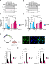
Fig. 3. Recognition of extrachromosomal DNA by Smc5/6 is transcription-dependent but does not require RNA polymerase II.
Western blots showing Smc5 and Nse4 levels in protein extracts of hTERT-RPE1 cells over-expressing HA-Smc6 treated for the indicated times with (A) 10 μg/ml Actinomycin D (ACTD) or (B) 10 μM Triptolide (TPT). Protein extract of GFP-tagged HBx expressing cells was used as a control for Smc5/6 complex degradation. *: Non-specific Nse4 band. Smc5 was used to assess the integrity of the Smc5/6 complex because only a small fraction of the overexpressed HA-Smc6 is assembled into the Smc5/6 complex that binds DNA and is consequently degraded by HBx,. hTERT-RPE1 cells (No HA) or HA-Smc6 hTERT-RPE1 cells transduced with an integrase-defective lentiviral luciferase reporter construct and treated with (C) ACTD or (D) TPT for the indicated times before anti-HA ChIP experiment (blues bars) or anti-H3 (pink bars). qPCR primers amplified the extrachromosomal Gluc. Data are expressed as a percentage relative to the input normalized to their corresponding 0 h time point set to 1 and are means ± SEM of 3 independent experiments. Statistical analysis was performed using one-way ANOVA with Tukey’s multiple comparisons. E Experimental design depiction (left panel). Immunofluorescence staining of HA-Smc6 hTERT-RPE1 cells expressing T7 RNA polymerase with a nuclear localization signal (NLS) (right panel). Nuclei were stained with DAPI. Scale bar, 50 μm. FH HA-Smc6 hTERT-RPE1 cells expressing or not the T7 RNA pol, co-transduced with lentiviruses containing either no gene insert (Mock) or HBx, together with an integrase-defective lentiviral construct carrying a GFP gene controlled by a T7 promoter and an IRES (Internal Ribosome Entry Site). Cells were treated with 10 μM Triptolide (TPT) for 24 h prior to anti-HA ChIP experiments. Three extrachromosomal regions - T7 promoter (F), IRES (G), GFP (H) - were tested and compared to their respective minus T7 RNA pol values. Data are expressed as a percentage relative to the input and are means ± SEM of 3 independent experiments. Statistical analysis was performed using one-way ANOVA with Tukey’s multiple comparisons. Source data are provided as a Source Data file.
Fig. 4
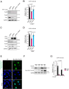
Fig. 4. Smc5/6 recognizes topological structures arising during transcription which are substrates for topoisomerases.
HA-Smc6-expressing hTERT RPE-1 cells transduced with an integrase-defective lentiviral luciferase reporter construct transfected with non-targeting control siRNA (siNTC) or with siRNAs against topoisomerase 1 (siTop1) and topoisomerases 2A and 2B (siTop2) before treatment or not with 10 μM Triptolide (TPT) prior to (A) Western blot analysis and (B) ChIP experiments using anti-HA (siTops: siTop1 and siTop2). ChiP data are expressed as a percentage relative to the input normalized to the siNTC alone (or not treated with TPT), which was set to 1. Data are means ± SEM of 3 independent experiments. Statistical analysis was performed using one-way ANOVA with Tukey’s multiple comparisons. C, D HA-Smc6-expressing hTERT RPE-1 cells co-transduced with lentiviruses containing either no gene insert (Mock) or encoding the c-Myc gene, together with an integrase-defective lentiviral luciferase reporter construct were transfected with siNTC or with siTop1 and siTop2. C Western blot and (D) ChIP using anti-HA were performed after 3 days (siTops: siTop1 and siTop2). The ChiP data are expressed as a percentage relative to the input normalized to the siNTC alone, which was set to 1. Data are means ± SEM of 3 independent experiments. Statistical analysis was performed using one-way ANOVA with Tukey’s multiple comparisons. E Immunofluorescence staining of HA-Smc6-expressing hTERT RPE-1 cells transduced or not (NT) with lentiviruses encoding a N-terminal 3xMyc-tag-NLS topoisomerase 1B from vaccinia virus (Myc-NLS-vTop1B). Nuclei were stained with DAPI. Scale bar, 50 μm. F, G HA-Smc6-expressing hTERT RPE-1 cells expressing or not Myc-NLS-vTop1B, transduced with an integrase-defective lentiviral luciferase reporter, were co-transduced with lentiviruses containing either no gene insert (Mock) or HBx. F Western blots using an anti-Myc confirmed the expression of Myc-NLS-vTop1B with no impact on Smc5/6 complex integrity as demonstrated by Smc5 protein levels. G ChIP with anti-HA were performed after 3 days. ChiP data are expressed as a percentage relative to the input normalized to no Topo1V mock, which was set to 1. Data are means ± SEM of 3 independent experiments. Statistical analysis was performed using one-way ANOVA with Tukey’s multiple comparisons. Source data are provided as a Source Data file.
Fig. 5
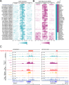
Fig. 5. The chromosomal association of Smc5/6 depends on DNA topological stress induced during transcription.
A Heatmaps of the ChIP-seq read depth around 41 identified Smc5/6 complex binding sites: in hTERT-RPE1 cells (No HA) (left panel); hTERT-RPE1 cells over-expressing a HA-tagged version of Smc6 treated either with DMSO (middle panel); and 10 μM Triptolide (TPT) (right panel) before HA-ChIP. Cells were transduced with an integrase-defective lentiviral luciferase reporter construct (GLuc). Rows represent Smc5/6 binding sites ±2 kb around the peak summit, ordered by chromosome number and according to the presence or absence of RNA Pol II as determined by RPB1-ChIP-seq (B). Peaks statistically detected using MACS2 software analysis (2.5 fold enrichment, 0.05 q-value) are depicted. Color scale represents the ChIP-seq normalized read depth (RPM) row-scaled identically across the 3 samples, with mapped reads virtually resized to 1 kb-length and looking at each genomic position for the amount of overlap between forward- and reverse-stranded reads. B Heatmaps of RNA pol II ChIP-seq peaks in hTERT-RPE1 cells over-expressing a HA-tagged version of Smc6 treated either with DMSO (DMSO), 10 μM Triptolide (TPT) or the corresponding input (NT), before RPB1-ChIP. Cells were transduced with an integrase-defective lentiviral luciferase reporter construct (GLuc). Rows: RNA pol II binding sites ±2 kb around the Smc5/6 peak summit, ordered by chromosome number and according to the presence or absence of RNA Pol II as determined by the RPB1-ChIP-seq results. Color scale represents the ChIP-seq normalized read depth (RPM) row-scaled identically across the 3 samples, with mapped reads virtually resized to 1 kb-length and looking at each genomic position for the amount of overlap between forward- and reverse-stranded reads. C Integrative Genomics Viewer (IGV) track screenshots from 2 representative genomic loci. Red line: Smc5/6 peaks location identified by MACS2. Tracks 1 to 3 (No Ha, DMSO, TPT) represent Smc5/6 ChIP-seq data for the corresponding samples, and tracks 4 to 6 (Input NT, DMSO, TPT) represent RNA pol II ChIP-seq data for the corresponding samples. The subsequent tracks, Top1 (GSE212468), RNA-seq (GSE89413), and Top2 (GSE136943) depict publicly available data obtained for hTERT-RPE1 cells. Scales refer to the signal range in individual genome tracks.
Fig. 6
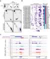
Fig. 6. In human cells, Smc5/6 binds to positively supercoiled DNA.
A Upper part: Schematic showing migration patterns in 2D gel electrophoresis of positively/negatively supercoiled circular DNA with/without chloroquine. Rel: relaxed, OC: open circular, *: topoisomer bands. Lower part: Southern blots of 2D gels on the control circles generated in vitro (panels 1 and 2) and on the circles recovered from cells (panels 3 and 4) with (panels 2 and 4) or without (panels 1 and 3) chloroquine. Blue line: main supercoiled population migration front. Red triangle: slightly less positively supercoiled population. B Heatmaps of GapR ChIP-seq peaks in hTERT-RPE1 cells No HA (left panel) or hTERT-RPE1 cells over-expressing a HA-tagged GapR at low (Low); and high levels (High). Cells were transduced with an integrase-defective lentiviral luciferase reporter construct (GLuc). Rows: GapR binding sites ±2 kb around the Smc5/6 peak summit, ordered by chromosome number according to the presence or absence of RNA Pol II as determined by the RPB1-ChIP-seq results. Peaks statistically detected using MACS2 software analysis (≥1.5-fold enrichment, ≤0.05 q-value) are indicated by a filled box on the right panel. The color scale represents the ChIP-seq normalized read depth (RPM) row-scaled identically across the 3 samples, with mapped reads virtually resized to 1 kb-length and looking at each genomic position for the amount of overlap between forward- and reverse-stranded reads. C Integrative Genomics Viewer (IGV) track screenshots from 2 representative genomic loci. Dark blue line: GapR peaks location identified by MACS2. Track 1 to 3 (No HA, Low GapR, High GapR) represent GapR ChIP-Seq data for the corresponding samples. Red line: Smc5/6 peaks location identified by MACS2. Tracks 4 to 5 (No HA, DMSO) represent Smc5/6 ChIP-seq data for the corresponding samples. Tracks 7 to 8 (Input NT, DMSO) represent RNA pol II ChIP-seq data for the corresponding samples. The subsequent tracks, Top1 (GSE212468), RNA-seq (GSE89413) depict publicly available data obtained for hTERT-RPE1 cells. Scales refer to the signal range in individual genome tracks. Source data are provided as a Source Data file.
Fig. 7
Fig. 7. Speculative model.
Upon transcription, the superhelical tension generated by the transcribing RNA polymerase II will be more significant on a covalently closed circular DNA molecule compared to a linear one, independently of the promoter strength. This will lead to the recruitment of Smc5/6 and to the topological DNA entrapment of the circular DNA while the complex will translocate along the linear DNA. On the chromosomes, an equivalent amount of superhelical tension can only be reached under conditions of a high transcriptional output, which also results in Smc5/6 recruitment. Based on previously reported data,, we hypothesize that, on chromosomes upon DNA entrapment, Smc5/6 stabilizes the plectonemic DNA structures formed. By preventing supercoil spreading, Smc5/6 would therefore act as a topological insulator and/or facilitate the resolution of such DNA structures. The same DNA entrapment on extrachromosomal circular DNA leads to ecDNA recruitment to PML bodies and restriction.

Similar articles

Cited by

References

    1. Jeppsson, K., Kanno, T., Shirahige, K. & Sjögren, C. The maintenance of chromosome structure: positioning and functioning of SMC complexes. Nat. Rev. Mol. Cell Biol.15, 601–614 (2014). 10.1038/nrm3857 - DOI - PubMed
    1. Hassler, M., Shaltiel, I. A. & Haering, C. H. Towards a unified model of SMC complex function. Curr. Biol.28, R1266–R1281 (2018). 10.1016/j.cub.2018.08.034 - DOI - PMC - PubMed
    1. Adamus, M. et al. Molecular insights into the architecture of the human SMC5/6 complex. J. Mol. Biol.432, 3820–3837 (2020). 10.1016/j.jmb.2020.04.024 - DOI - PubMed
    1. Aragón, L. The Smc5/6 complex: new and old functions of the enigmatic long-distance relative. Annu. Rev. Genet.52, 89–107 (2018). 10.1146/annurev-genet-120417-031353 - DOI - PubMed
    1. Roy, S., Adhikary, H. & D’Amours, D. The SMC5/6 complex: folding chromosomes back into shape when genomes take a break. Nucleic Acids Res.52, 2112–2129 (2024). 10.1093/nar/gkae103 - DOI - PMC - PubMed

Publication types

MeSH terms

Associated data