. 2024 Sep 6;20(1):394.
doi: 10.1186/s12917-024-04184-8.

Magnetic resonance imaging of the dromedary camel carpus

Affiliations

Magnetic resonance imaging of the dromedary camel carpus

Ayman El Nahas et al. BMC Vet Res. .

Abstract

Background: The dromedary camel (Camelus dromedarius) carpal joint presents multiple joints and constitutes several bones and soft tissues. Radiography and/or ultrasonography of the carpus are challenging due to structural superimposition. High-field magnetic resonance imaging (MRI) technique precludes superimposed tissues and offers high soft tissue contrast in multiple sequences and planes. Hence, understanding the normal MRI anatomy is crucial during clinical investigations. Magnetic resonance imaging is highly sensitive for investigation of soft tissues and articular cartilage; therefore, it is extensively used for outlining joint anatomy and evaluation of a wide range of musculoskeletal conditions. MRI images of a specific anatomical region acquired by using multiple sequences in various planes are necessary for a complete MRI examination. Given the dearth of information on the MRI features of the dromedary camel carpus, the current study demonstrates the MRI appearance of the clinically significant structures in the camel carpus in various sequences and planes using a high-field 1.5 Tesla superconducting magnet. For this purpose, twelve cadaveric forelimbs, obtained from 6 clinically sound lameness free adult dromedary camels, were examined.

Results: The cortex and medulla of the radius, carpal bones and metacarpus were evaluated. Articular cartilage of the carpal joints was depicted and showed intermediate intensity. Carpal tendons expressed lower signal intensity in all pulse sequences. The collateral and inter-carpal ligaments showed mixed signal intensity.

Conclusions: The obtained data outlines the validation of MRI for investigation of the camel carpus and could set as a reference for interpretation in clinical patients.

Keywords: Camel; Carpus; Imaging; MRI.


Conflict of interest statement

The authors declare no competing interests.

Figures
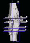
Fig. 1
Fig. 1
Three-dimensional reconstructed dorsolateral view of the normal dromedary camel carpus. Numbered sections (sagittal, 1; dorsal, 2; and transverse, 3–6) indicate the approximate levels of the selected magnetic resonance images. R, radius; Cu, ulnar carpal bone; Ci, intermediate carpal bone; Cr, radial carpal bone; C3, 3rd carpal bone; C4, fourth carpal bone; III, 3rd metacarpal bone; VI, fourth metacarpal bone
Fig. 2
Fig. 2
Sagittal T1 (A), T2 (B), STIR (C) and PD -weighted (D) high-filed MRI images obtained in the axial aspect of the carpus at the level of the intermediate carpal bone. Dorsal is to the left. R, radius; Ci, intermediate carpal bone; Ca, accessory carpal bone; III, 3rd metacarpal bone; F, fat pad; 1, common digital extensor tendon; 2, deep digital flexor tendon; 3, ulnar carpal flexor tendon; 4, radial carpal extensor tendon; 5, superficial digital flexor tendon; 6, articular cartilage of the antebrachiocarpal joint; 7, articular cartilage of the middle inter-carpal joint; 8, articular cartilage of the carpo-metacarpal joint; 9, synovial fluid; 10, accessory quartal ligament
Fig. 3
Fig. 3
Dorsal T1 (A) and T2-weighted (B) high-filed MRI images at the level of the collateral ligaments. R, radius; Rc, radial cortical bone; Cu, ulnar carpal bone; Ci, intermediate carpal bone; Cr, radial carpal bone; C2, 2nd carpal bone; C3, 3rd carpal bone; C4, fourth carpal bone; III, 3rd metacarpal bone; VI, fourth metacarpal bone; 6, articular cartilage of the antebrachiocarpal joint; 7, articular cartilage of the middle inter-carpal joint; 8, articular cartilage of the carpometacarpal joint; 11, short intercarpal ligaments; 12, long lateral collateral carpal ligament; 13, proximal part of the short lateral collateral carpal ligament; 13’, distal part of the short lateral collateral carpal ligament; 14, long medial collateral carpal ligament; 15, proximal part of the short medial collateral carpal ligament; 15’, distal part of the short medial collateral carpal ligament
Fig. 4
Fig. 4
Transverse T1 (A) and T2-weighted (B) high-filed MRI images at the level of the distal radius. Medial is to the left. R, radius; Ca, accessory carpal bone; 1, common digital extensor tendon; 2, deep digital flexor tendon; 4, radial carpal extensor tendon; 4’, tendon sheath of the radial carpal extensor tendon; 5’, origin of the superficial digital flexor tendon; 12, long lateral collateral carpal ligament; 13, proximal part of the short lateral collateral carpal ligament; 14, long medial collateral carpal ligament; 15, proximal part of the short medial collateral carpal ligament; 16, lateral digital extensor tendon; 17, extensor retinaculum; 18, radial carpal flexor tendon; 19, median artery; 20, median vein; 21, median nerve; 22, flexor retinaculum
Fig. 5
Fig. 5
Transverse T1 (A) and T2-weighted (B) and STIR (C) high-filed MRI images at the level of the proximal row of carpal bones. Cu, ulnar carpal bone; Ci, intermediate carpal bone; Cr, radial carpal bone; Ca, accessory carpal bone; 1, common digital extensor tendon; 2, deep digital flexor tendon; 4, radial carpal extensor tendon; 4’, radial carpal extensor tendon sheath; 5’, origin of the superficial digital flexor tendon; 12, long lateral collateral carpal ligament; 13, proximal part of the short lateral collateral carpal ligament; 14, long medial collateral carpal ligament; 15’, short medial collateral carpal ligament; 16, lateral digital extensor tendon; 17, extensor retinaculum; 18, radial carpal flexor tendon; 19, median artery; 20, median vein; 21, median nerve; 22, flexor retinaculum
Fig. 6
Fig. 6
Transverse T1-weighted (A) and STIR (B) high-filed MRI images at the level of the distal row of carpal bones. Medial is to the left. C2, 2nd carpal bone; C3, 3rd carpal bone; C4, 4th carpal bone; 1, common digital extensor tendon; 2, deep digital flexor tendon; 4, radial carpal extensor tendon; 5, superficial digital flexor tendon; 11, short inter-carpal ligament; 12, long lateral collateral carpal ligament; 13’, short lateral collateral carpal ligament; 14, long medial collateral carpal ligament; 15’, short medial collateral carpal ligament;16, lateral digital extensor tendon; 18, radial carpal flexor tendon; 19, median artery; 20, median vein; 23, radial artery; 24, radial vein; 25, accessoriometacarpal ligament
Fig. 7
Fig. 7
Transverse T1 (A) and PD-weighted (B) high-filed MRI images at the level of the proximal metacarpal region. Medial is to the left. III, 3rd metacarpal bone; VI, 4th metacarpal bone; 1, common digital extensor tendon; 2, deep digital flexor tendon; 4, radial carpal extensor tendon; 5, superficial digital flexor tendon; 12, long lateral collateral carpal ligament; 13’, distal part of the short lateral collateral carpal ligament; 14, long medial collateral carpal ligament; 15’, distal part of the short medial collateral carpal ligament; 16, lateral digital extensor tendon; 18, radial carpal flexor tendon; 19, median artery; 20, median vein; 23, radial artery; 24, radial vein; 25, accessoriometacarpal ligament

Similar articles

References

    1. Smuts MS, Bezuidenhout AJ. Anatomy of the Dromedary. Oxford: Clarendon; 1987. pp. 54–104.
    1. Kassab A. The normal anatomical, radiographical and ultrasonographic appearance of the carpal region of one-humped camel (camelus dromedarius). Anat Histol Embryol. 2008;37:24–9. 10.1111/j.1439-0264.2007.00790.x - DOI - PubMed
    1. Badawy AM, Marzok MA, Eshra EA. Computed tomographic arthrography of the normal dromedary camel carpus. Vet Comp Orthop Traumatol. 2016;29:188–94. 10.3415/VCOT-15-06-0112 - DOI - PubMed
    1. Kofler J. Ultrasonographic Examination of the Carpal region in cattle—normal appearance. Vet J. 2000;1:85–96.10.1053/tvjl.1998.0339 - DOI - PubMed
    1. Nagy A, Dyson S. Magnetic resonance anatomy of the carpus of the horse described from images acquired from low-field and high-field magnets. Vet Radiol Ultrasound. 2011;52:273–83. 10.1111/j.1740-8261.2010.01773.x - DOI - PubMed

MeSH terms

LinkOut - more resources