. 2024 Sep 6;8(1):61.
doi: 10.1038/s41538-024-00307-5.

A probiotic Limosilactobacillus fermentum GR-3 mitigates colitis-associated tumorigenesis in mice via modulating gut microbiome

Affiliations

A probiotic Limosilactobacillus fermentum GR-3 mitigates colitis-associated tumorigenesis in mice via modulating gut microbiome

Tuoyu Zhou et al. NPJ Sci Food. .

Abstract

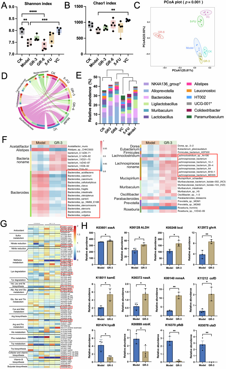
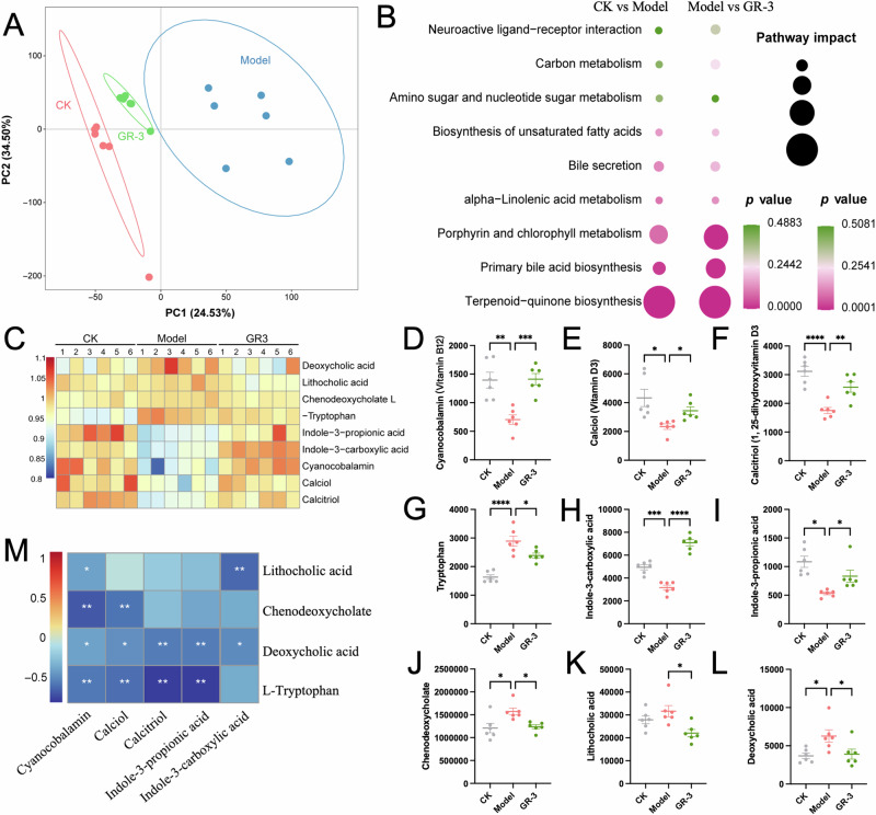
Bacterial therapy for colorectal cancer (CRC) represents a burgeoning frontier. The probiotic Limosilactobacillus fermentum GR-3, derived from traditional food "Jiangshui", exhibited superior antioxidant capacity by producing indole derivatives ICA and IPA. In an AOM/DSS-induced CRC mouse model, GR-3 treatment alleviated weight loss, colon shortening, rectal bleeding and intestinal barrier disruption by reducing oxidative stress and inflammation. GR-3 colonization in distant colon induced apoptosis and reduced tumor incidence by 51.2%, outperforming the control strain and vitamin C. The beneficial effect of GR-3 on CRC was associated with gut microbiome modulation, increasing SCFA producer Lachnospiraceae NK4A136 group and suppressing pro-inflammatory strain Bacteroides. Metagenomic and metabolic analyses revealed that GR-3 intervention upregulated antioxidant genes (xseA, ALDH) and butyrate synthesis gene (bcd), while increasing beneficial metabolites (SCFAs, ICA, IPA, VB12 and VD3) and reducing harmful secondary bile acids. Overall, GR-3 emerges as a promising candidate in CRC therapy, offering effective gut microbiome remediation.


Conflict of interest statement

The authors declare no competing interests.

Figures

Fig. 1
Fig. 1. Characterization of strains GR-3 and GR-6 in vitro.
A DPPH free radical scavenging capacity and total antioxidant capacity of different probiotics in vitro (mean ± SD; n = 3). T-AOC: total antioxidant capacity. B Cell viability of RKO and SW480 after incubation with different probiotics for 3 h (mean ± SD; n = 3). C Phylogenetic tree of strains GR-6, GR-3 and related bacteria. 16S rDNA sequences were aligned by ClustalW, and neighbor-Joining method was used to construct taxonomy tree. D Scanning electron microscope (SEM) morphology of strains GR-3 and GR-6. E Cell morphology of SW480 after incubation with strains GR-3 or GR-6 for 3 h (Bx53, Olympus; 40 × magnification). F The viability of SW480 cells was analyzed using flow cytometry following a 3-hour co-incubation with strains GR-3 or GR-6. Calcein-AM labeled cells exhibited green fluorescence and were classified into the FITC+ group, while the FITC- group represented the unlabeled cells. G mRNA expression levels of apoptotic markers p53, Bax and anti-apoptotic markers Bcl-2 in CT26 cells after co-incubation with GR-3 and GR-6 for 3 h (mean ± SD; n = 4). H Relative abundance of indole-3-carboxylic acid, indole-3-lactic acid, indole-3-propionic acid and indole-3-acetic acid in strains GR-3 or GR-6 (mean ± SD; n = 6). Different lowercase letters indicate significant differences between groups.
Fig. 2
Fig. 2. Effects of L. fermentum GR-3 on the tumor development in AOM/DSS-triggered CRC mouse model.
A Experimental procedure for development of the AOM/DSS-induced CRC model and drugs administration. Mice received a single injection of AOM, followed by a week of 2% DSS water and two weeks of regular water, repeated thrice. L. fermentum GR-3, P. acidilactici GR-6 (both at 1 × 10⁹ CFU), and vitamin C (VC, 100 mg/kg) were orally given to CRC mice. 5-FU (40 mg/kg) was injected intraperitoneally twice weekly. CK denotes the control group. B Variation in average mouse body weight across six groups (mean ± SEM). C Comparation of average body weight comparison on day 68 (mean ± SD; n = 5 for 5-FU group;n = 7 for Model group; n = 10 for other groups). D Weekly bleeding scores determined by fecal occult blood tests range from 0 (normal, negative result) to 3 (strongly positive). E Weekly assessment of fecal consistency with 0 representing normal texture and 1 representing soft and sticky feces. F Image showcasing colon lengths of different groups. G Mean colon lengths across six groups (mean ± SD). H Incidence of colon tumors in each group (mean ± SD). I H&E and Alcian blue-stained representative colon sections from the groups (100 × magnification).
Fig. 3
Fig. 3. Effect of L. fermentum GR-3 on apoptosis-related immune response in the colon of mice.
A Relative abundance of L. fermentum and P. acidilactici to total bacteria in per cm distal colonic mucosa (mean ± SD; n = 3). B Relative abundance of L. fermentum and P. acidilactici to total bacteria in per gram colon tumor (mean ± SD; n = 3). mRNA quantification of the pro-apoptosis markers p53 (C) and Bax (D), anti-apoptosis marker Bcl-2 (E), tumor cell proliferation markers NF-κB (F) andβ-catenin (G), CRC development related chemokines (Cxcl1, Cxcl2 and Cxcl3) (HJ) using qRT-PCR (mean ± SD; n = 5). The mRNA level was normalized with the mRNA level of GAPDH. K The expression levels of Bax and TLR4 in the mice colon were determined using immunohistochemistry. L Representative image of TUNEL staining on the mouse distal colon tissue. The positive rate of immunohistochemical images was analyzed using Aipathwell and marked in the lower left corner of the image.
Fig. 4
Fig. 4. Impact of L. fermentum GR-3 on oxidative stress, inflammatory response, and intestinal barrier in a CRC mouse model.
AC Protein levels of inflammatory cytokines TNF-α, IL-6 and IL-1β in colon and serum samples of mice in each group (mean ± SD; n = 4). mRNA quantification of anti-inflammatory cytokines IL-4 (D) and IL-10 (E), along with inflammatory related genes iNOS (F) and COX-2 (G) in colon tissues of mice in each group (mean ± SD; n = 5). The mRNA level was normalized with the mRNA level of GAPDH. H, I Serum and fecal total antioxidant capacity in each group (mean ± SD; n = 4). T-AOC: total antioxidant capacity. JL The contents of oxidative stress marker MDA, antioxidants reduced GSH and SOD in colon and serum samples of mice in each group (mean ± SD; n = 5). M The content of mice serum LPS in each group (mean ± SD; n = 5). N The content of serum FITC-dextran in the intestinal barrier integrity analysis (mean ± SD; n = 3). (O-R) mRNA expression levels of mucin-associated proteins MUC2 (O) and TFF3 (P), along with tight-junction structural proteins ZO-1 (Q) and Occludin (R) (mean ± SD; n = 5). The mRNA level was normalized with the mRNA level of GAPDH.
Fig. 5
Fig. 5. Impact of AOM/DSS and L. fermentum GR-3 on mouse gut microbiome.
A Evaluation of microbial diversity using the Shannon index (mean ± SD; n = 5). B Assessment of microbial richness using the Chao1 index (mean ± SD; n = 5). C PCoA plot based on Bray-Curtis dissimilarity. The p value is derived from the PERMANOVA. D Circos plot illustrating the relationship of gut microbiota at the family level in each group. E Composition analysis of gut microbiota of mice in each group at genus level. Correlation cluster analysis is performed based on Bray-Curtis distance. F Heatmap representing the abundance of microbial species. G Relative abundance of KO genes involved in representative microbial metabolism. H Significant differential KO genes between Model group and GR-3 group (mean ± SD; n = 3).
Fig. 6
Fig. 6. Impacts of AOM/DSS and L. fermentum GR-3 on gut microbiota metabolites.
A PLS-DA visualization distinguishing intestinal metabolomes of CK, Model, and GR-3 groups. B Varied metabolic pathways between CK and Model groups, and between Model and GR-3 groups. C Heatmaps showcasing the distinct metabolites modified by AOM/DSS and GR-3 treatment relative to CK. Relative abundance comparison of Vitamin B12 (D), Vitamin D3 (E), Calcitriol (F), Tryptophan (G), Indole-3-carboxylate (H), Indole-3-propionic acid (I), Chenodeoxycholate (J), Lithocholic acid (K) and Deoxycholic acid (L) in fecal contents of each group (mean ± SD; n = 6). M Correlation matrix among various metabolites were obtained using a pairwise Spearman’s rank correlation analysis. Correlations with adjusted p value less than 0.05 by the Benjamini-Hochberg FDR method are marked with white asterisk symbols.
Fig. 7
Fig. 7. The mechanism underlying the mitigation of AOM/DSS-induced CRC in mice by L. fermentum GR-3.
AOM/DSS induces CRC by triggering intestinal inflammation, oxidative stress, gut microbiota dysbiosis, metabolic disruptions, and impaired intestinal barrier function. GR-3 exerts protective effects by modulating the gut microbiome and enhancing the production of beneficial metabolites such as SCFAs, indole derivatives, and vitamins. Additionally, GR-3 alleviates oxidative stress, diminishes inflammatory response, and restores intestinal barrier integrity.

Similar articles

Cited by

References

    1. Patel, S. G., Karlitz, J. J., Yen, T., Lieu, C. H. & Boland, C. R. The rising tide of early-onset colorectal cancer: a comprehensive review of epidemiology, clinical features, biology, risk factors, prevention, and early detection. Lancet Gastroenterol. Hepatol.7, 262–274 (2022). 10.1016/S2468-1253(21)00426-X - DOI - PubMed
    1. Johnson, I. & Lund, E. Nutrition, obesity and colorectal cancer. Alimentary Pharmacol. therapeutics26, 161–181 (2007). 10.1111/j.1365-2036.2007.03371.x - DOI - PubMed
    1. Johdi, N. A. & Sukor, N. F. Colorectal Cancer Immunotherapy: Options and Strategies. Front Immunol.11, 1624 (2020). 10.3389/fimmu.2020.01624 - DOI - PMC - PubMed
    1. Weitz, J. et al. Colorectal cancer. Lancet365, 153–165 (2005). 10.1016/S0140-6736(05)17706-X - DOI - PubMed
    1. Marijnen, C. A. et al. Impact of short-term preoperative radiotherapy on health-related quality of life and sexual functioning in primary rectal cancer: report of a multicenter randomized trial. J. Clin. Oncol.23, 1847–1858 (2005). 10.1200/JCO.2005.05.256 - DOI - PubMed

LinkOut - more resources