. 2024 Sep 6;15(1):7812.
doi: 10.1038/s41467-024-52120-z.

Population genomics of Streptococcus mitis in UK and Ireland bloodstream infection and infective endocarditis cases

Affiliations

Population genomics of Streptococcus mitis in UK and Ireland bloodstream infection and infective endocarditis cases

Akuzike Kalizang'oma et al. Nat Commun. .

Abstract

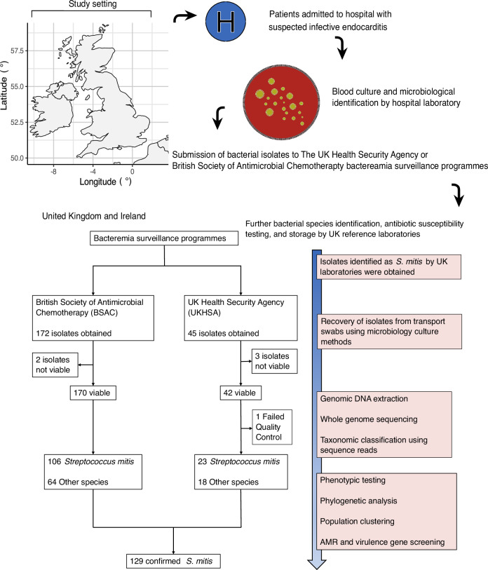
Streptococcus mitis is a leading cause of infective endocarditis (IE). However, our understanding of the genomic epidemiology and pathogenicity of IE-associated S. mitis is hampered by low IE incidence. Here we use whole genome sequencing of 129 S. mitis bloodstream infection (BSI) isolates collected between 2001-2016 from clinically diagnosed IE cases in the UK to investigate genetic diversity, antimicrobial resistance, and pathogenicity. We show high genetic diversity of IE-associated S. mitis with virtually all isolates belonging to distinct lineages indicating no predominance of specific lineages. Additionally, we find a highly variable distribution of known pneumococcal virulence genes among the isolates, some of which are overrepresented in disease when compared to carriage strains. Our findings suggest that S. mitis in patients with clinically diagnosed IE is not primarily caused by specific hypervirulent or antimicrobial resistant lineages, highlighting the accidental pathogenic nature of S. mitis in patients with clinically diagnosed IE.


Conflict of interest statement

The authors declare no competing interests.

Figures

Fig. 1
Fig. 1. Schematic of the study design and analysis workflow.
The S. mitis BSI isolates analysed were collected from clinically diagnosed IE cases, between 2001 and 2016, submitted to BSAC and UKHSA. The analysis involved WGS, population structure analysis, AMR genotyping and phenotyping, and identification of virulence genes using genotyping and bacterial genome-wide association analysis approaches to identify hypervirulent and dominant S. mitis lineages. The map of the UK and Ireland was generated by the authors in R software using the maps v4.0.0 package (https://cran.r-project.org/web/packages/maps/). The confirmed S. mitis isolates are described in Supplementary Data 1 and 2.
Fig. 2
Fig. 2. Genotypic and phenotypic characteristics of the Streptococcus mitis IE isolates.
Maximum-likelihood phylogenetic tree of 129 genotypically-confirmed IE S. mitis isolates. The phylogeny is built using 281,737 SNPs out of a total of 2,146,613 nucleotide bases from mapped sequencing reads, and is displayed next to metadata that includes GSC, ST, year of isolation, phenotypic susceptibility, and the presence or absence of acquired antibiotic resistance genes. Source data are provided as a Source Data file. The strain name in the same order as the phylogeny is shown in the source data file. The phylogenetic tree shows high genetic diversity of the IE-associated BSI S. mitis isolates, such that 98.4% of the isolates belong to different STs and GSCs. A limited number of resistance genes were explored as the analysis was limited to genes present in the curated public AMR databases. Unlike S. pneumoniae, no genomic tools and public databases are available for exploring resistance in S. mitis, to antibiotics such as beta-lactam antibiotics, that are determined through point mutations,. Phenotypic and genotypic non-susceptibility to multiple antibiotic classes was observed for several isolates throughout the surveillance period and across the phylogeny. There was good phenotypic to genotypic resistance concordance, as 96.9% (31/32) of phenotypically tetracycline-resistant isolates had a tet(M) gene, and 94.9% (74/78) of phenotypically macrolide-resistant isolates had a mef(A) or erm(B) gene. Source data are provided as a Source Data file.
Fig. 3
Fig. 3. Population structure, virulence, and antimicrobial resistance gene profiles of IE Streptococcus mitis in context of global isolates.
a Frequency plot of the STs identified across the combined IE and global S. mitis dataset. The plot shows that the UK IE S. mitis belonged to unique STs, however, two isolates isolated in 2014 and 2016 belonged to ST30, and another two isolates collected in 2007 and 2015 were assigned to ST36. Multiple carriage isolates belonging to the same ST were largely from a previous study that sampled multiple isolates from individuals. ST190 had the highest frequency of 23 isolates and was also likely due to sampling from the same individual. b Frequency plot of the GSCs identified across the combined IE and global S. mitis dataset. The plot shows that the UK IE S. mitis belonged to unique GSCs, however, two isolates isolated in 2014 and 2016 belonged to GSC27, and another two isolates collected in 2007 and 2015 were assigned to lineage GSC28. Multiple carriage isolates belonging to the same GSC were largely from a previous study that sampled multiple isolates from individuals. GSC1 had the highest frequency of 23 isolates and was also likely due to sampling from the same individual. c Maximum-likelihood phylogeny of IE and global S. mitis is built using 473,175 SNPs out of 1,237,113 core nucleotide bases. The coloured tips of the phylogeny and the first horizontal metadata bar show the isolation condition of the S. mitis isolates. From the second to fifteenth horizontal bars, the isolate metadata shows the IE UK S. mitis isolates, the GSC lineage, and virulence genes. The virulence gene matrix has 7 capsule genes (cps4Acps4F), autolysins (lytA and lytC), and pneumolysin (ply) genes. The phylogeny shows clustering of capsule genes among isolates in one region of the phylogeny, predominantly UK IE isolates. The last four horizontal bars show antimicrobial resistance (AMR) gene matrices. From the first to fourth matrix bar are the presence or absence of chloramphenicol (cat), macrolide (ermB and mefA), and tetracycline (tetM) genes. Source data are provided as a Source Data file.
Fig. 4
Fig. 4. Bacterial genome-wide association analysis.
The phylogeny of global confirmed S. mitis whole-genome sequences was built using 473,175 SNPs out of 1,237,113 nucleotide bases and annotated with disease status that was pruned to select for pairs of genetically closest carriage and invasive disease isolates. Source data are provided as a Source Data file. This phylogenetic-based approach provided an approximate matching of the isolates for the bacterial genome-wide association analysis. Source data are provided as a Source Data file.
Fig. 5
Fig. 5. Phylogenetic distribution of orthologous gene clusters differentially overrepresented between IE and non-IE S. mitis isolates among invasive isolates belonging to the genetically related and more pathogenic sister species pneumococcus.
Maximum-likelihood phylogeny of invasive bacteraemia S. pneumoniae isolates is built using 141,880 SNPs out of 1,520,986 core nucleotide bases. The coloured tips of the phylogeny and the first metadata row shows the Global Pneumococcal Sequence Cluster (GPSC), while the second row shows the serotype. The subsequent 12 rows show the presence or absence of the orthologous gene clusters (with prefix SCLS for sequence cluster locus sequence) that were overrepresented in invasive or carriage S. mitis. The phylogeny shows there are a few gene clusters that are also prevalent among the invasive pneumococcal isolates, however, most gene clusters are absent among the pneumococcal isolates. The largely absent S. mitis lytA gene was further investigated among the pneumococci using the virulence finder database, which identified the presence of pneumococcal lytA among 448 out of 493 (90.9%) isolates using pneumococcal specific lytA reference sequences in the virulence finder database. Source data are provided as a Source Data file.

References

    1. Holland, T. L. et al. Infective endocarditis. Nat. Rev. Dis. Primer2, 1–22 (2016). 10.1038/nrdp.2016.59 - DOI - PMC - PubMed
    1. Prendergast, B. D. The changing face of infective endocarditis. Heart92, 879–885 (2006). 10.1136/hrt.2005.067256 - DOI - PMC - PubMed
    1. Murdoch, D. R. et al. Clinical presentation, etiology and outcome of infective endocarditis in the 21st century: the international collaboration on endocarditis-prospective cohort study. Arch. Intern. Med.169, 463–473 (2009). 10.1001/archinternmed.2008.603 - DOI - PMC - PubMed
    1. Bin Abdulhak, A. A. et al. Global and regional burden of infective endocarditis, 1990–2010: a systematic review of the literature. Glob. Heart9, 131–143 (2014). 10.1016/j.gheart.2014.01.002 - DOI - PubMed
    1. Pant, S. et al. Trends in infective endocarditis incidence, microbiology, and valve replacement in the United States from 2000 to 2011. J. Am. Coll. Cardiol.65, 2070–2076 (2015). 10.1016/j.jacc.2015.03.518 - DOI - PubMed

Publication types

MeSH terms

LinkOut - more resources