. 2024 Sep 6;43(1):253.
doi: 10.1186/s13046-024-03177-7.

HSF1 is a prognostic determinant and therapeutic target in intrahepatic cholangiocarcinoma

Affiliations

HSF1 is a prognostic determinant and therapeutic target in intrahepatic cholangiocarcinoma

Antonio Cigliano et al. J Exp Clin Cancer Res. .

Abstract

Background: Intrahepatic cholangiocarcinoma (iCCA) is a lethal primary liver tumor characterized by clinical aggressiveness, poor prognosis, and scarce therapeutic possibilities. Therefore, new treatments are urgently needed to render this disease curable. Since cumulating evidence supports the oncogenic properties of the Heat Shock Factor 1 (HSF1) transcription factor in various cancer types, we investigated its pathogenetic and therapeutic relevance in iCCA.

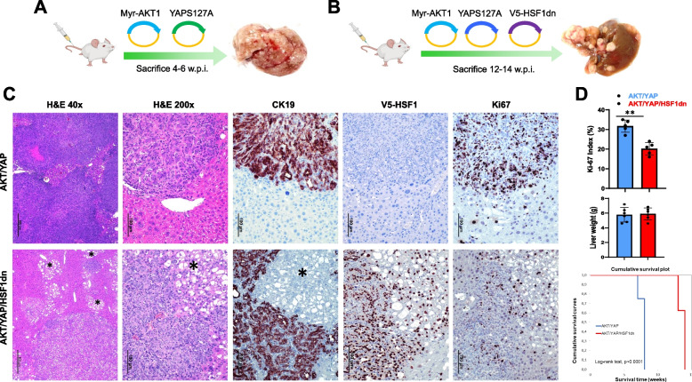
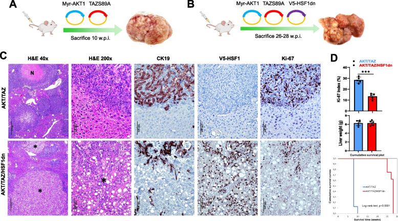
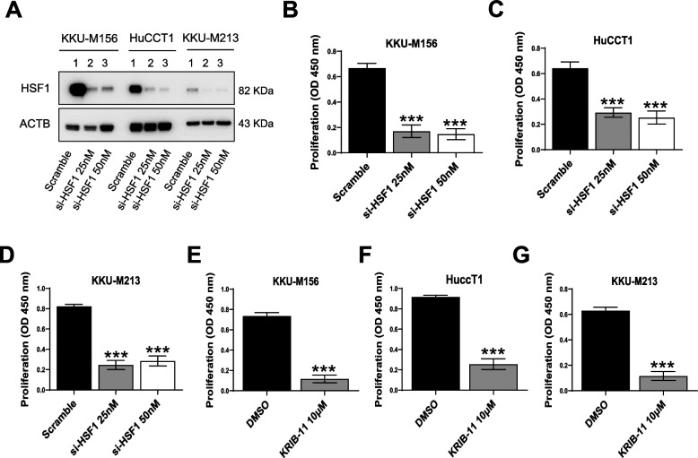
Methods: Levels of HSF1 were evaluated in a vast collection of iCCA specimens. The effects of HSF1 inactivation on iCCA development in vivo were investigated using three established oncogene-driven iCCA mouse models. In addition, the impact of HSF1 suppression on tumor cells and tumor stroma was assessed in iCCA cell lines, human iCCA cancer-associated fibroblasts (hCAFs), and patient-derived organoids.

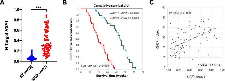
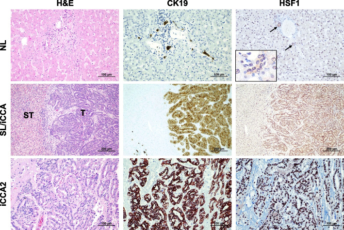
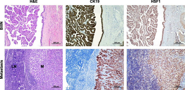
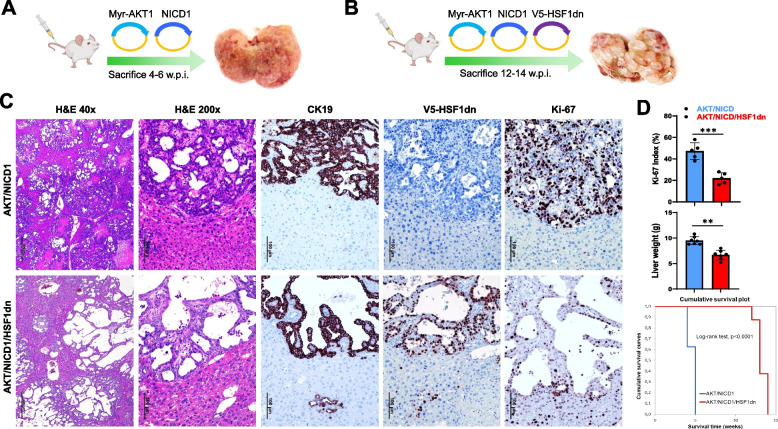
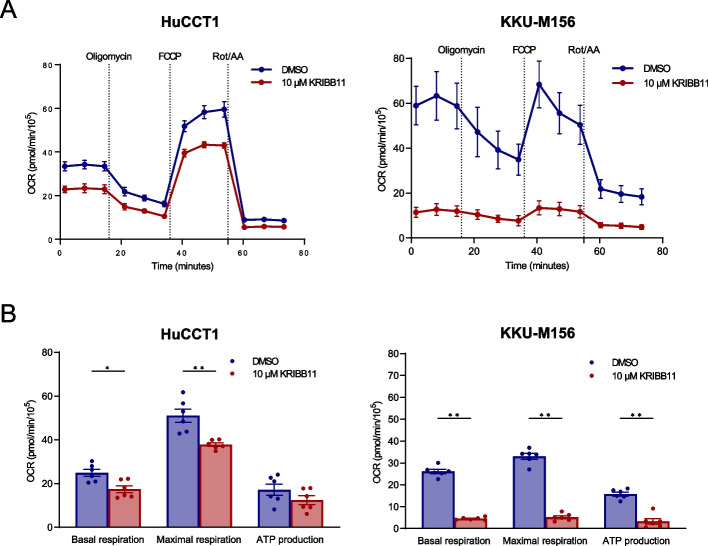
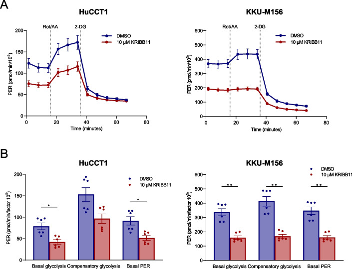
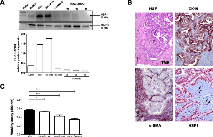
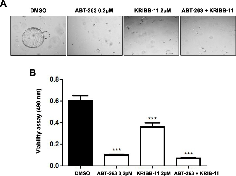
Results: Human preinvasive, invasive, and metastatic iCCAs displayed widespread HSF1 upregulation, which was associated with a dismal prognosis of the patients. In addition, hydrodynamic injection of a dominant-negative form of HSF1 (HSF1dn), which suppresses HSF1 activity, significantly delayed cholangiocarcinogenesis in AKT/NICD, AKT/YAP, and AKT/TAZ mice. In iCCA cell lines, iCCA hCAFs, and patient-derived organoids, administration of the HSF1 inhibitor KRIBB-11 significantly reduced proliferation and induced apoptosis. Cell death was profoundly augmented by concomitant administration of the Bcl-xL/Bcl2/Bcl-w inhibitor ABT-263. Furthermore, KRIBB-11 reduced mitochondrial bioenergetics and glycolysis of iCCA cells.

Conclusions: The present data underscore the critical pathogenetic, prognostic, and therapeutic role of HSF1 in cholangiocarcinogenesis.

Keywords: HSF1; Intrahepatic cholangiocarcinoma; KRIBB-11; Mouse models; Navitoclax.


Conflict of interest statement

The authors declare that they have no competing interests.

Figures

Fig. 1
Fig. 1
Upregulation of the HSF1 gene in human intrahepatic cholangiocarcinoma specimens and correlation with survival and proliferation. A Quantitative real-time RT-PCR values of HSF1 gene are significantly higher in the tumors (T) (n = 72) compared with corresponding non-tumorous counterparts (ST). B Kaplan-Meyer curve showing that HSF1 mRNA levels inversely correlate with patients’ survival in this disease. C Linear regression analysis revealing that HSF1 mRNA expression directly correlates with the tumors’ proliferation activity (as assessed with the Ki-67 index) in patients
Fig. 2
Fig. 2
Representative immunohistochemistry patterns of HSF1 protein in human intrahepatic cholangiocarcinoma (iCCA; n = 186) and corresponding non-tumorous livers. The upper panels show a human normal liver (NL) displaying weak to moderate HSF1 immunoreactivity in hepatocytes and biliary cells (indicated by arrows). Staining of cholangiocytes (indicated by arrows) is better appreciable in the inset. Middle panels show the immunohistochemical staining of HSF1 in a non-tumorous surrounding tissue and one human iCCA specimen. The staining pattern for HSF1 is significantly more pronounced in the tumor compartment (T) compared with the neighboring non-tumorous surrounding tissue (ST), which exhibits faint HSF1 staining. Lower panels depict a second tumor (iCC2), characterized by intense nuclear HSF1 immunoreactivity. CK19 staining was used as a biliary marker. Abbreviation: H&E, hematoxylin and eosin staining. Original magnifications: 200x in the upper and lower panels, 100x in the middle panels, and 400x in the inset. Scale bar: 100 µm in the top and low panels, 200 µm in the middle panels
Fig. 3
Fig. 3
HSF1 is upregulated in human intrahepatic cholangiocarcinoma preinvasive lesions and metastases. Upper panels: Representative immunohistochemistry of HSF1 protein in a preinvasive lesion (one biliary intraepithelial neoplasia or BilIN specimen is shown here). The lesion exhibits pronounced nuclear immunoreactivity for HSF1. CK19 staining was used as a biliary marker. Lower panels: an iCCA lymph node metastasis displaying pronounced HSF1 nuclear immunolabeling in the tumor metastasis (M) and scattered nuclear positiveness in the lymph node (LN) compartment. CK19 staining was used as a biliary marker. Original magnification: 100x; scale bar: 200 μm. Abbreviations: H&E, hematoxylin and eosin staining
Fig. 4
Fig. 4
Suppression of HSF1 activity delays cholangiocarcinogenesis and reduces the proliferation and aggressiveness of AKT/NICD1 mouse lesions. AB Study design. Briefly, wild-type FVB/N mice were subjected to hydrodynamic tail vein injection of either AKT/NICD1/pT3 (control) or AKT/NICD1/HSF1dn plasmids. HSF1dn is the dominant-negative form of the HSF1 transcription factor, whose overexpression effectively inhibits the endogenous HSF1 activity. C While liver lesions from AKT/NICD1 mice consisted of invasive and proliferative (as assessed by positive immunoreactivity for Ki-67) intrahepatic cholangiocarcinomas (upper panels), cystic lesions (denominated cystic adenomas and cystic adenocarcinomas) with low proliferation rate occupied the liver parenchyma of AKT/NICD1/HSF1dn mice. As expected, V5-tagged-HSF1dn staining was observed only in AKT/NICD1/HSF1dn mice. D Liver weight and proliferative activity were significantly higher in AKT/NICD1 mice than in AKT/NICD1/HSF1dn mice. Moreover, the survival curve showed significantly longer survival of AKT/NICD1/HSF1dn mice. Student’s t-test: ***, P < 0.0001; **, P < 0.001. Original magnifications: 40x and 200x; scale bar: 500 μm in 40x and 100 μm in 200x. Abbreviations: H&E, hematoxylin and eosin staining
Fig. 5
Fig. 5
Suppression of HSF1 activity delays cholangiocarcinogenesis and reduces the proliferation and aggressiveness of AKT/YAP mouse lesions. A, B Study design. Briefly, wild-type FVB/N mice were subjected to hydrodynamic tail vein injection of either AKT/YAPS127A/pT3 (control; AKT/YAP mice) or AKT/YAPS127A/HSF1dn (AKT/YAP/HSF1dn mice) plasmids. In particular, HSF1dn is the dominant-negative form of the HSF1 transcription factor, whose overexpression effectively inhibits the endogenous HSF1 activity. C Liver lesions from AKT/YAP mice consisted of invasive and proliferative (as assessed by positive immunoreactivity for Ki-67) intrahepatic cholangiocarcinomas (upper panels). In contrast, neoplastic lesions from AKT/YAP/HSF1dn consisted of both hepatocellular, clear-cell (indicated by an asterisk), and cholangiocellular lesions with low proliferation rates. The hepatocellular lesions were CK19-negative, whereas the cholangiocellular lesions were CK19-positive. As expected, V5-tagged-HSF1dn staining was observed only in AKT/YAP/HSF1dn mice. D While liver weight was equivalent in the two mouse cohorts, the proliferative activity was significantly higher in AKT/YAP mice than in AKT/YAP/HSF1dn mice. Moreover, the survival curve showed significantly longer survival of AKT/YAP/HSF1dn mice. Student’s t-test: **, P < 0.001. Original magnifications: 40x and 200x; scale bar: 500 μm in 40x and 100 μm in 200x. Abbreviations: H&E, hematoxylin and eosin staining
Fig. 6
Fig. 6
Suppression of HSF1 activity delays cholangiocarcinogenesis and reduces the proliferation and aggressiveness of AKT/TAZ mouse lesions. A, B Study design. Briefly, wild-type FVB/N mice were subjected to hydrodynamic tail vein injection of either AKT/TAZS89A/pT3 (control; AKT/TAZ mice) or AKT/TAZS89A/HSF1dn (AKT/TAZ/HSF1dn mice) plasmids. HSF1dn is the dominant-negative form of the HSF1 transcription factor, whose overexpression effectively inhibits the endogenous HSF1 activity. C Liver lesions from AKT/TAZ mice consisted of invasive and proliferative (as assessed by positive immunoreactivity for Ki-67) intrahepatic cholangiocarcinomas (upper panels) with frequent necrotic areas (N). In contrast, neoplastic lesions from AKT/TAZ/HSF1dn consisted of both hepatocellular, clear-cell (indicated by asterisks), and cholangiocellular lesions with low proliferation rates. The hepatocellular lesions were CK19-negative, whereas the cholangiocellular lesions were CK19-positive. As expected, V5-tagged-HSF1dn staining was observed only in AKT/TAZ/HSF1dn mice. D While liver weight was equivalent in the two mouse cohorts, the proliferative activity was significantly higher in AKT/TAZ mice than in AKT/TAZ/HSF1dn mice. Moreover, the survival curve showed significantly longer survival of AKT/TAZ/HSF1dn mice. Student’s t-test: **, P < 0.0001. Original magnifications: 40x and 200x; scale bar: 500 μm in 40x and 100 μm in 200x. Abbreviations: H&E, hematoxylin and eosin staining
Fig. 7
Fig. 7
Effect of HSF1 inhibition on the growth of intrahepatic cholangiocarcinoma cell lines. A Inhibition of HSF1 via specific small-interfering RNA in KKU-M156, HuCCT1, and KKU-M213 human intrahepatic cholangiocarcinoma (iCCA) cell lines, as assessed by Western blot analysis. B-D BrdU incorporation assay was conducted on B KKU-M156, C HuCCT1, and D KKUM-213 cells. Analysis was conducted 48 h after siRNA administration. Two siRNA concentrations (25 and 50 nm) were applied. Scramble-treated cells served as control. EG KKU-M156, HuCCT1, and KKUM-213 cells were also treated for 48 h with 10 µM KRIBB-11, an HSF1 inhibitor, and BrdU incorporation was assessed. DMSO-treated cells served as controls. The values in optical densities (OD) at 450 nm are presented. All results are expressed as mean ± SD of three independent experiments in triplicate. For statistical analysis, Tukey’s multiple comparisons test was performed (*** p < 0.0001, vs. scramble and DMSO). Abbreviation: si-HSF1, small-interfering RNA against HSF1
Fig. 8
Fig. 8
The HSF1 inhibitor KRIBB11 decreases the mitochondrial bioenergetics in cholangiocarcinoma cell lines. A Cell Mito Stress test profiles of human HuCCT1 and KKU-M156 cell lines treated with 10 µM KRIBB11 and matching DMSO concentration for 24 h. Seahorse XF Cell Mito Stress Tests (Agilent) were performed employing serial injections of oligomycin, FCCP, and Rot/AA; Hoechst 33342 was injected last for nuclei staining required for normalization. OCR was measured in real-time using the Seahorse XF HS mini analyzer. B Changes in basal respiration, maximal respiration and ATP production. Data were background corrected and normalized to the mean fluorescent intensity per well; a factor 105 was applied. Graphs depict the mean ± SEM of two independent experiments each performed in technical triplicates (multiple Mann–Whitney tests; *p < 0.05; **p < 0.01). Abbreviations: OCR, oxidative consumption rate (in pmol/min); FCCP, carbonyl cyanide-p-trifluoromethoxyphenylhydrazone; Rot/AA, rotenone/antimycin A
Fig. 9
Fig. 9
The HSF1 inhibitor KRIBB11 reduces the glycolytic metabolism of cholangiocarcinoma cell lines. A Glycolytic rate profiles of human HuCCT1 and KKU-M156 cell lines treated with 10 µM KRIBB11 and matching DMSO concentration for 24 h. Seahorse XF Glycolytic Rate assays (Agilent) were performed employing serial injections of Rot/AA, and 2-DG and Hoechst 33342 for nuclei staining; the PER reflecting the glycolytic function was measured using the Seahorse XF HS mini analyzer. B Changes in basal glycolysis, compensatory glycolysis, and basal PER. Data were background corrected and normalized to cell number as determined by Hoechst 33342 nuclei staining; a factor of 105 was applied. Graphs depict the mean ± SEM of two independent experiments each performed in technical triplicates (multiple Mann–Whitney tests; *p < 0.05). Abbreviations: PER, proton efflux rate (in pmol/min); Rot/AA, rotenone/antimycin A; 2-DG, 2-deoxy-D-glucose
Fig. 10
Fig. 10
The HSF1 inhibitor KRIBB-11 reduces the growth in vitro of human cancer-associated fibroblasts. A Levels of HSF1 in human HuCCT1, RBE, KKU-M156, and KKU-M213 intrahepatic cholangiocarcinoma (iCCA) cell lines, and in human cancer-associated fibroblasts (hCAFs), as determined by Western blot analysis. GAPDH was used as a housekeeping protein and protein values of each sample are reported below. B Representative immunohistochemistry showing immunoreactivity for HSF1 in the tumor (T) and the tumor microenvironment (TME) compartments. Arrows indicate fibroblasts showing moderate immunolabeling for HSF1 protein. Alpha-smooth-muscle (α-SMA) staining was used as a tumor stroma marker, while CK19 staining was used as a biliary marker. Original magnification: 200x for H&E, CK19, and α-SMA; 400x for HSF1; scale bar: 100 μm for H&E, CK19, and α-SMA; 50 μm for HSF1. Abbreviations: H&E, hematoxylin and eosin staining. C The co-treatment with ABT-263 and KRIBB-11 inhibits the cell viability in iCCA hCAFs. Effect of ABT-263, KRIBB-11, and their co-administration on the viability of iCCA hCAFs, grown for 72 h in culture medium supplemented with 10% FBS. Note the higher anti-growth effects of the two combined drugs than single treatments. Experiments were repeated three times in triplicate. *P < 0.05 and ***P < 0.001, as calculated with a One-way ANOVA test
Fig. 11
Fig. 11
The HSF1 inhibitor KRIBB-11 and the apoptosis inducer ABT-263 reduce the viability of iCCA organoids. A, B Effect of ABT-263, KRIBB-11, and their combination on the viability of iCCA organoids, treated for 10 days in the specific medium of the hepatic organoid. Experiments were repeated three times in triplicate. ***P < 0.001, as calculated with a One-way ANOVA test

References

    1. Banales JM, Cardinale V, Carpino G, Marzioni M, Andersen JB, Invernizzi P, et al. Expert consensus document: cholangiocarcinoma: current knowledge and future perspectives consensus statement from the European Network for the Study of Cholangiocarcinoma (ENS-CCA). Nat Rev Gastroenterol Hepatol. 2016;13:261–80. 10.1038/nrgastro.2016.51 - DOI - PubMed
    1. Banales JM, Marin JJG, Lamarca A, Rodrigues PM, Khan SA, Roberts LR, et al. Cholangiocarcinoma 2020: the next horizon in mechanisms and management. Nat Rev Gastroenterol Hepatol. 2020;17:557–88. 10.1038/s41575-020-0310-z - DOI - PMC - PubMed
    1. Ilyas SI, Khan SA, Hallemeier CL, Kelley RK, Gores GJ. Cholangiocarcinoma - evolving concepts and therapeutic strategies. Nat Rev Clin Oncol. 2018;15:95–111. 10.1038/nrclinonc.2017.157 - DOI - PMC - PubMed
    1. El-Diwany R, Pawlik TM, Ejaz A. Intrahepatic cholangiocarcinoma. Surg Oncol Clin N Am. 2019;28:587–99. 10.1016/j.soc.2019.06.002 - DOI - PubMed
    1. Acher AW, Paro A, Elfadaly A, Tsilimigras D, Pawlik TM. Intrahepatic cholangiocarcinoma: a summative review of biomarkers and targeted therapies. Cancers. 2021;13:5169. 10.3390/cancers13205169 - DOI - PMC - PubMed

Substances