. 2025 Jun;16(3):817-830.
doi: 10.1007/s12975-024-01266-5. Epub 2024 Sep 7.

Extracellular Vesicles Obtained from Hypoxic Mesenchymal Stromal Cells Induce Neurological Recovery, Anti-inflammation, and Brain Remodeling After Distal Middle Cerebral Artery Occlusion in Rats

Affiliations

Extracellular Vesicles Obtained from Hypoxic Mesenchymal Stromal Cells Induce Neurological Recovery, Anti-inflammation, and Brain Remodeling After Distal Middle Cerebral Artery Occlusion in Rats

Mihaela Abuzan et al. Transl Stroke Res. 2025 Jun.

Abstract

Small extracellular vesicles (sEVs) obtained from mesenchymal stromal cells (MSCs) have shown considerable promise as restorative stroke treatment. In a head-to-head comparison in mice exposed to transient proximal middle cerebral artery occlusion (MCAO), sEVs obtained from MSCs cultured under hypoxic conditions particularly potently enhanced long-term brain tissue survival, microvascular integrity, and angiogenesis. These observations suggest that hypoxic preconditioning might represent the strategy of choice for harvesting MSC-sEVs for clinical stroke trials. To test the efficacy of hypoxic MSCs in a second stroke model in an additional species, we now exposed 6-8-month-old Sprague-Dawley rats to permanent distal MCAO and intravenously administered vehicle, platelet sEVs, or sEVs obtained from hypoxic MSCs (1% O2; 2 × 10⁶ or 2 × 10⁷ cell equivalents/kg) at 24 h, 3, 7, and 14 days post-MCAO. Over 28 days, motor-coordination recovery was evaluated by rotating pole and cylinder tests. Ischemic injury, brain inflammatory responses, and peri-infarct angiogenesis were assessed by infarct volumetry and immunohistochemistry. sEVs obtained from hypoxic MSCs did not influence infarct volume in this permanent MCAO model, but promoted motor-coordination recovery over 28 days at both sEV doses. Ischemic injury was associated with brain ED1+ macrophage infiltrates and Iba1+ microglia accumulation in the peri-infarct cortex of vehicle-treated rats. Hypoxic MSC-sEVs reduced brain macrophage infiltrates and microglia accumulation in the peri-infarct cortex. In vehicle-treated rats, CD31+/BrdU+ proliferating endothelial cells were found in the peri-infarct cortex. Hypoxic MSC-sEVs increased the number of CD31+/BrdU+ proliferating endothelial cells. Our results provide evidence that hypoxic MSC-derived sEVs potently enhance neurological recovery, reduce neuroinflammation. and increase angiogenesis in rat permanent distal MCAO.

Keywords: Angiogenesis; Exosome; Hypoxic preconditioning; Ischemic stroke; Macrophage; Permanent focal cerebral ischemia.


Conflict of interest statement

Declarations. Competing interests: DMH and BG hold patents for the application of extracellular vesicles for the treatment of inflammatory conditions (EP2687219A1; US9877989B2). Ethics Approval: The experiments conducted at the University of Medicine and Pharmacy Craiova received approval from the Institutional Animal Care and Use Committee (#112–15-11–2017). The experiments were carried out in accordance with national regulations in accordance with the EU Directive 2010/63/EU on the care and use of laboratory animals. Randomization of experiments was strictly implemented following the ARRIVE criteria to ensure unbiased data analysis, including behavioral tests, while the examiners involved were kept completely blinded to the experimental conditions. The authors will provide supporting data upon reasonable request to support the conclusions derived from this study. Informed Consent: N/A. Conflict of Interest: DMH and BG hold patents for the application of extracellular vesicles for the treatment of inflammatory conditions (EP2687219A1; US9877989B2).

Figures

Fig. 1
Fig. 1
Experimental design and time course of body weight changes in rats subjected to permanent distal middle cerebral artery occlusion (MCAO). A total of 52 animals were included in this study. A Temporal sequence of experimental interventions. Rats exposed to permanent distal MCAO received intravenous injections of vehicle, platelet-derived small extracellular vesicles (sEVs), or sEVs obtained from hypoxic (1%O2) mesenchymal stromal cells (MSCs; 2 × 106 or 2 × 107 cell equivalents/kg) at four different time points, that is 1, 3, 7, and 14 days after MCAO. Motor-coordination deficits were assessed through behavioral analysis from 3 to 28 days post-MCAO (that is, from 2 to 27 days post-treatment onset). Rats were sacrificed at 28 days post-MCAO for histochemical analysis of brain tissue. B Body weight development of rats exposed to permanent distal MCAO (n = 10 animals per group). The weight of rats receiving vehicle modestly decreased by ~ 5% subsequent to MCAO. Hypoxic MSC-sEV administration nominally, but not statistically significantly reduced this weight reduction. The body weight of all groups almost completely recovered within 14–21 days post-MCAO. Data did not statistically differ between groups. Data are mean ± standard error of the mean (SEM) values
Fig. 2
Fig. 2
Hypoxic MSC-sEV delivery enhances post-ischemic motor-coordination recovery. Motor-coordination deficits in A the rotating pole test at 3 rpm, B the rotating pole test at 6 rpm, and C the cylinder test performance of rats exposed to permanent distal MCAO, which were intravenously treated with vehicle, platelet-derived sEVs or sEVs obtained from hypoxic MSCs (1% O2; 2 × 106 or 2 × 107 cell equivalents/kg) at 1, 3, 7, and 14 days post-MCAO (n = 10 animals per group). The rotating pole test measures the duration required to cross a pole rotating at a defined speed of 3 or 6 rpm and the cylinder test evaluates asymmetry in forelimb usage, with more negative results indicating a decreased use of the left affected limb. Note that hypoxic MSC-sEV delivery improved motor coordination recovery in rats exposed to permanent distal MCAO at both doses. *p < 0.05/**p < 0.01 compared with vehicle; #p < 0.05 compared with platelet-sEVs. Data are shown as mean ± SEM values
Fig. 3
Fig. 3
Hypoxic MSC-sEVs do not influence infarct volume after permanent distal MCAO. Infarct volume assessed by methyl green/pyronine Y staining in the brains of permanent distal MCAO rats, which obtained vehicle, platelet-derived sEVs or sEVs obtained from hypoxic MSCs (2 × 106 or 2 × 107 cell equivalents/kg) at 1, 3, 7, and 14 days post-MCAO and which were sacrificed at 28 days post-MCAO (that is, 27 days post-treatment onset). Representative microphotographs are also shown. Note that reproducible brain infarcts covering the lateral parietal cortex were obtained. Infarct volume was not influenced by MSC-sEV delivery at both doses (2 × 106 or 2 × 107 MSC equivalents/kg). Data did not statistically differ between groups. Data are mean ± SEM values. Data of individual animals are depicted as dots. Scale bar, 1 mm
Fig. 4
Fig. 4
Hypoxic MSC-sEVs decrease peri-infarct brain macrophage infiltrates. Density of activated ED1+ macrophages in the peri-infarct tissue of rats, which were treated with vehicle, platelet-derived sEVs or sEVs obtained from hypoxic MSCs (2 × 106 or 2 × 107 cell equivalents/kg) at 1, 3, 7, and 14 days post-MCAO and which were sacrificed at 28 days post-MCAO. Macrophages were discriminated from monocytes through their size, which is ~ 50% larger than that of monocytes. Hence, monocytes were excluded from cell countings. Representative microphotographs are depicted. Note that the MSC-sEVs at a dose of 2 × 107 MSC equivalents/kg reduce peri-infarct ED1+ macrophage infiltrates. IC, ischemic cortex. **p < 0.01 compared with vehicle. Data are mean ± SD values. Data of individual animals are depicted as dots. Scale bar, 20 µm
Fig. 5
Fig. 5
Hypoxic MSC-sEVs decrease peri-infarct microglia accumulation. Density of Iba1+ microglia in the peri-infarct brain tissue of rats, which were treated with vehicle, platelet-derived sEVs or sEVs obtained from hypoxic MSCs (2 × 106 or 2 × 107 cell equivalents/kg) at 1, 3, 7, and 14 days post-MCAO and which were sacrificed at 28 days post-MCAO. Representative microphotographs are shown. Note that the MSC-sEVs at a dose of 2 × 106 MSC equivalents/kg reduce peri-infarct Iba1+ microglia accumulation. PI, peri-infarct cortex. **p < 0.01 compared with vehicle. Data are mean ± SD values. Data of individual animals are depicted as dots. Scale bar, 20 µm
Fig. 6
Fig. 6
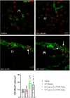
Hypoxic MSC-sEVs enhance post-ischemic angiogenesis in the peri-infarct cortex. Density of CD31+/BrdU+ proliferating endothelial cells in the peri-infarct cortex of rats, which were treated with vehicle, platelet-derived sEVs or hypoxic MSC-sEVs (2 × 106 or 2 × 107 cell equivalents/kg) at 1, 3, 7, and 14 days post-MCAO, followed by animal sacrifice at 28 days post-MCAO. For proliferating endothelial cell labeling, BrdU (50 mg/kg) was intraperitoneally administered from 8 to 18 days post-MCAO. Representative microphotographs are shown. Arrows depict double-labeled cells. Note that MSC-sEVs at doses of 2 × 106 and 2 × 107 MSC equivalents/kg increase endothelial proliferation in the peri-infarct cortex. PI, peri-infarct cortex. *p < 0.05/**p < 0.01 compared with vehicle/#p < 0.05 compared with platelet-sEVs. Data are mean ± SD values. Data of individual animals are depicted as dots. Scale bar, 20 µm

Similar articles

Cited by

References

    1. Buchhold B, Mogoanta L, Suofu Y, Hamm A, Walker L, Kessler C, Popa-Wagner A. Environmental enrichment improves functional and neuropathological indices following stroke in young and aged rats. Restor Neurol Neurosci. 2007;25(5–6):467–84. - PubMed
    1. Buga AM, Margaritescu C, Scholz CJ, Radu E, Zelenak C, Popa-Wagner A. Transcriptomics of post-stroke angiogenesis in the aged brain. Front Aging Neurosci. 2014;6:44. 10.3389/fnagi.2014.00044. - PMC - PubMed
    1. Doeppner TR, Herz J, Gorgens A, Schlechter J, Ludwig AK, Radtke S, de Miroschedji K, Horn PA, Giebel B, Hermann DM. Extracellular vesicles improve post-stroke neuroregeneration and prevent postischemic immunosuppression. Stem Cells Transl Med. 2015;4(10):1131–43. 10.5966/sctm.2015-0078. - PMC - PubMed
    1. Dumbrava DA, Surugiu R, Börger V, Ruscu M, Tertel T, Giebel B, Hermann DM, Popa-Wagner A. Mesenchymal stromal cell-derived small extracellular vesicles promote neurological recovery and brain remodeling after distal middle cerebral artery occlusion in aged rats. Geroscience. 2022;44(1):293–310. 10.1007/s11357-021-00483-2. - PMC - PubMed
    1. Florian B, Vintilescu R, Balseanu AT, Buga AM, Grisk O, Walker LC, Kessler C, Popa-Wagner A. Long-term hypothermia reduces infarct volume in aged rats after focal ischemia. Neurosci Lett. 2008;438(2):180–5. 10.1089/neu.2008.0806. - PubMed

MeSH terms

LinkOut - more resources