. 2024 Nov:57:101050.
doi: 10.1016/j.neo.2024.101050. Epub 2024 Sep 6.

FABP4 facilitates epithelial-mesenchymal transition via elevating CD36 expression in glioma cells

Affiliations

FABP4 facilitates epithelial-mesenchymal transition via elevating CD36 expression in glioma cells

Zhongsheng You et al. Neoplasia. 2024 Nov.

Abstract

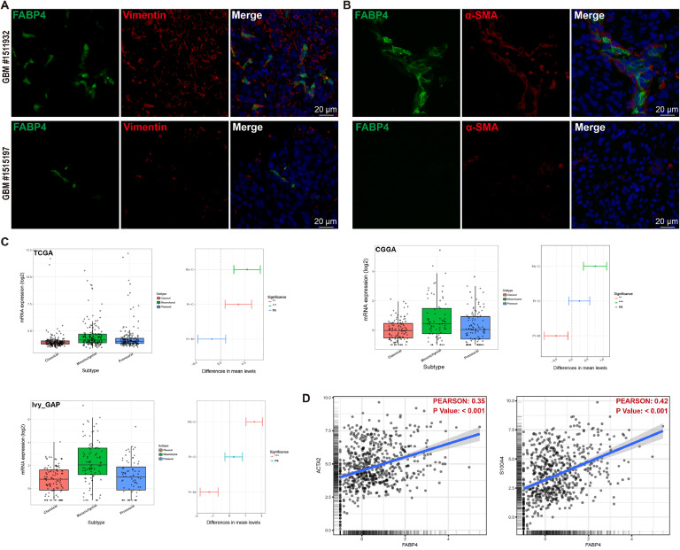
Glioblastoma multiforme (GBM) is the most aggressive brain tumor with poor prognosis. A better understanding of mechanisms concerned in glioma invasion might be critical for treatment optimization. Given that epithelial-mesenchymal transition in tumor cells is closely associated with glioma progression and recurrence, identifying pivotal mediators in GBM EMT process is urgently needed. As a member of Fatty acid binding protein (FABP) family, FABP4 serves as chaperones for free fatty acids and participates in cellular process including fatty acid uptake, transport, and metabolism. In this study, our data revealed that FABP4 expression was elevated in human GBM samples and correlated with a mesenchymal glioma subtype. Gain of function and loss of function experiments indicated that FABP4 potently rendered glioma cells increased filopodia formation and cell invasiveness. Differential expression genes analysis and GSEA in TCGA dataset revealed an EMT-related molecular signature in FABP4-mediated signaling pathways. Cell interaction analysis suggested CD36 as a potential target regulated by FABP4. Furthermore, in vitro mechanistic experiments demonstrated that FABP4-induced CD36 expression promoted EMT via non-canonical TGFβ pathways. An intracranial glioma model was constructed to assess the effect of FABP4 on tumor progression in vivo. Together, our findings demonstrated a critical role for FABP4 in the regulation invasion and EMT in GBM, and suggest that pharmacological inhibition of FABP4 may represent a promising therapeutic strategy for treatment of GBM.

Keywords: CD36; Epithelial-mesenchymal transition; FABP4; Glioblastoma; Tumor invasion.


Conflict of interest statement

Declaration of competing interest The authors declare that they have no known competing financial interests or personal relationships that could have appeared to influence the work reported in this paper.

Figures

Fig 1
Fig. 1
FABP4 is abundantly expressed in GBM samples. (a) Representative MRI, H&E and FABP4 IHC staining images of specimens from patients with GBM and lower grade glioma. Fluorescence staining for FABP4 of GBM and lower grade glioma patients. (b) IHC images of high and low grade glioma from Human Protein Atlas datasets. Scale bar= 50/20 μm. (c) IHC score of FABP4 in human glioma specimens of different grades. (d) Correlation of FABP4 expression with gender, age, tumor size and tumor grade. (e) Protein level of FABP4 in primary glioma cells and cell lines. (f) Fluorescence staining of FABP4 in glioma cells. (g) Secretion level of FABP4 in glioma cells. Data are expressed as Mean ± SD.**p < 0.01, ***p < 0.001. H&E, hematoxylin and eosin. IHC, Immunohistochemistry.
Fig 2
Fig. 2
FABP4 is closely associated with mesenchymal subtype in GBM. (a) Fluorescence staining of FABP4 and vimentin in GBM samples. (b) Fluorescence staining of FABP4 and α-SMA in GBM samples. (c) FABP4 mRNA expression in different molecular subtypes of GBM samples from TCGA, CGGA and Ivy database. (d) Correlation between the expression of FABP4 and EMT-related markers (ACTA2 and S100A4) in TCGA GBM dataset was evaluated. Results are represented as Mean ± SD of biologically triplicate assays. *p < 0.05, **p < 0.01, ***p < 0.001.
Fig 3
Fig. 3
FABP4 confers increased invasiveness in glioma cells. (a) primary glioma cells were transfected with empty vector or FABP4-expressing lentiviral vector, fluorescent staining of FABP4 and Phalloidin were assessed. White arrowheads indicate filopodia on glioma cells. (b) Glioma cells were treated with PBS or rhFABP4, FABP4 and Phalloidin expressions were evaluated. (c) primary glioma cells with scrambled shRNA or shFABP4 were stained with FABP4 and Phalloidin. (d) Invasion abilities of glioma cells with FABP4 expression were assessed. (e) Gene set enrichment analysis (GSEA) was performed according to FABP4 expression in TCGA GBM dataset showed enriched pathways associated with cell invasiveness. Results are represented as Mean ± SD of biologically triplicate assays. *p < 0.05, **p < 0.01, ***p < 0.001.
Fig 4
Fig. 4
FABP4 induces EMT process in GBM. (a) differential gene expression analysis between high and low FABP4 groups from TCGA GBM dataset. Several key molecules related to EMT are indicated. (b) GSEA analysis reveals a significant correlation between FABP4 expression and EMT process in TCGA GBM dataset. (c) Expression of EMT-related molecules in glioma cells that transfected with FABP4-lentiviral vector or treated with recombinant human FABP4 protein. (d) Immunofluorescence analysis of N-Cadherin and Vimentin in glioma cells with FABP4 over-expression. (e) Immunoblot analysis of N-Cadherin and Vimentin in glioma cells when FABP4 is downregulated. (f) Immunofluorescence analysis of N-Cadherin in glioma cells treated with shRNA targeting FABP4. Scale bar=20 μm. Results are represented as Mean ± SD of biologically triplicate assays. *p < 0.05, **p < 0.01, ***p < 0.001. ns, not significant.
Fig 5
Fig. 5
FABP4-induced EMT is mediated in CD36-dependent manner. (a) Protein-protein interaction analysis of FABP4 via using STRING database. (b) CD36 mRNA expression in high or low FABP4 group from TCGA GBM dataset. (c) Immunoblot analysis of CD36 and PPARG in glioma cells with FABP4 upregulation or downregulation. (d) Transcriptional levels of CD36 and PPARG in glioma cells upon FABP4 overexpression. (e) CD36 mRNA expression in glioma samples with different grades. (f) CD36 mRNA expression in three GBM subtypes. (g) Kaplan-Meier survival analysis for CD36 in patients with glioma from TCGA dataset. (h) Correlation between FABP4 and CD36 in TCGA glioma dataset. (i) Immunofluorescence staining of CD36 in glioma cells with FABP4 upregulation. (j) Indicated proteins in glioma cells treated with exogenous FABP4 followed by CD36 knockdown. (k) Immunofluorescence staining of N-Cadherin and Vimentin in FABP4-expressed glioma cells treated with CD36 downregulation. (l) Glioma cells with stable FABP4 expression are treated with CD36 knockdown, F-actin staining is detected with phalloidin. White arrowheads indicate filopodia on glioma cells. Scale bar=50 μm. Results are represented as Mean ± SD of biologically triplicate assays. *p < 0.05, **p < 0.01, ***p < 0.001. ns, not significant.
Fig 6
Fig. 6
CD36 enhances EMT process by activating non-canonical TGFβ pathways. (a) Association between CD36 mRNA level and EMT proteins (PAI.1, fibronectin) in TCPA. (b) Primary glioma cells with empty vector or CD36 transfection were treated with 100 pM TGFβ1 for 6 h, indicated proteins of canonical and non-canonical TGFβ pathways were examined. (c) Immunofluorescence staining of ROCK1 and CD36 in glioma cells with EV or CD36 transfection. (d) PGCs with (without) CD36 knockdown were treated with recombinant human FABP4 proteins at different time point, expressions of indicated proteins were analyzed by immunoblotting. (e) PGCs with CD36 downregulation were stimulated with rhFABP4, immunofluorescence stainings of ROCK1 and p-Smad3 were evaluated. (f) PGC-shScr or PGC-shCD36 was added with rhFABP4, cell invasiveness and filopodia formation were measured. Results are represented as Mean ± SD of biologically triplicate assays. *p < 0.05, **p < 0.01, ***p < 0.001.
Fig 7
Fig. 7
Targeting FABP4 effectively reduces tumor progression and EMT in a xenograft model. (a) Luminescent imaging of representative nude mice xenografts from mCherry-LUC-labeled shScr (n = 5) or shFABP4 PGC#2 (n = 5) at day 3 and 25. (b) Luminescent signal intensity of GBM-bearing mice in two groups were evaluated. (c) Representative immunofluorescence images of FABP4, N-Cadherin, Vimentin and α-SMA. Scale bar=20 μm. (d) Sample sections from control (upper) and shFABP4 tumors (bottom) were immunofluorescently labeled with mCherry. (e) Schematic diagram illustrating the FABP4-CD36 signaling pathway in EMT regulation. Results are represented as Mean ± SD of biologically triplicate assays. *p < 0.05, **p < 0.01, ***p < 0.001.

References

    1. Ohgaki H., Kleihues P. Epidemiology and etiology of gliomas. Acta Neuropathol. 2005;109:93–108. - PubMed
    1. Louis D.N., Perry A., Reifenberger G., et al. The 2016 World Health Organization classification of tumors of the central nervous system: a summary. Acta Neuropathol. 2016;131:803–820. - PubMed
    1. Louis D.N., Perry A., Wesseling P., et al. The 2021 WHO classification of tumors of the central nervous system: a summary. Neuro Oncol. 2021;23:1231–1251. - PMC - PubMed
    1. Tiwari N., Gheldof A., Tatari M., Christofori G. EMT as the ultimate survival mechanism of cancer cells. Semin. Cancer Biol. 2012;22:194–207. doi: 10.1016/j.semcancer.2012.02.013. - DOI - PubMed
    1. Lah T.T., Novak M., Breznik B. Brain malignancies: Glioblastoma and brain metastases. Semin. Cancer Biol. 2020;60:262–273. - PubMed

Publication types

MeSH terms