. 2024 Oct 17;187(21):5981-5997.e14.
doi: 10.1016/j.cell.2024.08.009. Epub 2024 Sep 6.

Divergent sensory pathways of sneezing and coughing

Affiliations

Divergent sensory pathways of sneezing and coughing

Haowu Jiang et al. Cell. .

Abstract

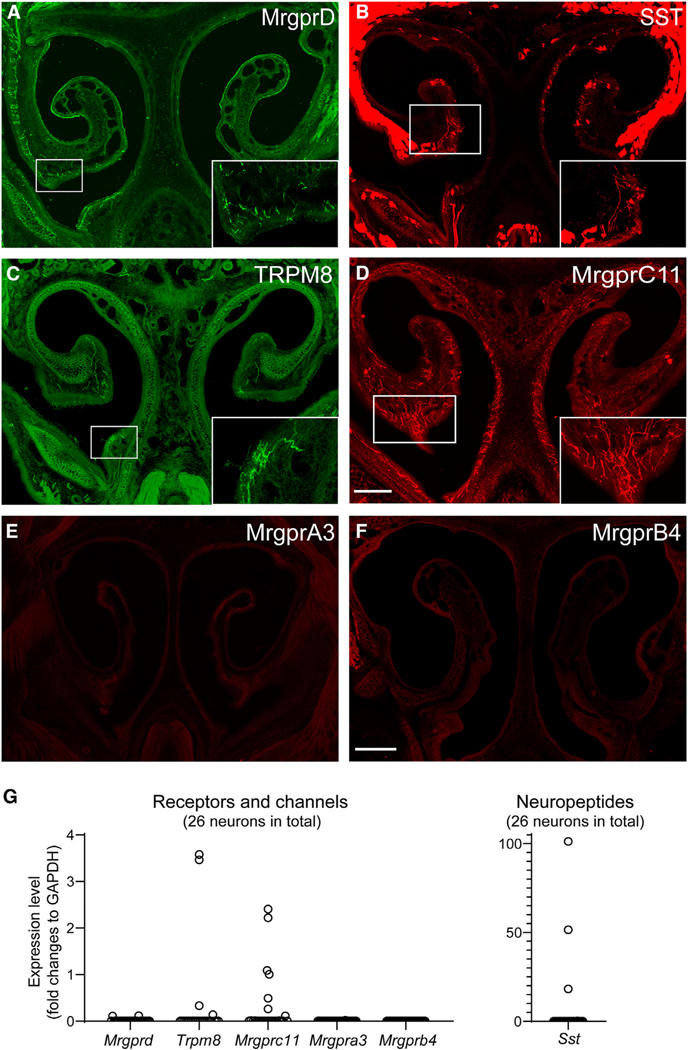
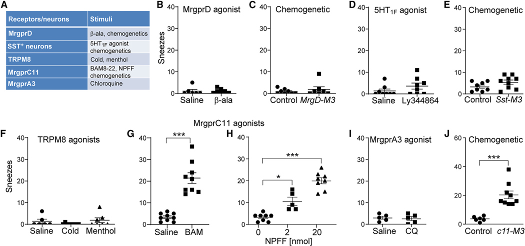
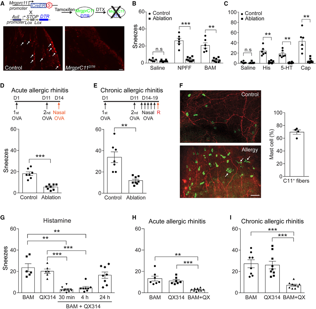
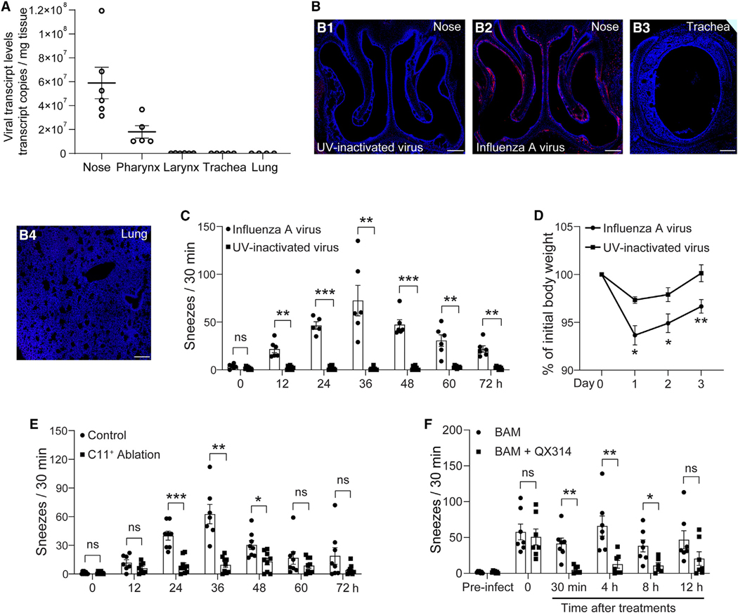
Sneezing and coughing are primary symptoms of many respiratory viral infections and allergies. It is generally assumed that sneezing and coughing involve common sensory receptors and molecular neurotransmission mechanisms. Here, we show that the nasal mucosa is innervated by several discrete populations of sensory neurons, but only one population (MrgprC11+MrgprA3-) mediates sneezing responses to a multitude of nasal irritants, allergens, and viruses. Although this population also innervates the trachea, it does not mediate coughing, as revealed by our newly established cough model. Instead, a distinct sensory population (somatostatin [SST+]) mediates coughing but not sneezing, unraveling an unforeseen sensory difference between sneezing and coughing. At the circuit level, sneeze and cough signals are transmitted and modulated by divergent neuropathways. Together, our study reveals the difference in sensory receptors and neurotransmission/modulation mechanisms between sneezing and coughing, offering neuronal drug targets for symptom management in respiratory viral infections and allergies.

Keywords: allergy; coughing; respiratory viral infection; sensory neurons; sneezing.


Conflict of interest statement

Declaration of interests M.J.H. has noncompeting financial interests unrelated to this work and is the Founder of NuPeak Therapeutics Inc.

Figures

Figure 1.
Figure 1.. The mouse nasal mucosa is innervated by multiple populations of sensory neurons
(A) MrgprD-expressing polymodal C fibers (green) innervate the nasal turbinate and the septum in the nasal anterior region of MrgprdEGFPf/+ mice. The inset shows a higher-magnification view of the boxed area. (B) Somatostatin (SST)-expressing itch-sensing fibers (red) innervate the nasal turbinate in SstCre/+; ROSA26tdTomato/+ mice. (C) TRPM8-expressing cold-sensitive fibers (green) sparsely innervate the nasal turbinate and septum in TRPM8EGFPf/+ mice. (D) MrgprC11+ sensory fibers (red) densely innervate the nasal wall, turbinate, and septum in Mrgprc11CreERT2; ROSA26tdTomato/+ mice. Notably, MrgprC11+ innervation of the nasal turbinate is particularly high. (E) MrgprA3-expressing itch-sensing fibers (red) do not innervate the nasal mucosa in Mrgpra3GFP-Cre; ROSA26tdTomato/+ mice. (F) MrgprB4-expressing C-fiber tactile afferents (red) do not innervate the nasal mucosa in Mrgprb4tdTomato-Cre/+ mice. (G) Single-cell RT-qPCR of nasal sensory neurons retrogradely labeled from the anterior ethmoidal nerve. Each dot represents one sensory neuron. All images shown are representative of three biological replicates. The scale bars represent 500 μm. See also Figure S1 and Table S1.
Figure 2.
Figure 2.. In vivo functional screening for the neuronal population(s) mediating sneezing
(A) Specific agonists for various populations of nasal sensory neurons. (B) Aerosolized β-alanine solution (a specific agonist for MrgprD, 100 mM) did not elicit significant sneezing in WT mice compared with the vehicle control saline. (C) Nasal application of clozapine-N-oxide (CNO, 2 nmol in 2 μL/nostril) did not elicit significant sneezing responses in MrgprDCreERT2/+; TgCAG-LSL-Gq-DREADD(hM3Dq) (MrgprD-M3) mice compared with littermate control mice. (D) Aerosolized LY344864 solution (a 5HT1F agonist that selectively activates SST+ neurons, 1 mM) did not elicit significantly more sneezing than saline (vehicle control) in WT mice. (E) Nasal application of CNO (2 nmol in 2 μL/nostril) did not elicit significant sneezing responses in SstCre/+; TgCAG-LSL-Gq-DREADD(hM3Dq) (SST-M3) mice compared with littermate control SstCre/+ mice. (F) Neither cold air (10°C) nor aerosolized menthol solution (a TRPM8 agonist, 1 mM) induced significant sneezing in WT mice. (G and H) Nasal application of MrgprC11 agonists BAM 8–22 peptide (20 nmol in 2 μL/nostril) or NPFF peptide (2 and 20 nmol in 2 μL/nostril) elicited significant sneezing responses in WT mice compared with saline control. (I) Aerosolized chloroquine solution (a specific agonist for MrgprA3, 12 mM) did not elicit significant sneezing compared with the vehicle control (saline) in WT mice. (J) Nasal application of CNO (2 nmol in 2 μL/nostril) induced significant sneezing in Mrgprc11CreERT2; TgCAG-hM3Dq-mCitrine mice (c11-M3) compared with control Mrgprc11CreERT2 mice. Each dot represents an individual mouse tested (n = 5–11 mice/group). Data are presented as mean ± SEM. * p ≤ 0.05; ***p ≤ 0.001. See also Figures S2, S3, and S4.
Figure 3.
Figure 3.. Nasal MrgprC11+ sensory neurons comprise a core sneeze population
(A) Genetic ablation of MrgprC11+ sensory neurons. Diagram showing the genetic strategy for specific ablation of MrgprC11+ sensory neurons. Representative images show that MrgprC11+ sensory neurons were ablated by diphtheria toxin (DTX) in the trigeminal ganglia from tamoxifen-treated Mrgprc11CreERT2; AviliDTR mice (called MrgprC11DTR mice), but not from control AviliDTR mice (indicated by arrows), as revealed by immunostaining for MrgprC11. (B) Genetic ablation of MrgprC11+ sensory neurons abolished the sneezing responses to MrgprC11 agonists NPFF (20 nmol in 2 μL/nostril) and BAM 8–22 (20 nmol in 2 μL/nostril) compared with control AviliDTR mice. (C) Sneezing responses to aerosolized histamine solution (His, 100 mM), serotonin (5-HT, 1 mM), and capsaicin (Cap, 12 μM) were virtually abolished in MrgprC11+ neuron-ablated mice compared with control AviliDTR mice. (D and E) In the mouse models of acute and chronic allergic rhinitis, sneezing responses to allergen (ovalbumin, 0.2 mg in 2 μL PBS/nostril) challenge was significantly reduced in MrgprC11+ neuron-ablated mice compared with control AviliDTR mice. (F) Representative images of the whole-mount nasal mucosa from normal and allergic Mrgprc11CreERT2; ROSA26tdTomato/+ mice. Note the accumulation and degranulation of avidin-stained mast cells (green, indicated by arrows) in the allergic mice. The bar graph shows the proportion of mast cells closely associated with MrgprC11+ sensory fibers (red) in allergic mice (n = 4 nasal mucosa explants). (G) Sneezing responses induced by aerosolized histamine solution (100 mM) were inhibited by nasal application of QX-314 (1%, 2 μL) with BAM (2 nmol), compared with the control groups pretreated with either BAM (2 nmol in 2 μL) or QX-314 (1%, 2 μL) alone. (H and I) In both acute and chronic allergic rhinitis models, allergen ovalbumin-induced sneezing responses were significantly suppressed by pretreatment of QX-314 (1%, 2 μL) with BAM (2 nmol) compared with the control groups pretreated with either BAM (2 nmol in 2 μL) or QX-314 (1%, 2 μL) alone. Each dot represents an individual mouse (n = 6–11 mice/group). Data are presented as mean ± SEM. ** p ≤ 0.01; *** p ≤ 0.001; ns, not significant. All images shown are representative of at least three biological replicates. Scale bars, 50 μm. See also Figures S5 and S6.
Figure 4.
Figure 4.. MrgprC11+ sensory neurons mediate influenza-associated sneezing
(A) In our mouse model of influenza infection, influenza virus A/PR/8/34 mainly infected the nasal mucosa and pharynx, as revealed by digital PCR for viral transcript. (B) Immunostaining shows the influenza viral infection (red) of the nasal mucosa (B2) but not the lower airway (trachea and lung, B3 and B4). As a negative control, UV-inactivated virus failed to infect the nasal mucosa (B1). Tissues were counterstained with DAPI (blue). (C) Influenza-infected mice exhibited significant sneezing, which peaked at 36 h after viral inoculation. As a negative control, mice infected with UV-inactivated virus did not sneeze. (D) Influenza-infected mice lost body weight moderately and survived well. As a control, UV-inactivated virus induced significantly less body-weight loss. (E) Genetic ablation of MrgprC11+ sensory neurons in Mrgprc11CreERT2; AviliDTR mice significantly decreased influenza-associated sneezing compared with AviliDTR littermate controls. (F) Pharmacological silencing of nasal MrgprC11+ neurons with 1% QX-314 solution containing low-dose BAM (2 nmol in 2 μL; applied at 24 h after viral inoculation) significantly alleviated influenza-associated sneezing compared with controls treated with BAM (2 nmol in 2 μL) alone. Each dot represents an individual mouse tested (n = 4–9 mice/group). Data are presented as mean ± SEM. * P ≤ 0.05, ** P ≤ 0.01, *** P ≤ 0.001. All images shown are representative of three biological replicates. Scale bars, 200 μm.
Figure 5.
Figure 5.. Establishment of a coughing model in mice
(A) Representative respiratory patterns of sneeze and cough-like responses in influenza-infected mice. (B) Diagram showing a noninvasive intratracheal delivery approach. Intratracheal delivery of ammonia (0.2%, 10 μL) induced cough-like respiratory responses. (C) Cough-like responses to intratracheal delivery of tussive agents, including ammonia (0.2%, 10 μL), citric acid (0.5 M, 10 μL), and bradykinin (25 μg in 10 μL). The vehicle saline control induced mild coughing via mechanical stimulation of the trachea. (D) Representative audio patterns of sneeze and cough-like responses. The cough-like sound lacks sharp audio peaks observed in sneezing (indicated by an arrow). Audio spectrum analysis indicates that the sneeze sound displays many more ultrasonic components (indicated by an arrow) than the cough-like sound. (E) Cough-like responses effectively expelled dye-containing ammonia solution from the trachea to the pharynx and mouth, as indicated by arrows (n = 5 mice). As a control, dye-containing saline does not induce such potent expelling (n = 3 mice). (F) Lidocaine (1%) applied to the trachea, but not the nose, significantly reduced subsequent cough-like responses to intratracheal ammonia (0.2%, 10 μL). (G) Pulmonary fibrosis after viral infection induces spontaneous coughing but not sneezing. Each dot represents an individual mouse tested (n = 5–8 mice/group). Data are presented as mean ± SEM. * P ≤ 0.05, *** P ≤ 0.001. All images shown are representative of three biological replicates. See also Video S1.
Figure 6.
Figure 6.. SST+ sensory neurons rather than MrgprC11+ neurons mediate coughing
(A and B) MrgprC11+ and SST+ sensory neurons innervate the trachea (indicated by arrows), as revealed by Mrgprc11CreERT2; ROSA26tdTomato/+ and SstCre/+; ROSA26tdTomato/+ reporter lines, respectively. (C and D) MrgprD+ polymodal C-fiber afferent neurons and TRPM8+ cold-sensing neurons do not innervate the trachea, as revealed by MrgprdCreERT2/+; ROSA26tdTomato and Trpm8EGFPf/+ reporter lines. (E) Airway retrograde tracing and single-cell RT-qPCR confirmed the expression of Mrgprc11 and Sst but no Mrgprd or Trpm8 by airway vagal sensory neurons. Each dot represents an individual sensory neuron. (F) Genetic ablation of MrgprC11+ sensory neurons did not reduce influenza-associated coughing compared with AviliDTR littermate controls. (G) Intratracheal application of MrgprC11 agonist BAM 8–22 peptide (100 nmol in 10 μL) did not induce significantly more coughs than vehicle control (saline) in WT mice. (H) Intratracheal application of CNO (10 nmol in 10 μL) did not induce significant coughing in Mrgprc11CreERT2; TgCAG-hM3Dq-mCitrine (c11-M3) mice compared with vehicle control and Mrgprc11CreERT2 littermate controls. (I) Single-cell RT-qPCR confirmed the expression of Htr1f and Il31r mRNA by tracheal SST+ vagal neurons. (J) Pharmacological activation of tracheal SST+ neurons by LY344864 (a 5HT1F agonist that selectively activates SST+ neurons, 10 nmol in 10 μL) and IL-31 (0.04 nmol in 10 μL) induced significantly more coughs than vehicle control (saline). (K) Chemogenetic activation of tracheal SST+ neurons with CNO (10 nmol in 10 μL) induced significant coughing in SstCre/+; TgCAG-hM3Dq-mCitrine (Sst-M3) mice compared with SstCre/+ littermate controls. (L) MrgprC11+ sensory fibers (red, indicated by arrows) densely innervate the whole-mount pharynx and larynx, as revealed by the Mrgprc11CreERT2; ROSA26tdTomato/+ reporter line. (M) In a mouse model of oropharyngeal aspiration, aspiration of saline solution (25 μL) evoked significant coughing via mechanical stimulation of the airway. Aspiration of BAM 8–22 solution (250 nmol in 25 μL) did not induce more coughs than saline. As a positive control, aspiration of Ly344864 solution (25 nmol in 25 μL) induced significantly more coughs than saline. Each dot in (F)–(H), (J), (K), and (M) represents an individual mouse tested (n = 5–9 mice/group). Data are presented as mean ± SEM. *** P ≤ 0.001; ns, not significant. All images shown are representative of three biological replicates. Scare bars, 100 μm. See also Figure S7.
Figure 7.
Figure 7.. Sneezing and coughing are mediated and modulated by divergent sensory pathways
(A) Representative images showing that MrgprC11+ afferents (red) express the presynaptic marker synaptophysin 1 (blue) and synapse with NMBR+ neurons (green) in the brainstem’s sneeze-evoking region. The synaptic connections were indicated by arrows in higher-magnification view of the boxed region. The graph shows the proportion of NMBR+ neurons postsynaptic to MrgprC11+ afferents in the sneeze-evoking region. Each dot represents an individual mouse (n = 3). (B) Representative traces showing that activation of MrgprC11+ sensory neurons by BAM 8–22 peptide (100 μM) induced significant excitatory postsynaptic potentials (EPSPs) and led to action potential discharge in 7 out of 12 NMBR-GFP neurons recorded within the sneeze-evoking region. As a control, menthol (1 mM) activation of TRPM8+ cold-sensing neurons failed to induce EPSP in NMBR-GFP neurons (0 out of 6 neurons recorded). (C) Mrgprc11CreERT2; Nmbflox/flox mice display significantly reduced sneezing responses to BAM (20 nmol in 2 μL saline) and neuropeptide FF (NPFF; 20 nmol in 2 μL saline). (D) Representative coronal brainstem section of NmbreGFP mice showing that few to no NMBR-GFP neurons (green) in the central projection zone of airway sensory neurons (red, indicated by arrows) within the nucleus tractus solitarius (NTS), as revealed by retrograde tracing from the mouse airway using WGA-Alexa Fluor 555. The graph shows the number of NMBR-GFP neurons within the airway central projection zone (each dot represents a brainstem section). DMX: dorsal motor nucleus. XII: hypoglossal nucleus. (E) Microinjection of NMB-saporin into the NTS did not significantly change the coughing responses to intratracheal delivery of LY344864 (10 nmol in 10 μL) or IL-31 (0.04 nmol in 10 μL). (F) Microinjection of SST-saporin into the NTS induced significant spontaneous coughing compared with the blank saporin. Each dot in (C), (E), and (F) represents an individual mouse (n = 5–9 mice/group). Data are presented as mean ± SEM. ** P ≤ 0.01. *** P ≤ 0.001; ns, not significant. All images shown are representative of three biological replicates. Scale bars, 100 μm. See also Figure S7.

Similar articles

Cited by

References

    1. Bourouiba L. (2020). Turbulent Gas Clouds and Respiratory Pathogen Emissions: Potential Implications for Reducing Transmission of COVID-19. JAMA 323, 1837–1838. 10.1001/jama.2020.4756. - DOI - PubMed
    1. Tang JW, Li Y, Eames I, Chan PK, and Ridgway GL (2006). Factors involved in the aerosol transmission of infection and control of ventilation in healthcare premises. J. Hosp. Infect. 64, 100–114. 10.1016/j.jhin.2006.05.022. - DOI - PMC - PubMed
    1. Cole EC, and Cook CE (1998). Characterization of infectious aerosols in health care facilities: an aid to effective engineering controls and preventive strategies. Am. J. Infect. Control 26, 453–464. 10.1016/s0196-6553(98)70046-x. - DOI - PMC - PubMed
    1. Songu M, and Cingi C. (2009). Sneeze reflex: facts and fiction. Ther. Adv. Respir. Dis. 3, 131–141. 10.1177/1753465809340571. - DOI - PubMed
    1. Wallois F, Macron JM, Jounieaux V, and Duron B. (1991). Trigeminal afferences implied in the triggering or inhibition of sneezing in cats. Neurosci. Lett. 122, 145–147. - PubMed

Substances

LinkOut - more resources