. 2024 Dec;57(12):e13723.
doi: 10.1111/cpr.13723. Epub 2024 Sep 8.

Single-cell atlas of healthy vocal folds and cellular function in the endothelial-to-mesenchymal transition

Affiliations

Single-cell atlas of healthy vocal folds and cellular function in the endothelial-to-mesenchymal transition

Danling Liu et al. Cell Prolif. 2024 Dec.

Abstract

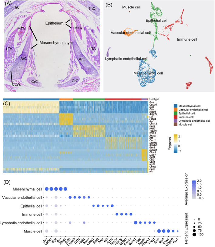
The vocal fold is an architecturally complex organ comprising a heterogeneous mixture of various layers of individual epithelial and mesenchymal cell lineages. Here we performed single-cell RNA sequencing profiling of 5836 cells from the vocal folds of adult Sprague-Dawley rats. Combined with immunostaining, we generated a spatial and transcriptional map of the vocal fold cells and characterized the subpopulations of epithelial cells, mesenchymal cells, endothelial cells, and immune cells. We also identified a novel epithelial-to-mesenchymal transition-associated epithelial cell subset that was mainly found in the basal epithelial layers. We further confirmed that this subset acts as intermediate cells with similar genetic features to epithelial-to-mesenchymal transition in head and neck squamous cell carcinoma. Finally, we present the complex intracellular communication network involved homeostasis using CellChat analysis. These studies define the cellular and molecular framework of the biology and pathology of the VF mucosa and reveal the functional importance of developmental pathways in pathological states in cancer.


Conflict of interest statement

The authors declare no conflicts of interest.

Figures

FIGURE 1
FIGURE 1
Cellular classification by scRNA‐seq in healthy rat vocal fold (VF) tissue. (A) HE staining of the rat VF in the coronal position. ArC, arytenoid cartilage; CrC, cricoid cartilage; LTA, lateral thyroarytenoid muscle; MTA, medial thyroarytenoid muscle. (B) Uniform Manifold Approximation and Projection showing all separate cell clusters for the VF mucosa, including epithelial cells (green), mesenchymal cells (blue), lymphatic ECs (yellow), vascular ECs (purple), immune cells (red) and muscle cells (brown). (C) Heatmap showing genes (rows) that are differentially expressed across five main cell populations (columns) clustered according to gene expression patterns. (D) DotPlot of example genes identified in each cell cluster. The size of the dot indicates the percentage of cells within a cell type, and the colour indicates the average expression level.
FIGURE 2
FIGURE 2
Heterogeneity and distribution of vocal fold (VF) epithelial cells (EPs). (A) Uniform Manifold Approximation and Projection clustering (left) and velocity plot (right) of VF EPs. (B) Violin plot of mucin gene expression and the distribution in VF EPs. (C1) Immunofluorescent staining for KRT15, KRT17 and B2M showing EP1 (basal cells) and EP4 (late suprabasal cells). Red indicates MUC1, green indicates KRT17 and white indicates KRT15. (C2) Immunofluorescent staining for KRT15, MUC1 and MUC20 showing mucin expression in EP1 and EP4. Red indicates B2M, green indicates KRT17 and white indicates KRT15. (C3) Immunofluorescent staining for KRT15, LMO4 and KRT13 showing EP3 (early and mid‐suprabasal cells). Red indicates LMO4, green indicates KRT15 and white indicates KRT13. (C4) Immunofluorescent staining for KRT15, MUC5b and SCGB1A1 showing EP2 (club cells). Red indicates SCGB1A1, green indicates MUC5b and white indicates KRT15. (D) Dot plot of the top gene markers expressed by EPs. The size and colour are illustrated in the same way as in Figure 1. (E) Scatter plot for the distribution of the top gene markers (upper half) and lowly expressed markers (lower half) in EPs.
FIGURE 3
FIGURE 3
Identification and localization of EP5 cells and epithelial‐to‐mesenchymal transition (EMT) cells. (A, B) EP5 cells at the bottom of the vocal fold epithelia among the EP1 cells, which are morphologically similar. (C–F) Four states of EMT cells, including KRT15highCOL3A1low cells with a round shape, elongated KRT15highCOL3A1high cells with enlarged or mitotic nuclei, KRT15lowCOL3A1high cells with higher mesenchymal marker expression and narrow nuclei, and LMOD1high cells with a mesenchymal phenotype with higher mesenchymal marker expression. (G, H) Mesenchymal cells in the para‐cancerous area with flatter and smaller spindle‐like shapes; (I) The ratio of KRT15high and COL3A1high EMT cells in the High EMT area in L‐HNSCC, the Low EMT area in L‐HNSCC, the W‐HNSCC stroma and the proliferative verrucous leukoplakia stroma. (J) A schematic diagram depicting the levels of EMT in the Low EMT area and High EMT area. (K) A schematic diagram of the EMT process from the epithelial phenotype to the hybrid EMT state and finally to the mesenchymal phenotype. (L) The Low EMT area (tumour stroma) showing a higher ratio of KRT15+ EMT cells with erythrocyte infiltration; (M) The High EMT area (tumour parenchyma) showing a higher ratio of COL3A1+ EMT cells.
FIGURE 4
FIGURE 4
Cell types of the vocal fold (VF) endothelial cells. (A) Uniform Manifold Approximation and Projection of vascular endothelial cells (VECs) and lymphatic endothelial cells (LECs). (B) Violin plot of the top gene markers in VECs and LECs. (C) Dot plot of the gene features expressed by LECs and VECs. The size of the dot indicates the percentage of cells within a cell type, and the red colour represents a high expression level and the blue colour represents a low expression level. (D) Chord plot of 39 cell–cell signalling pathways in VF tissue. (E–G) VECs and LECs form blood vessels and lymphatic vessels, respectively, within muscles and cartilage.
FIGURE 5
FIGURE 5
Profiles and spatial location of vocal fold (VF) mesenchymal cells (MSs). (A) Uniform Manifold Approximation and Projection of VF MSs (upper) and a schematic diagram of the distribution of MS1–3. (B) Violin plots of the overlapping gene markers in MS cells. (C) Dot plot of the top gene markers expressed by MSs. The size and colour are illustrated in the same way as in Figure 1. (D–F) Mapping of MS subpopulations. (D1) The MS1 cells with irregular shape in the centre of the ring‐like structure. (D2) The MS2 cells in the outer layer of the rings. (E, E1) The MS3 cells in the middle circle of the rings. (F, F1). The pericyte MS4 cells together with the ECs form the blood vessels in the anterior commissure of the VF. (G) Scatter plot of the expression intensity of the top gene markers in MS cells (upper half) and lowly expressed markers in MSs (lower half).
FIGURE 6
FIGURE 6
Cellular interactions and microstructure of rat vocal fold (VF). (A) Uniform Manifold Approximation and Projection for VF epithelial cells, VF mesenchymal cells (MSs) and VF endothelial cells. (B1) Chord plot for Collagen signalling with the relative contribution of its ligand‐receiver pairs below. (B2) Chord plot for Laminin signalling with the relative contribution of its ligand‐receiver pairs below. (B3) Chord plot for WNT signalling with the relative contribution of its ligand‐receiver pairs below. (B4) Chord plot for NOTCH signalling with the relative contribution of its ligand‐receiver pairs below. (C) Diagram of VF structure, including the anatomy of rat VFs (coronal position), and a schematic of the anterior commissure, anterior Macula Flava, posterior Macula Flava and the interstitial space between the thyroarytenoid muscle and the arytenoid cartilage. (C) Diagram of VF structure, including the anatomy of rat VFs (coronal position) and a schematic of cell arrangement in the anterior commissure, anterior Macula Flava, posterior Macula Flava and the interstitial space between the thyroarytenoid muscle and the arytenoid cartilage.

Similar articles

References

    1. Lungova V, Thibeault SL. Mechanisms of larynx and vocal fold development and pathogenesis. Cell Mol Life Sci. 2020;77(19):3781‐3795. - PMC - PubMed
    1. Li L, Stiadle JM, Lau HK, et al. Tissue engineering‐based therapeutic strategies for vocal fold repair and regeneration. Biomaterials. 2016;108:91‐110. - PMC - PubMed
    1. Kumai Y. Pathophysiology of fibrosis in the vocal fold: current research, future treatment strategies, and obstacles to restoring vocal fold pliability. Int J Mol Sci. 2019;20(10):1‐18. - PMC - PubMed
    1. Smith G. Structure of the normal rat larynx. Lab Anim. 1977;11(4):223‐228. - PubMed
    1. Li X, Wang CY. From bulk, single‐cell to spatial RNA sequencing. Int J Oral Sci. 2021;13(1):36. - PMC - PubMed

LinkOut - more resources