Review
. 2024 Sep 8;29(1):120.
doi: 10.1186/s11658-024-00640-3.

Glucokinase (GCK) in diabetes: from molecular mechanisms to disease pathogenesis

Affiliations
Review

Glucokinase (GCK) in diabetes: from molecular mechanisms to disease pathogenesis

Yasmin Abu Aqel et al. Cell Mol Biol Lett. .

Abstract

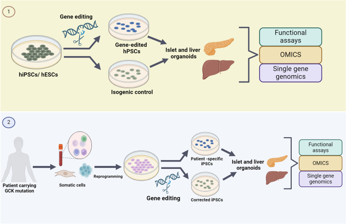
Glucokinase (GCK), a key enzyme in glucose metabolism, plays a central role in glucose sensing and insulin secretion in pancreatic β-cells, as well as glycogen synthesis in the liver. Mutations in the GCK gene have been associated with various monogenic diabetes (MD) disorders, including permanent neonatal diabetes mellitus (PNDM) and maturity-onset diabetes of the young (MODY), highlighting its importance in maintaining glucose homeostasis. Additionally, GCK gain-of-function mutations lead to a rare congenital form of hyperinsulinism known as hyperinsulinemic hypoglycemia (HH), characterized by increased enzymatic activity and increased glucose sensitivity in pancreatic β-cells. This review offers a comprehensive exploration of the critical role played by the GCK gene in diabetes development, shedding light on its expression patterns, regulatory mechanisms, and diverse forms of associated monogenic disorders. Structural and mechanistic insights into GCK's involvement in glucose metabolism are discussed, emphasizing its significance in insulin secretion and glycogen synthesis. Animal models have provided valuable insights into the physiological consequences of GCK mutations, although challenges remain in accurately recapitulating human disease phenotypes. In addition, the potential of human pluripotent stem cell (hPSC) technology in overcoming current model limitations is discussed, offering a promising avenue for studying GCK-related diseases at the molecular level. Ultimately, a deeper understanding of GCK's multifaceted role in glucose metabolism and its dysregulation in disease states holds implications for developing targeted therapeutic interventions for diabetes and related disorders.

Keywords: Beta cells; Diabetes; Glucokinase; Glucose; Insulin; Liver; Mutations; Pancreas; Stem cells.


Conflict of interest statement

A.K.K.T. is a co-founder and shareholder of BetaLife Pte Ltd but is not employed by BetaLife Pte Ltd. All other authors declare no conflict of interest.

Figures

Fig. 1
Fig. 1
Surface representation of overall structure of glucokinase (GCK). The complex structure of glucose-bound GCK in the presence of the non-hydrolysable ATP analogue adenosine 50-(β,-γ-imino) triphosphate (AMP-PNP) and allosteric activator N-thiazol-2-yl-2-amino-4-fluoro5-(1-methylimidazol-2-yl)thiobenzamide (TAFMT) (PDBID: 3ID8). The complex comprises glucose (green, sphere), AMP-PNP (magenta, ball, and stick), and TAFMT (orange, sphere) with GCK in an active conformation. The conformational relationship of the large domain (light blue) and the small domain (salmon) exhibited a closed form
Fig. 2
Fig. 2
Surface representation of the overall structure of GCK/GKRP complex (PDBID: 4LC9). GCK is shown in light blue (large domain) and salmon (small domain). The structure of GKRP consists of two sugar isomerase (SIS) superfamily domains and a C-terminal extended all-helical motif (CTM). GKRP is depicted in green (SIS I), wheat (SIS II), and magenta (CTM). Fructose 6-phosphate (F6P) is shown as a sphere representation and binds at the interface of SIS (I and II) and CTM
Fig. 3
Fig. 3
GCK role in pancreatic β-cells. Glucose enters pancreatic β-cells via low affinity glucose transporters. GCK then catalyzes the ATP-dependent phosphorylation of glucose into G6P. G6P starts the glycolysis and the Krebs cycle, which elevate the adenosine diphosphate (ADP) ATP/ADP ratio. Raised ATP/ADP levels result in K+ efflux, causing cell membrane depolarization and the opening of the voltage sensitive Ca2+ channels. The opening of Ca2+ channels elevate cytosolic Ca2+ levels, which, with other vital coupling factors, activate the endoplasmic reticulum and Golgi apparatus to secrete insulin granules from pancreatic β-cells
Fig. 4
Fig. 4
Regulation of GCK activity in pancreatic β-cells. In pancreatic β-cells, GCK activity is regulated by several binding partners: (1) GCK activity can be inhibited when it binds to insulin granules. The interaction between GCK and insulin granules is partly mediated by NOS. To reverse this interaction and stimulate GCK activity, NOS performs S-nitrosylation of GCK. (2) PFK-2/ FBPase-2. The regulation of GCK by PFK-2/ FBPase-2 involves direct binding of GCK and activation, depending on the phosphorylation status of FBPase-2. (3) Activation of GCK occurs via the BAD protein at the mitochondrial membrane. When phosphorylated at the BH3 domain, BAD binds to GCK near its active site, leading to GCK activation and subsequent insulin secretion. Furthermore, the interaction between BAD and GCK can provide protection against apoptosis
Fig. 5
Fig. 5
Schematic representation of the hPSC-based approaches for modeling monogenic diabetes caused by GCK mutations. Modeling monogenic diabetes (MD) caused by GCK mutations can be achieved by utilizing human pluripotent stem cells (hPSCs). One approach entails introducing GCK mutations or knocking out the GCK gene using gene editing tools on preexisting hPSC lines (1). Alternatively, induced pluripotent stem cells (iPSCs) can be generated from patients with GCK mutations (patient-iPSCs), followed by correcting the mutation using genome editing tools (2). These hPSC lines can then be differentiated into β-cells and liver cells to study the impact of the mutation or edited gene on the development and function of the β-cells and liver cells

Similar articles

Cited by

References

    1. Iynedjian PB. Molecular physiology of mammalian glucokinase. Cell Mol Life Sci. 2009;66(1):27–42. 10.1007/s00018-008-8322-9 - DOI - PMC - PubMed
    1. Matschinsky FM, Magnuson MA, Zelent D, Jetton TL, Doliba N, Han Y, et al. The network of glucokinase-expressing cells in glucose homeostasis and the potential of glucokinase activators for diabetes therapy. Diabetes. 2006;55(1):1–12. 10.2337/diabetes.55.01.06.db05-0926 - DOI - PubMed
    1. Matschinsky FM, Wilson DF. The central role of glucokinase in glucose homeostasis: a perspective 50 years after demonstrating the presence of the enzyme in islets of langerhans. Front Physiol. 2019;10:148. 10.3389/fphys.2019.00148 - DOI - PMC - PubMed
    1. Matschinsky FM, Ellerman JE. Metabolism of glucose in the islets of Langerhans. J Biol Chem. 1968;243(10):2730–6. 10.1016/S0021-9258(18)93432-0 - DOI - PubMed
    1. Salas M, Vinuela E, Sols A. Insulin-dependent synthesis of liver glucokinase in the rat. J Biol Chem. 1963;238:3535–8. 10.1016/S0021-9258(19)75303-4 - DOI - PubMed

LinkOut - more resources