. 2024 Sep 8;15(1):7853.
doi: 10.1038/s41467-024-52149-0.

Human cell surface-AAV interactomes identify LRP6 as blood-brain barrier transcytosis receptor and immune cytokine IL3 as AAV9 binder

Affiliations

Human cell surface-AAV interactomes identify LRP6 as blood-brain barrier transcytosis receptor and immune cytokine IL3 as AAV9 binder

Timothy F Shay et al. Nat Commun. .

Abstract

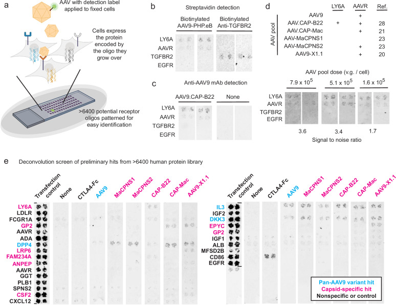
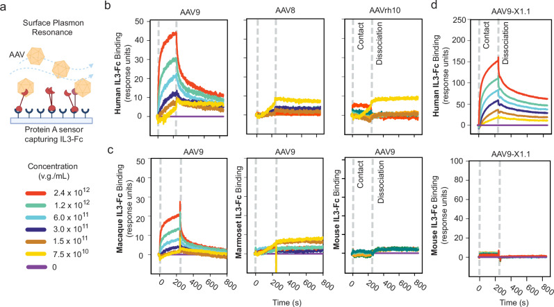
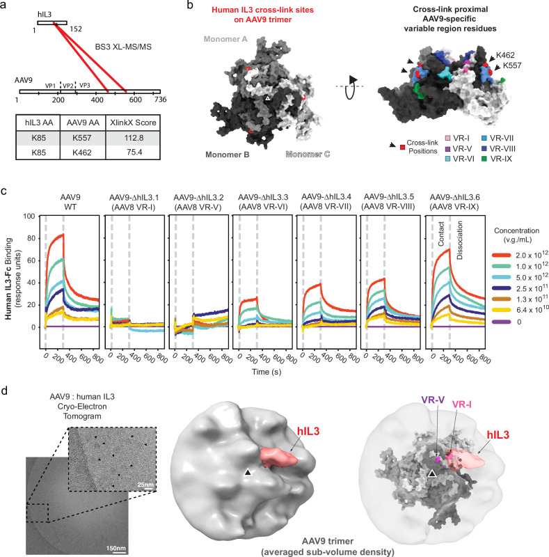
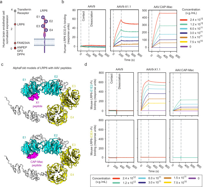
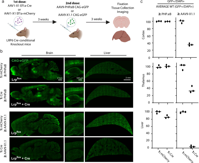
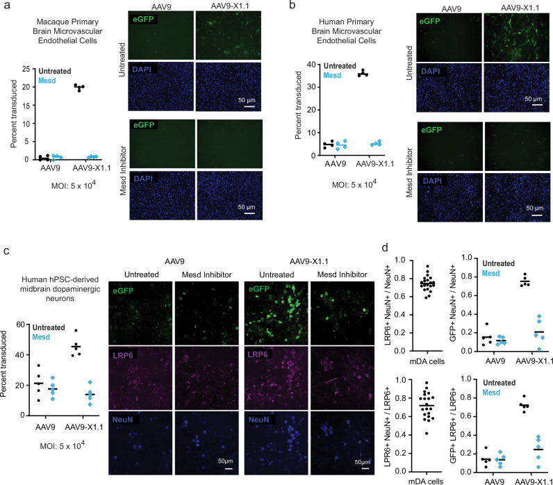
Adeno-associated viruses (AAVs) are foundational gene delivery tools for basic science and clinical therapeutics. However, lack of mechanistic insight, especially for engineered vectors created by directed evolution, can hamper their application. Here, we adapt an unbiased human cell microarray platform to determine the extracellular and cell surface interactomes of natural and engineered AAVs. We identify a naturally-evolved and serotype-specific interaction between the AAV9 capsid and human interleukin 3 (IL3), with possible roles in host immune modulation, as well as lab-evolved low-density lipoprotein receptor-related protein 6 (LRP6) interactions specific to engineered capsids with enhanced blood-brain barrier crossing in non-human primates after intravenous administration. The unbiased cell microarray screening approach also allows us to identify off-target tissue binding interactions of engineered brain-enriched AAV capsids that may inform vectors' peripheral organ tropism and side effects. Our cryo-electron tomography and AlphaFold modeling of capsid-interactor complexes reveal LRP6 and IL3 binding sites. These results allow confident application of engineered AAVs in diverse organisms and unlock future target-informed engineering of improved viral and non-viral vectors for non-invasive therapeutic delivery to the brain.


Conflict of interest statement

The California Institute of Technology has a patent pending for the delivery methods identified in this manuscript, with T.F.S, X.C., S.J., and V.G. listed as inventors (PCT Patent Application No: PCT/US2024/0139329) and a provisional patent for the sequences described in this manuscript, with S.J., T.J.B., T.F.S., and V.G listed as inventors. V.G. is a co-founder and board of directors member of Capsida Therapeutics, a fully integrated AAV engineering and gene therapy company. T.F.S and V.G. are co-founders and X.C. and X.D. are co-founders and employees of Receptive Biotherapeutics. B.W. and C.T. are employees of Charles River Laboratories. The remaining authors declare no competing interests.

Figures

Fig. 1
Fig. 1. High-throughput screen identifies AAV-binding human proteins.
a Schematic of AAV cell microarray screen. DNA oligos that encode individual membrane proteins are chemically coupled to slides in a known pattern, reverse transfecting the cells that grow on them and thereby creating spots of cells overexpressing a particular, known protein. Each protein is expressed in duplicate at two different locations on the slide. When AAVs are applied to the slides, enhanced binding can be detected from duplicate cell spots overexpressing cognate AAV receptors. b Known AAV capsid receptor interactions, such as AAVR and LY6A with AAV-PHP.eB, were used to optimize conditions for streptavidin-based detection of biotinylated capsids with two sets of replicate spots. Anti-TGFBR2 antibody was used as a non-AAV positive control. Uncropped blots in source data. c AAVR and LY6A interaction with AAV9.CAP-B22 were used to optimize conditions for anti-AAV9 antibody direct detection of unmodified capsids with two sets of replicate spots. Anti-TGFBR2 antibody was used as a non-AAV control. Uncropped blots in source data. d Pooled AAV capsid screening conditions were optimized by varying the concentrations of individual capsids within the pool to maximize signal to noise after direct detection with anti-AAV9 antibody, with two sets of replicate spots. v.g.: viral genomes. Uncropped blots in source data. e Pooled screening identified preliminary hits which were deconvoluted by individual-capsid screens, identifying previously-unreported potential capsid-binding proteins by direct detection with anti-AAV9 antibody. Transfection control condition detected fluorescent protein reverse transfected along with each receptor. None condition was treated only with anti-AAV9 antibody. Proteins in cyan were identified in all individual AAV screens, and likely represent interactions outside the engineered regions of AAV9. Proteins in magenta specifically bind to at least one engineered capsid. Uncropped blots in source data. Panel a created with BioRender.com released under a Creative Commons Attribution-NonCommercial-NoDerivs 4.0 International license.
Fig. 2
Fig. 2. Species and serotype-specific interaction between AAV9 and the human immunomodulatory cytokine IL3.
a Schematic of surface plasmon resonance (SPR) experiments where IL3-Fc is captured on a protein A sensor chip and AAV analyte flows over the sensor. v.g.: viral genomes. b SPR confirms serotype-specific interaction of AAV9 with the human immunomodulatory cytokine IL3. c SPR confirms AAV9 binding with macaque but not marmoset or mouse IL3. d SPR confirms that the VR-IV and VR-VIII modified AAV9-X1.1 capsid binds to human but not mouse IL3. Panel (a) created with BioRender.com released under a Creative Commons Attribution-NonCommercial-NoDerivs 4.0 International license.
Fig. 3
Fig. 3. Structural characterization of AAV9 interaction with human IL3.
a Schematic depicting intermolecular cross-links between human IL3 (hIL3) and AAV9 with XlinkX scores above 40, indicating high confidence. b Structure of AAV9 (PDB ID: 3UX1) trimer indicating human IL3 cross-linking amino acids (red) and the capsid variable regions (VR) within reach of the cross-linker (VR-I: pink, VR-V: purple, VR-VI: teal, VR-VII: cyan, VR-VIII: blue, VR-IX: green). c SPR of human IL3 binding for AAV9 and chimeric capsids containing AAV8 amino acid identity at the indicated variable regions. v.g.: viral genomes. d Left: A central slice from a cryo-electron tomogram of AAV9 with human IL3. Arrows in magnification indicate AAV9 capsids. 46 3D tomograms, composed of 41 such images each, yielded 85,135 symmetry-expanded particles for sub-tomogram averaging. Middle: Map of AAV9 trimer bound by human IL3-Fc obtained after symmetry expansion and particle subtraction followed by multiple rounds of refinement. Human IL3-Fc density is segmented in red. Right: The same map overlaid with a model of the AAV9 trimer (PDB ID: 3UX1) highlighting the variable regions relevant for human IL3 binding (VR-I: pink, VR-V: purple).
Fig. 4
Fig. 4. Primate brain-enhanced AAVs gain interaction with LRP6.
a Left: Arraying AAV capsid-specific hits by human brain endothelial cell expression levels reveals highly-conserved LRP6 as a potential receptor for BBB crossing. Right: the LRP6 extracellular domain contains 4 YWTD domains (E1-E4). b SPR confirms that the engineered capsids AAV9-X1.1 and CAP-Mac gained direct binding interactions with human LRP6. v.g.: viral genomes. c Representative AlphaFold models, from 5 structural models each of X1 and CAP-Mac peptides with up to 20 recycles, predict selective interaction with human LRP6 domain E1 (E1 and E2: teal, E3 and E4: yellow, AAV peptides: purple). d SPR of mouse LRP6-E1E2 and LRP6-E3E4 (the minimal stable extracellular domain fragments due to cooperative folding) confirms that AAV9-X1.1 and CAP-Mac bind only to LRP6-E1E2. Panel a created with BioRender.com released under a Creative Commons Attribution-NonCommercial-NoDerivs 4.0 International license.
Fig. 5
Fig. 5. LRP6 modulates CNS function of engineered AAVs in mice.
a Schematic of Lrp6 conditional knockout by sequential AAV injection. Cre-conditional Lrp6 knockout mice were systemically injected with AAV1-X1 packaging either Cre or mCherry, creating cohorts of mice that differ in their Lrp6 expression. After allowing time for expression, these cohorts were each injected with AAV9-PHP.eB or AAV9-X1.1 packaging eGFP. By switching serotypes, neutralizing antibodies are evaded and vector dependence on Lrp6 in vivo may be assessed. b Representative sagittal brain images (left) and liver images (right). Imaging parameters were optimized independently for AAV9-X1.1 and AAV9-PHP.eB second dose conditions. c Quantification of AAV potency demonstrating that conditional knockout of Lrp6 in mouse selectively and potently reduces AAV9-X1.1 brain and liver gene delivery. Data points are the average of two technical replicate sections per tissue region for each of 3 biological replicate animals, with consistent physiological regions of interest across the four experimental cohorts. Bars represent the mean value. Panel a created with BioRender.com released under a Creative Commons Attribution-NonCommercial-NoDerivs 4.0 International license.
Fig. 6
Fig. 6. LRP6 enhances engineered AAV potency in primate BBB and neuronal cell culture.
AAV9-X1.1 has enhanced potency in (a) macaque and (b) human primary brain microvascular endothelial cell culture (black data points), which decreases to AAV9 levels with Mesd inhibition of LRP6 (blue data points). 4 biological replicates were performed and quantified for each condition. Bars indicate the mean value. MOI: multiplicity of infection. c AAV9-X1.1 has enhanced potency in human pluripotent stem cell (hPSC)-derived midbrain dopaminergic neuronal culture (black data points), which decreases to AAV9 levels with Mesd inhibition of LRP6 (blue data points). 5 biological replicates were performed and quantified for each condition. d Quantification of LRP6 and NeuN immunohistochemistry confirms that LRP6 expression is largely restricted to mature neurons (20 biological replicates) and that the LRP6-dependent enhanced potency of AAV9-X1.1 is enriched in this population (5 biological replicates for each condition, Mesd inhibition condition in blue).

References

    1. Haery, L. et al. Adeno-associated virus technologies and methods for targeted neuronal manipulation. Front Neuroanat.13, 93 (2019). 10.3389/fnana.2019.00093 - DOI - PMC - PubMed
    1. Kuzmin, D. A. et al. The clinical landscape for AAV gene therapies. Nat. Rev. Drug Discov. d41573-021-00017–7 10.1038/d41573-021-00017-7 (2021). - PubMed
    1. Foust, K. D. et al. Intravascular AAV9 preferentially targets neonatal neurons and adult astrocytes. Nat. Biotechnol.27, 59–65 (2009). 10.1038/nbt.1515 - DOI - PMC - PubMed
    1. Mendell, J. R. et al. Single-dose gene-replacement therapy for spinal muscular atrophy. N. Engl. J. Med. 377, 1713–1722 (2017). 10.1056/NEJMoa1706198 - DOI - PubMed
    1. Al-Zaidy, S. A. & Mendell, J. R. From clinical trials to clinical practice: Practical considerations for gene replacement therapy in SMA type 1. Pediatr. Neurol.100, 3–11 (2019). 10.1016/j.pediatrneurol.2019.06.007 - DOI - PubMed

Publication types

Substances

LinkOut - more resources