

Case study of CD19 CAR T therapy in a subject with immune-mediate necrotizing myopathy treated in the RESET-Myositis phase I/II trial
- PMID: 39245937
- PMCID: PMC11573600
- DOI: 10.1016/j.ymthe.2024.09.009
Case study of CD19 CAR T therapy in a subject with immune-mediate necrotizing myopathy treated in the RESET-Myositis phase I/II trial
Abstract
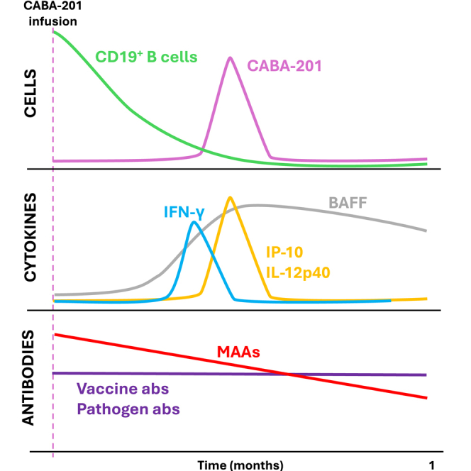
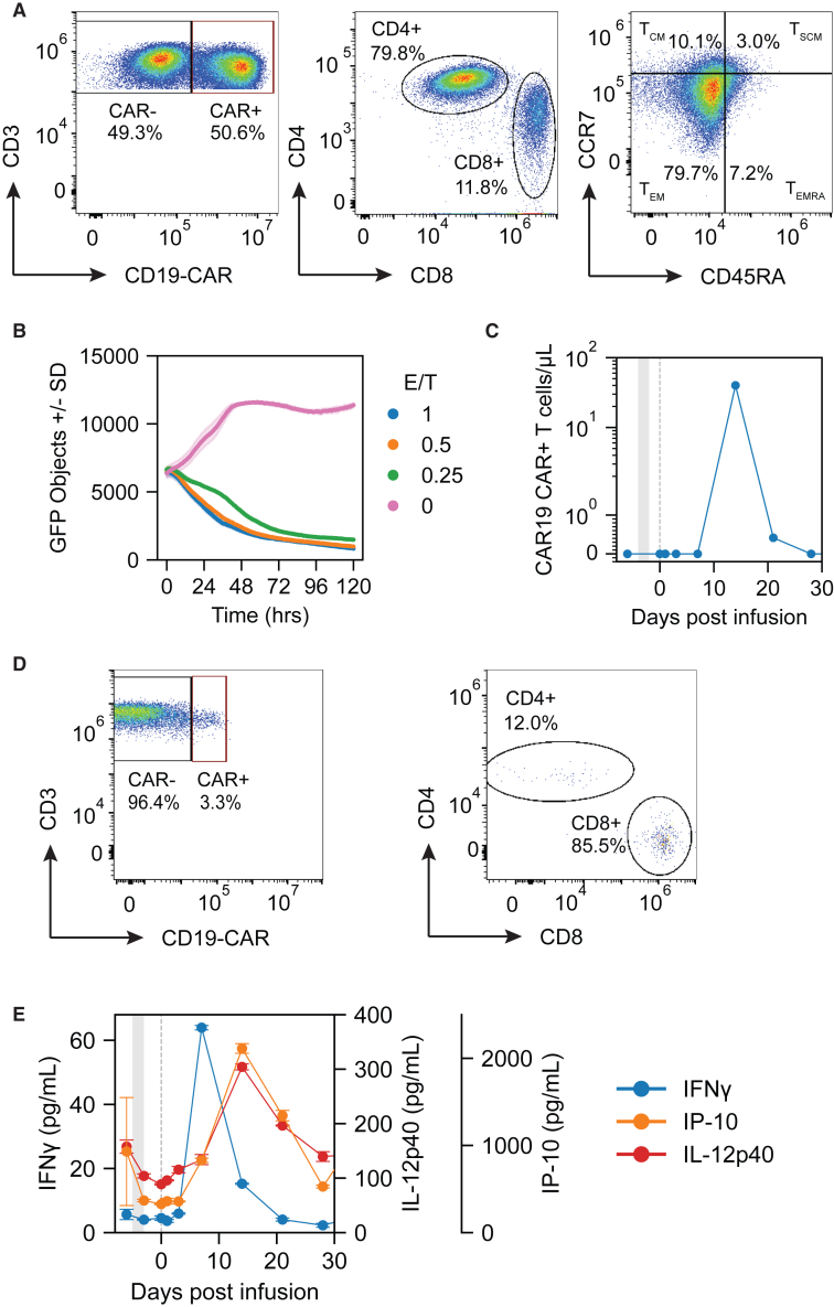
Under compassionate use, chimeric antigen receptor (CAR) T cells have elicited durable remissions in patients with refractory idiopathic inflammatory myopathies (IIMs). Here, we report on the safety, efficacy, and correlative data of the first subject with the immune-mediated necrotizing myopathy (IMNM) subtype of IIM who received a fully human, 4-1BBz anti-CD19-CAR T cell therapy (CABA-201) in the RESET-Myositis phase I/II trial (NCT06154252). CABA-201 was well-tolerated following infusion. Notably, no evidence of cytokine release syndrome or immune effector cell-associated neurotoxicity syndrome was observed. Creatine kinase levels decreased, and muscular strength improved post-infusion. Peripheral B cells were depleted rapidly following infusion, and the subject achieved peripheral B cell aplasia by day 15 post-infusion. Peripheral B cells returned at 2 months post-infusion and were almost entirely transitional. Autoantibodies to SRP-9, SRP-72, SRP-54, and Ro-52, decreased relative to baseline, whereas antibodies associated with pathogens and vaccinations remained stable. The infusion product consisted of predominantly CD4+ effector memory T cells and exhibited in vitro cytolytic activity. Post-infusion, CABA-201 expansion peaked at day 15 and was preceded by a serum IFN-γ peak on day 8 with peaks in serum IL-12p40 and IP-10 on day 15. These data detail the safety, efficacy, and pharmacodynamics of CABA-201 in the first IMNM subject.
Copyright © 2024 Cabaletta Bio. Published by Elsevier Inc. All rights reserved.
Conflict of interest statement
Declaration of interests J.V., D.N., J.S., M.W., Z.V., A.E., J.W., J.C., Q.L., T.F., C.S., F.H.-N., D.T., C.M., C.L., D.C., and S.B. are employees of Cabaletta Bio.
Figures

References
-
- Lundberg I.E., Fujimoto M., Vencovsky J., Aggarwal R., Holmqvist M., Christopher-Stine L., Mammen A.L., Miller F.W. Idiopathic inflammatory myopathies. Nat. Rev. Dis. Primers. 2021;7:86. - PubMed
-
- Moghadam-Kia S., Oddis C.V. Current and new targets for treating myositis. Curr. Opin. Pharmacol. 2022;65 - PubMed
-
- Dalakas M.C. Inflammatory Muscle Diseases. N. Engl. J. Med. 2015;373:393–394. - PubMed
-
- Espitia-Thibault A., Masseau A., Néel A., Espitia O., Toquet C., Mussini J.M., Hamidou M. Sjögren's syndrome-associated myositis with germinal centre-like structures. Autoimmun. Rev. 2017;16:154–158. - PubMed
-
- Piper C.J.M., Wilkinson M.G.L., Deakin C.T., Otto G.W., Dowle S., Duurland C.L., Adams S., Marasco E., Rosser E.C., Radziszewska A., et al. CD19(+)CD24(hi)CD38(hi) B Cells Are Expanded in Juvenile Dermatomyositis and Exhibit a Pro-Inflammatory Phenotype After Activation Through Toll-Like Receptor 7 and Interferon-alpha. Front. Immunol. 2018;9:1372. - PMC - PubMed
Publication types
MeSH terms
Substances
LinkOut - more resources
Full Text Sources
Medical
Research Materials
