. 2024 Dec;15(6):2361-2374.
doi: 10.1002/jcsm.13570. Epub 2024 Sep 8.

Improved health by combining dietary restriction and promoting muscle growth in DNA repair-deficient progeroid mice

Affiliations

Improved health by combining dietary restriction and promoting muscle growth in DNA repair-deficient progeroid mice

Wilbert P Vermeij et al. J Cachexia Sarcopenia Muscle. 2024 Dec.

Abstract

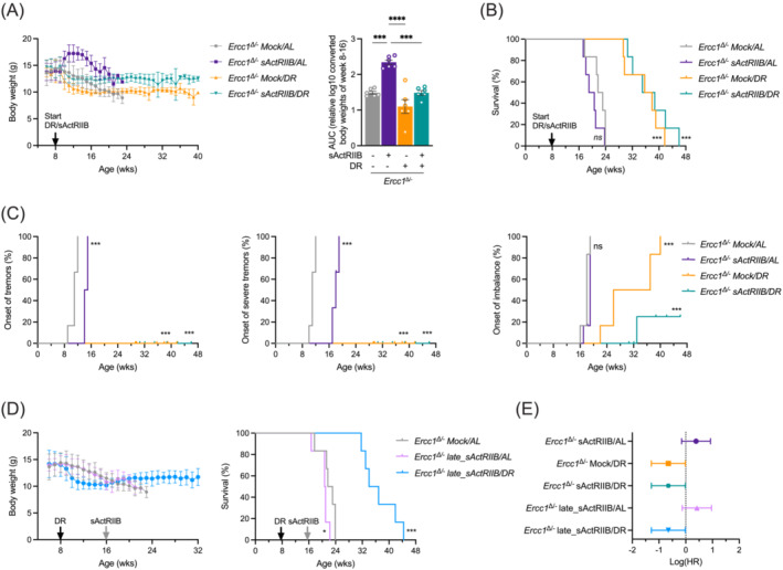
Background: Ageing is a complex multifactorial process, impacting all organs and tissues, with DNA damage accumulation serving as a common underlying cause. To decelerate ageing, various strategies have been applied to model organisms and evaluated for health and lifespan benefits. Dietary restriction (DR, also known as caloric restriction) is a well-established long-term intervention recognized for its universal anti-ageing effects. DR temporarily suppresses growth, and when applied to progeroid DNA repair-deficient mice doubles lifespan with systemic health benefits. Counterintuitively, attenuation of myostatin/activin signalling by soluble activin receptor (sActRIIB), boosts the growth of muscle and, in these animals, prevents muscle wasting, improves kidney functioning, and compresses morbidity.

Methods: Here, we investigated a combined approach, applying an anabolic regime (sActRIIB) at the same time as DR to Ercc1Δ/- progeroid mice. Following both single treatments and combined, we monitored global effects on body weight, lifespan and behaviour, and local effects on muscle and tissue weight, muscle morphology and function, and ultrastructural and transcriptomic changes in muscle and kidney.

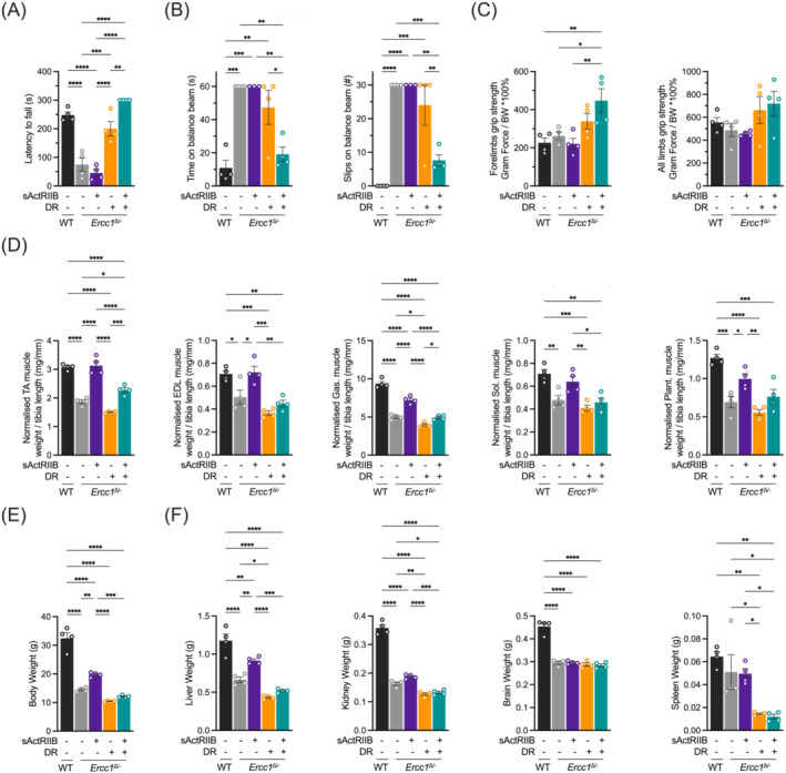
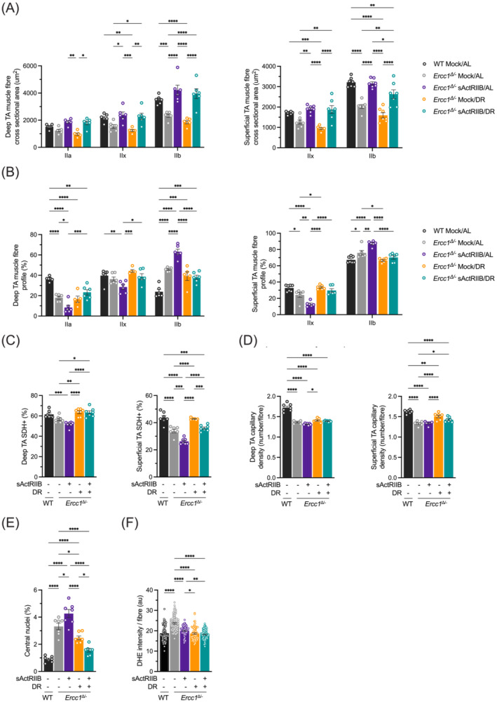
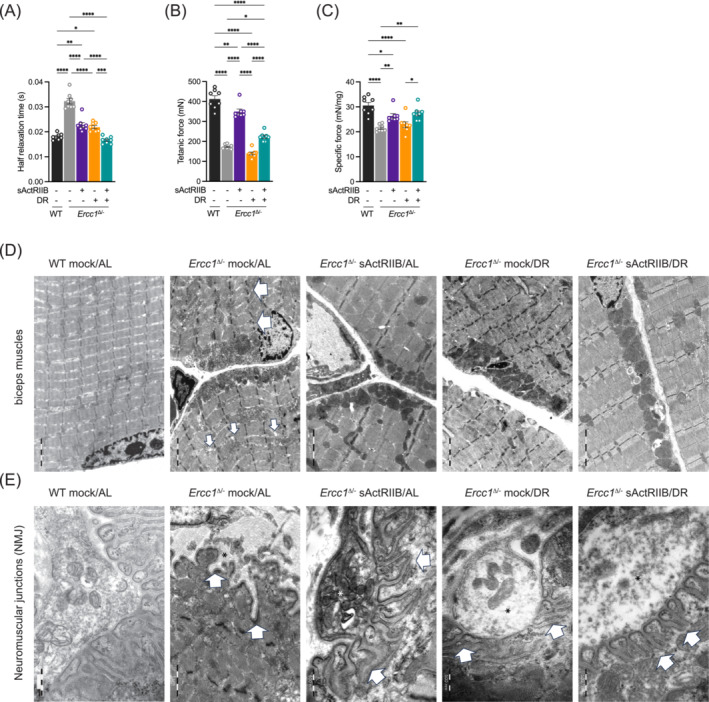
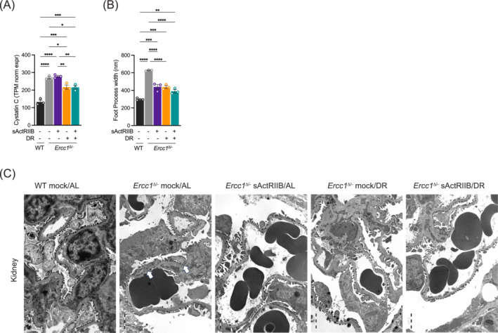
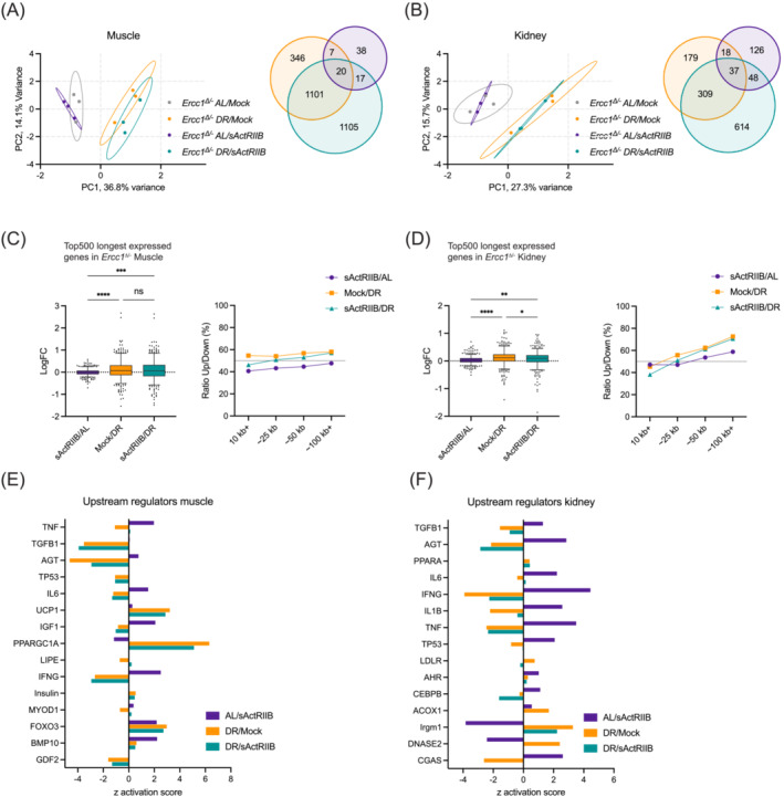
Results: Lifespan was mostly influenced by DR (extended from approximately 20 to 40 weeks; P < 0.001), with sActRIIB clearly increasing muscle mass (35-65%) and tetanic force (P < 0.001). The combined regime yielded a stable uniform body weight, but increased compared with DR alone, synergistically improved motor coordination and further delayed the onset and development of balance problems. sActRIIB significantly increased muscle fibre size (P < 0.05) in mice subjected to DR and lowered all signs of muscle damage. Ercc1Δ/- mice showed abnormal neuromuscular junctions. Single interventions by sActRIIB treatment or DR only partially rescued this phenotype, while in the double intervention group, the regularly shaped junctional foldings were maintained. In kidney of Ercc1Δ/- mice, we observed a mild but significant foot process effacement, which was restored by either intervention. Transcriptome analysis also pointed towards reduced levels of DNA damage in muscle and kidney by DR, but not sActRIIB, while these levels retained lower in the double intervention.

Conclusions: In muscle, we found synergistic effects of combining sActRIIB with DR, but not in kidney, with an overall better health in the double intervention group. Crucially, the benefits of each single intervention are not lost when administered in combination, but rather strengthened, even when sActRIIB was applied late in life, opening opportunities for translation to human.

Keywords: Ageing; Dietary restriction; Kidney; Muscle; Myostatin; Progeria.


Conflict of interest statement

The authors declare no competing interests.

Figures

Figure 1
Figure 1
Whole body assessment of sActRIIB, dietary restriction and dual intervention on Ercc1 Δ/− mice. (A) Mean body weights development (±SD) and area under the curve (AUC) of body weight (as mean ± SE and individual data points) of Ercc1 Δ/− mice under sActRIIB and/or dietary restriction (DR) conditions versus mock‐treated ad libitum (AL) fed. All treatments were initiated from 8 weeks of age (black arrow). (B) Survival curves of Ercc1 Δ/− mice across the different intervention groups. (C) Onset of neurological abnormalities tremors, severe tremors, and imbalance with age. (D) Body weight changes and lifespan curves of Ercc1 Δ/− mice under AL and DR conditions when sActRIIB was administered late in life from 16 weeks of age (grey arrow). (E) Forest plot of effect size for the logarithm of the hazard ratio (HR; with 95% confidence interval as error bars) for changes in survival of all cohorts. n = 6 animals per group. *P < 0.05, ***P < 0.001, ****P < 0.0001.
Figure 2
Figure 2
Exercise profiling of Ercc1 Δ/− mice and organ weights following sActRIIB, dietary restriction and combined at 16 weeks of age. (A) Average time spent on an accelerating rotarod of wild‐type and Ercc1 Δ/− mice on different interventions at 16 weeks of age. (B) Motor coordination on the balance beam indicated by time to cross the beam and the number of missteps made during. (C) Grip strength measure of forelimbs and all limbs normalized to body weight. (D) Dissected muscle weights for tibialis anterior (TA), extensor digitorum longus (EDL), gastrocnemius (Gas.), soleus (Sol.), and plantaris (Plant.) normalized to tibia length. (E) Body weight and (F) indicated organ weights at week 16. n = 4 animals per group. Mean ± SE and individual data points are indicated. *P < 0.05, **P < 0.01, ***P < 0.001, ****P < 0.0001.
Figure 3
Figure 3
Profiling of Ercc1 Δ/− muscle fibres following dual intervention. (A) MHCIIA/IIB fibre size profile of deep and superficial regions of TA muscle. (B) MHCIIA/IIB fibre frequency profile of deep and superficial regions of TA muscles. Between 60–80 fibres were counted from each mouse before being averaged per cohort. (C) SDH profiling of deep and superficial regions of TA muscles. (D) Capillary density profile, through CD31 staining, of deep and superficial regions of TA muscles. (E) Quantification of regenerating fibres in the EDL through counting of centrally located nuclei. (F) Measure of oxidate damage in EDL. All muscles are from mice at 16 weeks of age. n = 8 WT Mock/AL, n = 7/8 Ercc1 Δ/− Mock/AL, n = 7/8, Ercc1 Δ/− sActRIIB/AL, n = 7/8, Ercc1 Δ/− Mock/DR, n = 7/8 Ercc1 Δ/− sActRIIB/DR. *P < 0.05, **P < 0.01, ***P < 0.001, ****P < 0.0001.
Figure 4
Figure 4
Impact of dual intervention on Ercc1 Δ/− muscle function and ultrastructure. Ex‐vivo assessment of (A) half relaxation time and (B) tetanic and (C) specific force. All muscles are from mice at 16 weeks of age. n = 8 WT Mock/AL, n = 7/8 Ercc1 Δ/− Mock/AL, n = 7/8, Ercc1 Δ/− sActRIIB/AL, n = 7/8, Ercc1 Δ/− Mock/DR, n = 7/8 Ercc1 Δ/− sActRIIB/DR. (D) sActRIIB but not dietary restriction prevents Ercc1 Δ/− muscle ultrastructural abnormalities. All transmission electron microscopy (TEM) images are from biceps muscle. In Ercc1 Δ/− mice, biceps muscles display disorganized, split sarcomeres of high variable width frequently containing a disrupted M‐Line. Moreover, the sarcoplasmic reticulum of these muscles was dilated. sActRIIB‐treatment alone as well as in combination with dietary restriction rescues this phenotype completely. In contrast, dietary restriction alone has no effect on any ultrastructural changes observed in Ercc1 Δ/− mice. Large arrows indicate disorganized sarcomeres and small arrows dilated sarcoplasmatic reticulum. (E) Neuromuscular junctions (NMJ) of biceps muscles in Ercc1 Δ/− mice display and obvious phenotype at the postsynaptic side. Namely in Ercc1 Δ/− mice the postsynaptic junctional folds of the basal lamina are almost completely absent or appear rudimentary. sActRIIB or dietary restriction could only partially rescue this phenotype, that is, the distance between the junctional foldings is still highly variable as well as their depth and width. Only double intervention by sActRIIB and dietary restriction fully restores the NMJ phenotype observed in Ercc1 Δ/− mice. *P < 0.05, **P < 0.01, ***P < 0.001, ****P < 0.0001.
Figure 5
Figure 5
Impact of dual intervention on Ercc1 Δ/− kidney function and ultrastructure. (A) In Ercc1 Δ/− kidney the expression of cystatin C doubled as compared with WT mice. DR alone or in combination with sActRIIB‐treatment partly rescued this phenotype while sActRIIB‐treatment alone does not. (B, C) As a morphological correlate for the functional alteration the renal filtration barrier of Ercc1 Δ/− mice displays a significant foot process effacement of about 600 nm (B, C, arrows), which is partly rescued by sActRIIB‐treatment or dietary restriction alone as well as by the combination of both interventions. n = 3 per intervention. *P < 0.05, **P < 0.01, ***P < 0.001, ****P < 0.0001.
Figure 6
Figure 6
Transcriptome analysis of the various interventions in muscle and kidney by PCA, gene lengths, and upstream regulators. (A, B) Principal component analysis (PCA; left) and Venn diagrams of differently expressed genes (DEGs; right) in quad muscle (A) and kidney (B) of 16‐week‐old Ercc1 Δ/− mice subjected to sActRIIB or mock injections and under DR or AL feeding regimens. (C, D) Mean log fold‐change of the 500 longest expressed genes (left) and the ratio up:down within bins of 500 expressed genes across various gene length categories (right) for muscle (C) and kidney (D). Whiskers indicate 5–95 percentile. (E, F) Upstream regulator analysis of the effects of sActRIIB, DR, and double intervention in both organs. *P < 0.05, **P < 0.01, ***P < 0.001, ****P < 0.0001.

References

    1. Lopez‐Otin C, Galluzzi L, Freije JMP, Madeo F, Kroemer G. Metabolic Control of Longevity. Cell 2016;166:802–821. - PubMed
    1. Pluvinage JV, Wyss‐Coray T. Systemic factors as mediators of brain homeostasis, ageing and neurodegeneration. Nat Rev Neurosci 2020;21:93–102. - PubMed
    1. Schumacher B, Pothof J, Vijg J, Hoeijmakers JHJ. The central role of DNA damage in the ageing process. Nature 2021;592:695–703. - PMC - PubMed
    1. Lopez‐Otin C, Blasco MA, Partridge L, Serrano M, Kroemer G. The hallmarks of aging. Cell 2013;153:1194–1217. - PMC - PubMed
    1. van den Boogaard WMC, Komninos DSJ, Vermeij WP. Chemotherapy Side‐Effects: Not All DNA Damage Is Equal. Cancers (Basel) 2022;14. - PMC - PubMed