. 2024 Nov;11(42):e2407619.
doi: 10.1002/advs.202407619. Epub 2024 Sep 9.

Novel Endogenous Engineering Platform for Robust Loading and Delivery of Functional mRNA by Extracellular Vesicles

Affiliations

Novel Endogenous Engineering Platform for Robust Loading and Delivery of Functional mRNA by Extracellular Vesicles

Antje M Zickler et al. Adv Sci (Weinh). 2024 Nov.

Abstract

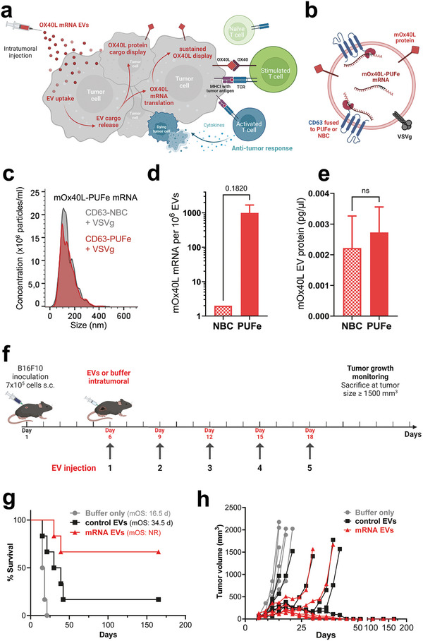
Messenger RNA (mRNA) has emerged as an attractive therapeutic molecule for a plethora of clinical applications. For in vivo functionality, mRNA therapeutics require encapsulation into effective, stable, and safe delivery systems to protect the cargo from degradation and reduce immunogenicity. Here, a bioengineering platform for efficient mRNA loading and functional delivery using bionormal nanoparticles, extracellular vesicles (EVs), is established by expressing a highly specific RNA-binding domain fused to CD63 in EV producer cells stably expressing the target mRNA. The additional combination with a fusogenic endosomal escape moiety, Vesicular Stomatitis Virus Glycoprotein, enables functional mRNA delivery in vivo at doses substantially lower than currently used clinically with synthetic lipid-based nanoparticles. Importantly, the application of EVs loaded with effective cancer immunotherapy proves highly effective in an aggressive melanoma mouse model. This technology addresses substantial drawbacks currently associated with EV-based nucleic acid delivery systems and is a leap forward to clinical EV applications.

Keywords: bioengineering; cancer immunotherapy; drug delivery; extracellular vesicles; nanotechnology; nucleic acid therapeutics.


Conflict of interest statement

The authors declare the following competing interests: S.E.A. is a co‐founder, consultant, and stakeholder of Evox Therapeutics Ltd. J.N., D.G., O.W., and A.G. are consultants and/or stakeholders of Evox Therapeutics Ltd. V.C.L., M.D.L., L.E., J.H., and T.S. are current or former employees of Evox Therapeutics Ltd. This work is protected by patent families WO2019092145 and WO2020225392 owned by Evox Therapeutics Ltd. Patent information WO2019092145: applicant – Evox Therapeutics Ltd; inventors – J.N., D.G., L.E., and J.H.; international application number – PCT/EP2018/08 0681. Patent information WO2020225392: applicant – Evox Therapeutics Ltd; inventors – V.C.L, A.Z., X.L., M.D.L., and L.E.; international application number – PCT/EP2020/06 2791. D.M., G.C., O.E., N.K., Z.N., G.Z., H.Z., and S.R. declare no conflict of interest.

Figures

Figure 1
Figure 1
Engineering strategy for endogenous mRNA loading into EVs. a) EV sorting strategy: four novel RNA‐binding domains (RBD), the non‐cleaving mutant Cas6f and three engineered versions of the designer Pumilio and FBF homology domain (PUF), termed PUFm, PUFe, and PUFx2, were fused via a glycine‐rich linker peptide to the C‐terminus of the EV sorting protein CD63. As control for passive loading (non‐binding control, NBC), CD63 was fused to the MS2 coat protein (MCP, as published by Hung et al., 2016). b) mRNA‐binding strategy: mRNA coding sequences were codon‐optimized and engineered to contain 6–10 repeats of the specific high‐affinity motif recognized by the respective RNA‐binding domain in their 3′UTR. Figure was created using BioRender.
Figure 2
Figure 2
Active mRNA loading into EVs from engineered mRNA single‐stable producer cells. a) Illustration showing mRNA stable cell line generation using the ϕC31 integrase system, and subsequent EV production. For both mRNA EV and control EV production, the same mRNA stable EV producer cell line was used. Cells were either transiently expressing the compatible CD63‐RBD or incompatible CD63‐NBC (non‐binding control). This approach ensured the same biological pre‐requisites during EV biogenesis for passive loading of engineered mRNA and protein. Figure created using BioRender. b) Average particle size determination by NTA of mRNA EVs and respective control EVs harvested from transfected Nanoluc‐RBD mRNA stable producer cells (RBD motif as indicated). c) Negative stain Transmission Electron Microscopy images of control EVs and mRNA EVs loaded with Nanoluc‐PUFe mRNA. Scale bar: 300 nm d) Western Blot analysis of EVs produced from Nanoluc‐RBD mRNA stable EV producer cells as indicated on top. The expression of the CD63‐RBD fusion proteins, either CD63‐Cas6f (C), CD63‐PUFm (Pm), CD63‐PUFe (Pe), CD63‐PUFx2 (Px2), or CD63‐NBC (NBC), respectively, was validated by probing for CD63, sizes are indicated with colored arrowheads. As loading reference, expression of the EV marker SDCBP (Syntenin) was detected. Protein loaded per lane: 3 µg e) Absolute quantification by RT‐qPCR of Nanoluc mRNA molecules per 1 × 106 control EVs (NBC) or mRNA EVs (C, Pm, Pe, Px2) averaged from three representative experiments. CD63‐PUFe repeatedly showed significant enrichment of Nanoluc mRNA in EVs. Data presented as mean ± SD with n = 2 (Cas6) or n = 3 (all others); P‐values were calculated by two‐way ANOVA using Tukeys Multiple Comparisons test; α = 0.05; ns (non‐significant) p > 0.05, *p < 0.05, **p < 0.01, ***p < 0.001, ****p < 0.0001. f) RNAse challenge assay to determine the efficiency of intraluminal EV mRNA cargo encapsulation in 4 independently produced EV batches. g) Uptake of Nanoluc mRNA EVs at a dose of 0.6 pg Nanoluc mRNA per 1 × 104 cells or particle count‐matched control EVs in Huh7 recipient cells. Cellular Nanoluc protein activity was measured at indicated timepoints and normalized to the signal measured at 2 h, which corresponds to the signal from passively loaded Nanoluc protein. Uptake of Nanoluc mRNA EVs led to a significantly higher Nanoluc protein accumulation over time compared to control EVs, demonstrating EV‐mediated engineered mRNA delivery and functional translation. Data presented as mean ± SD with n = 3; P‐values were calculated by two‐way ANOVA using Šìdàks Multiple Comparisons test; α = 0.05; ns (non‐significant) p > 0.05, *p < 0.05, **p < 0.01, ***p < 0.001, ****p < 0.0001.
Figure 3
Figure 3
Co‐expression of the viral fusogen VSVg induces endosomal escape and substantially increased EV cargo delivery. a) Illustration of VSVg co‐expression in Nanoluc‐PUFe mRNA stable EV producer cells, and subsequent EV production. Figure created using BioRender. b) Average particle size determination by NTA of Nanoluc‐PUFe mRNA EVs and control EVs with or without VSVg co‐expression. c) Western Blot analysis to validate CD63‐PUFe and VSVg expression in Nanoluc‐PUFe mRNA EV producer cells (left) and corresponding EVs (right). d) Absolute quantification by RT‐qPCR of Nanoluc‐PUFe mRNA loading into EVs with or without VSVg co‐expression. Data presented as mean ± SD with n = 3; P‐values were calculated by two‐way ANOVA using Šìdàks Multiple Comparisons test; α = 0.05; ns (non‐significant) p > 0.05, *p < 0.05, **p < 0.01, ***p < 0.001, ****p < 0.0001. e) In vitro uptake analysis of Huh7 recipient cells treated with equal amounts of Nanoluc‐PUFe mRNA EVs at a dose of 0.3 pg and 3 pg mRNA per 1 × 104 cells, or particle count‐matched control EVs, both with and without VSVg expression. Co‐expression of VSVg on the EVs significantly enhanced Nanoluc protein signal in treated cells after 24 h, indicating a general improvement of EV cargo release by VSVg‐mediated endosomal escape. Data presented as mean ± SD with n = 4; P‐values were calculated by two‐way ANOVA using Tukeys Multiple Comparisons test; α = 0.05; ns (non‐significant) p > 0.05, *p < 0.05, **p < 0.01, ***p < 0.001, ****p < 0.0001. f) Co‐expression of VSVg enhanced EV cargo delivery in vivo. Mice were injected intraperitoneally with Nanoluc mRNA EVs with or without VSVg co‐expression at a Nanoluc mRNA dose of 50 ng kg−1 body weight, or particle count‐matched control EVs with VSVg co‐expression. Data presented as mean ± SD with n = 4; P‐values were calculated by two‐way ANOVA using Šìdàks Multiple Comparisons test; α = 0.05; ns (non‐significant) p > 0.05, *p < 0.05, **p < 0.01, ***p < 0.001, ****p < 0.0001. Nanoluc protein signal was measured in the liver and extrahepatic organs at 24 h post injection and showed extrahepatic mRNA delivery as well as higher protein expression in mice treated with EVs co‐expressing VSVg.
Figure 4
Figure 4
Engineered EVs efficiently delivered Cre recombinase mRNA in vitro. a) Illustration of triple co‐transfection methodology for Cre‐PUFe mRNA EV production from HEK293T producer cells, and subsequent EV production. Figure created using BioRender. b) Average particle size determination by NTA of Cre‐PUFe mRNA EVs and control EVs with VSVg co‐expression. c) In vitro uptake analysis of T47D Traffic Light Cre reporter cells treated with equal amounts of Cre‐PUFe mRNA EVs at a dose of 2 pg and 20 pg mRNA per 1 × 104 cells, or particle count‐matched control EVs, both with VSVg expression. Frequencies of genomically edited Cre reporter cells were assessed at 24, 48, and 72 h post treatment by flow cytometry. Cells treated with mRNA loaded EVs show a time‐ and dose‐dependent increase in genomically edited cell frequencies, even exceeding efficiencies achieved by Cre plasmid transfection (black). Data presented as single data points and mean (column), n = 4; P‐values were calculated by two‐way ANOVA using Šìdàks Multiple Comparisons test; α = 0.05; ns (non‐significant) p > 0.05, *p < 0.05, **p < 0.01, ***p < 0.001, ****p < 0.0001.
Figure 5
Figure 5
Efficient EV‐mediated delivery of the immunomodulatory molecule mOx40L showed significant therapeutic impact in murine tumor model in vivo. a) Illustration of the proposed therapeutic short‐term and long‐term dual effect of EV‐mediated delivery of mOx40L protein and mRNA. Co‐loaded mOx40L protein is displayed immediately at the surface of targeted tumor cells, while the delivered mRNA is translated into protein for a sustained co‐stimulatory immunotherapeutic effect leading to an anti‐tumor response. Figure created using BioRender. b) Cargo composition of engineered mOx40L mRNA and protein‐loaded EVs. c) Average particle size determination by NTA of mOx40L‐PUFe mRNA EVs and control EVs with VSVg co‐expression. d) Absolute quantification of mOx40L‐PUFe mRNA molecules per 106 EVs. Data presented as mean ± SD with n = 2; P‐value calculated using the two‐tailed unpaired t‐test; P = 0.05; ns (non‐significant) p > 0.05. e) Concentration of mOx40L protein in mOx40L‐PUFe mRNA and control EVs as measured by murine Ox40L sandwich ELISA. Data presented as mean ± SD with n = 2; P‐value calculated using the two‐tailed unpaired t‐test; p = 0.05; ns (non‐significant) p > 0.05. f) Injection scheme for intratumoral injections of mRNA EVs, control EVs, or suspension buffer (PBS‐HAT) at a dose of 2 ng mRNA per kg bodyweight into B16F10 melanoma‐bearing mice (n = 6 per group). After tumor engraftment, mice were injected 5 times and monitored regularly for tumor growth. Figure created using BioRender. g) Kaplan‐Meier survival analysis of mice treated with mOx40L mRNA EVs, control EVs, or buffer only with assessment of median overall survival (mOS). All curves are significantly different from each other (Log‐rank (Mantel‐Cox) test, P value 0.0003. NR – not registered. h) Tumor volumes measured regularly after the last injection. Each line represents one mouse of the respective group. Four out of six of the mRNA EV‐treated mice and one out of six of the control EV treated mice went into complete remission and lost their tumor beyond palpability for the duration of the experiment (165 days). Curve analysis (Wilcoxon Signed Rank Test): Buffer only group P value (two‐tailed) 0.0312, Control EV group P value (two‐tailed) 0.0002, mRNA EV group P value (two‐tailed) 0.0002, α = 0.05, all curves significant.

References

    1. Chaudhary N., Weissman D., Whitehead K. A., Nat. Rev. Drug Discovery 2021, 20, 817. - PMC - PubMed
    1. Weide B., Pascolo S., Scheel B., Derhovanessian E., Pflugfelder A., Eigentler T. K., Pawelec G., Hoerr I., Rammensee H. G., Garbe C., J. Immunother. 2009, 32, 498. - PubMed
    1. Sahin U., Karikó K., Türeci Ö., Nat. Rev. Drug Discovery 2014, 13, 759. - PubMed
    1. Vallazza B., Petri S., Poleganov M. A., Eberle F., Kuhn A. N., Sahin U., Wiley Interdiscip. Rev. RNA 2015, 6, 471. - PubMed
    1. Xiong Q., Lee G. Y., Ding J., Li W., Shi J., Nano Res. 2018, 11, 5281. - PMC - PubMed

LinkOut - more resources