. 2024 Aug 27;15(37):15367-15376.
doi: 10.1039/d4sc03856h. Online ahead of print.

Antitumor effects of a Sb-rich polyoxometalate on non-small-cell lung cancer by inducing ferroptosis and apoptosis

Affiliations

Antitumor effects of a Sb-rich polyoxometalate on non-small-cell lung cancer by inducing ferroptosis and apoptosis

Jie-Wei Lin et al. Chem Sci. .

Abstract

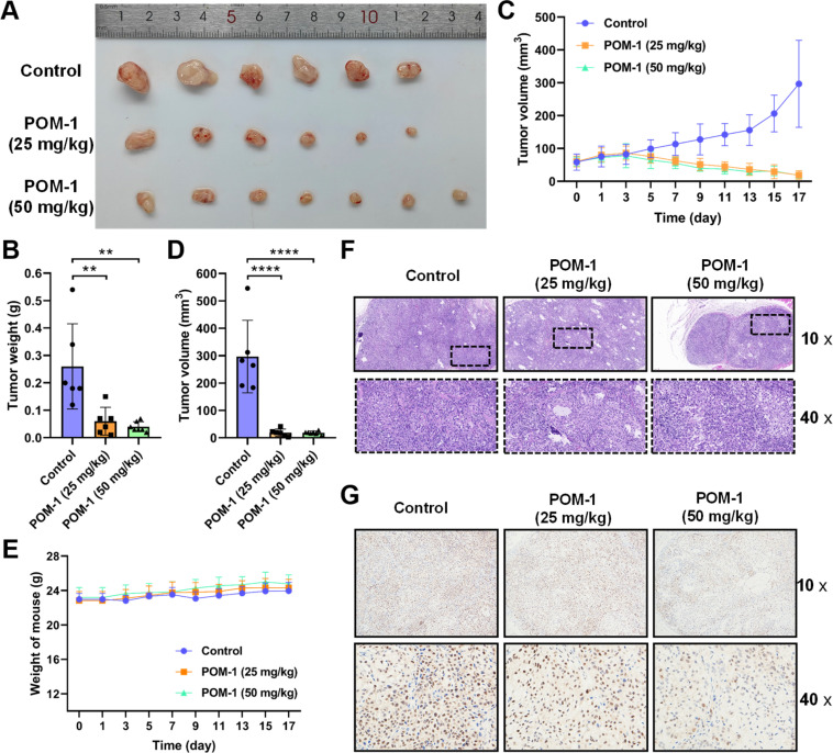
Polyoxometalates (POMs) are a class of anionic metal-oxygen clusters with versatile biological activities. Over the past decade, an increasing number of POMs, especially Sb-rich POMs, have been proven to exert antitumor activity. However, the antitumor effects and mechanisms of POMs in the treatment of non-small cell lung cancer (NSCLC) remain largely unexplored. This study employed a Sb-rich {Sb21Tb7W56} POM (POM-1) for NSCLC therapy and investigated its mechanism of action. Our results demonstrated that POM-1 exhibited cytotoxicity against H1299 and A549 cells with IC50 values of 3.245 μM and 3.591 μM, respectively. The migration and invasion were also inhibited by 28.05% and 76.18% in H1299 cells, as well as 36.88% and 36.98% in A549 cells at a concentration of 5 μM. In a tumor xenograft mouse model, POM-1 suppressed tumor growth by 76.92% and 84.62% at doses of 25 and 50 mg kg-1, respectively. Transcriptomic analysis indicated the alteration of ferroptosis and apoptosis signaling pathways in POM-treated NSCLC cells. Subsequent experimentation confirmed the induction of ferroptosis, evidenced by 5.6-fold elevated lipid peroxide levels with treatment of 5 μM POM-1, alongside increased expression of ferroptosis-associated proteins. Additionally, the apoptosis induced by POM-1 was also validated by the 19.67% and 30.1% increase in apoptotic cells in H1299 and A549 cells treated with 5 μM POM-1, respectively, as well as the upregulated activation of caspase-3. In summary, this study reveals, for the first time, ferroptosis as the antitumor mechanism of Sb-rich POM, and that synergism with ferroptosis and apoptosis is a highly potent antitumor strategy for POM-based antitumor therapy.


Conflict of interest statement

There are no conflicts to declare.

Figures

Fig. 1
Fig. 1. Polyhedral and ball-stick representation of POM-1. Polyhedral key: WO6, blue.
Fig. 2
Fig. 2. POM-1 inhibited proliferation, migration and invasion of NSCLC cells in vitro. (A) Dose-dependent cytotoxicity of POM-1 against H1299 and A549 cells. (B) Time-dependent cytotoxicity of POM-1 against H1299 and (C) A549 cells. Cell viability was evaluated by CCK-8 assay. (D) Colony formation of H1299 and A549 cells with treatment with 2.5 μM or 5 μM POM-1 for 24 hours. Colonies were visualized by crystal violet staining. (E) Quantitation of colony formation rates in D. (F) Representative images of H1299 and A549 cells treated with 5 μM POM-1 in wound healing assay at 0 hours and 24 hours. (G) The proportion of wound scratch area at 24 h was analyzed and calculated with the software of Image J by setting the area at 0 h as 100%. (H) Representative images of H1299 and A549 cells treated with 5 μM POM-1 for 24 hours in transwell invasion assay. (I) The invasion rate of cells treated with POM-1 was calculated by determining A570 nm in crystal violet stained cells. The values are represented as mean ± SD (n = 3). *P < 0.05, **P < 0.01, ***P < 0.001, and ****P < 0.0001.
Fig. 3
Fig. 3. POM-1 suppressed tumor growth in an NSCLC xenograft mouse model. (A) Photographs of tumor tissues resected from the mice on day 17 days after the daily treatment with saline (control), 25 mg kg−1, or 50 mg kg−1 POM-1. (B) Tumor weights in panel A. (C) Tumor volumes determined every second day in tumor-bearing mice in the above groups. (D) Tumor volumes on day 17. (E) Body weights were determined every second day in tumor-bearing mice treated with saline or POM-1. (F) Representative images of histopathological sections of tumor tissue with H&E staining and (G) Ki-67 staining. The values are represented as mean ± SD (n = 6–7). **P < 0.01 and ****P < 0.0001.
Fig. 4
Fig. 4. Transcriptomic analysis on H1299 cells treated with saline or 5 μM POM-1. (A) GSEA enrichment of all genes and Top15 enriched pathways. (B) Normalized enrichment score (NES) of ferroptosis using GSEA analysis. (C) Up-regulated and (D) down-regulated DEGs of ferroptosis from POM-1 treated cells.
Fig. 5
Fig. 5. Ferroptosis induced by POM-1 in H1299 cells. (A and B) Dose-dependent cytotoxicity of POM-1 (A) and RSL3 (B) against H1299 cells in the presence or absence of ferrostatin-1 (Fer-1). (C) Protein expression of HMOX1, GPX4, and FTH1 identified by immunoblot. (D) Fluorescence microscopy imaging of LPO induced by POM-1 visualized by C11-BODIPY 581/591 in H1299 cells. (E and F) C11-BODIPY 581/591 oxidized probe was quantitatively analyzed using a flow cytometer after treatment with different concentrations of POM-1. The values are represented as mean ± SD (n = 3). **P < 0.01 and ****P < 0.0001.
Fig. 6
Fig. 6. Apoptosis induced by POM-1 in NSCLC cells. (A) Fluorescence imaging of phosphatidylserine by FITC-Annexin V in H1299 and A549 cells treated with POM-1. (B) Flow cytometry-based Annexin V/PI apoptosis analysis in H1299 and A549 cells treated with 5 μM POM-1. (C) Quantitation of apoptosis rates in panel B. (D) Expression of cleaved caspase-3 in H1299 and A549 cells treated with 5 μM POM-1 determined by western blot. The values are represented as mean ± SD (n = 3). ****P < 0.0001.
Scheme 1
Scheme 1. Illustration of the antitumor mechanism of POM-1.

References

    1. Chen S. Li M. Weng T. Wang D. Geng J. J. Mater. Chem. B. 2023;11:5715–5747. doi: 10.1039/D2TB02277J. - DOI - PubMed
    1. Yin H. Ma J. Chen L. Piao S. Zhang Y. Zhang S. Ma H. Li Y. Qu Y. Wang X. Xu Q. Cell. Physiol. Biochem. 2018;46:471–481. doi: 10.1159/000488615. - DOI - PubMed
    1. Thai A. A. Solomon B. J. Sequist L. V. Gainor J. F. Heist R. S. Lancet. 2021;398:535–554. doi: 10.1016/S0140-6736(21)00312-3. - DOI - PubMed
    1. Zhu H. Liu H. Zhu J. H. Wang S. Y. Zhou S. S. Kong M. Mao Q. Long F. Fang Z. J. Li S. L. Food Funct. 2021;12:2225–2241. doi: 10.1039/D0FO03341C. - DOI - PubMed
    1. Granadeiro C. M. Julião D. Ribeiro S. O. Cunha-Silva L. Balula S. S. Coord. Chem. Rev. 2023;476:214914. doi: 10.1016/j.ccr.2022.214914. - DOI

LinkOut - more resources