. 2024 Aug 22;9(35):37076-37085.
doi: 10.1021/acsomega.4c03959. eCollection 2024 Sep 3.

Optical Detection of Interleukin-6 Using Liquid Janus Emulsions Using Hyperthermophilic Affinity Proteins

Affiliations

Optical Detection of Interleukin-6 Using Liquid Janus Emulsions Using Hyperthermophilic Affinity Proteins

Michelle Chen et al. ACS Omega. .

Erratum in

Abstract

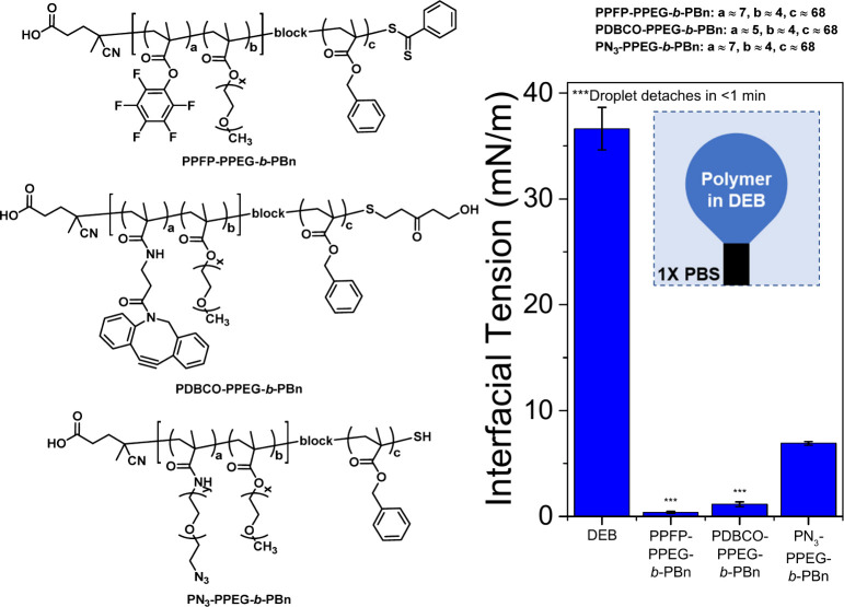
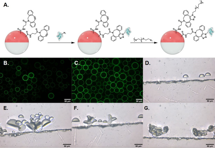
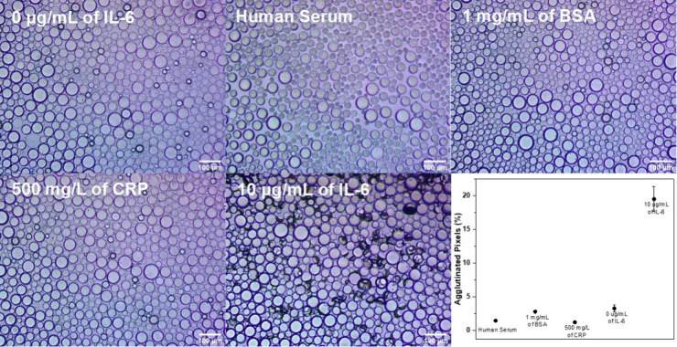
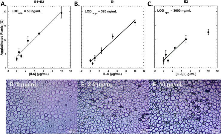
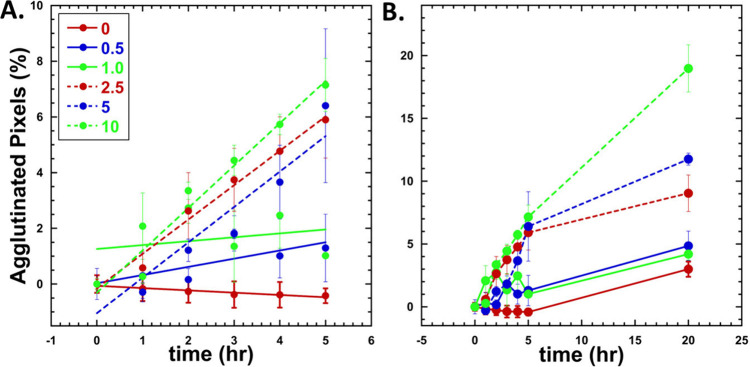
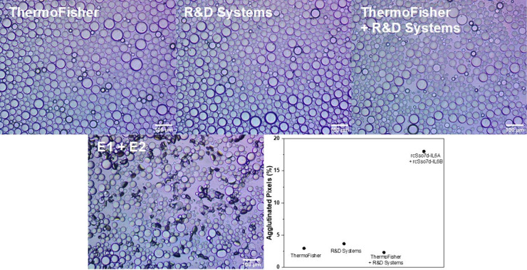
When equal volumes of two immiscible liquids are mixed (e.g., a hydrocarbon and a fluorocarbon), Janus droplets can form in an aqueous solution. In a gravity-aligned Janus droplet, the boundary between the two phases is flat and thus optically transparent when viewed from above. When tipped due to interactions with an analyte (i.e., agglutination), the resulting change in refraction and reflection yields an optical signal that can be detected and quantified. This study reports the detection and quantitation of interleukin-6 (IL-6) using emulsions functionalized at the hydrocarbon:aqueous interface with engineered proteins that bind IL-6 at high affinity and specificity. Hyperthermophilic affinity proteins (rcSso7d) are derived from thermophiles, giving them excellent thermal stability. Two rcSso7d affinity protein variants were synthesized with a noncanonical azide-functionalized amino acid to enable click chemistry to novel polymeric anchors embedded in the hydrocarbon phase. The two binding proteins recognize different epitopes, enabling the detection of both monomeric and dimeric IL-6 via agglutination. It is noteworthy that the rsSso7d protein variants, in addition to having superior thermal stability and facile recombinant synthesis in E. coli, show superior performance when compared to commercial antibodies for IL-6.


Conflict of interest statement

The authors declare no competing financial interest.

Figures

Figure 1
Figure 1
Chemical structures of the atactic amphiphilic diblock copolymers and pendant drop analysis of the polymers in diethylbenzene (DEB). An inverted pendant drop is utilized, because DEB is less dense than the buffer. Having copolymers dissolved in the DEB lowers the DEB-buffer interfacial tension, which indicates that the diblock copolymers are surface active.
Figure 2
Figure 2
A. Droplet bioconjugation scheme. PDBCO-PPEG-b-PBn-functionalized droplets were sequentially reacted with azide-functionalized rcSso7d proteins, followed by poly(ethylene glycol) methyl ether azide. Images are not drawn to scale; droplets are orders of magnitude larger than protein components. Fluorescence microscopy images of droplets reacted with B. FITC-labeled E1 and C. FITC-labeled E2. Side-view images of D. E1+PEG-functionalized Janus droplets, E. and F. 1:1 E1+PEG- and E2+PEG-functionalized droplets incubated with 5 μg/mL of IL-6, and G. DNA-functionalized droplets in 0.1 wt % of 4:6 CTAB:Zonyl that are agglutinated due to electrostatic interactions between the DNA and CTAB. All images are of polydisperse droplets.
Figure 3
Figure 3
Inverted microscope images of 1:1 E1+PEG- and E2+PEG-functionalized droplets incubated under different conditions overnight (0 μg/mL of IL-6, human serum, 1 mg/mL of BSA, 500 mg/L of CRP, and 10 μg/mL of IL-6). The agglutinated droplets appear on their side and create darkened spots in the images as a result of their efficient scattering of transmitted light. At the bottom right is a plot of the percent of agglutinated pixels according to image processing for each control relative to the 10 μg/mL of IL-6 assay.
Figure 4
Figure 4
Dose response curves for A. 1:1 ratio of E1+PEG- and E2+PEG- functionalized Janus droplets (k = 1.591, R = 0.98), B. E1+PEG-functionalized droplets (k = 1.445, R = 0.99), and C. E2+PEG-functionalized droplets (k = 1.559, R = 0.95) after overnight incubation with IL-6. Data are fit to linear responses, and standard errors are reported. The last data point for C. is excluded from the linear fit. Data represent n ≥ 3 replicates. Representative images of agglutinated droplets (1:1 ratio of E1+PEG- and E2+PEG- functionalized) after incubation with D. 0 μg/mL, E. 2.5 μg/mL, and F. 10 μg/mL of IL-6.
Figure 5
Figure 5
Kinetics of agglutination at various concentrations (0–10 ug/mL) A. First 5 h after addition of IL-6. Lines indicate linear fit of data (0 μg/mL = −0.082, R = 0.95, 0.5 μg/mL = 0.29, R = 0.76, 2.5 μg/mL = 1.23, R = 0.99, 5 μg/mL = 1.27, R = 0.94, and 10 μg/mL = 1.52, R = 0.98. 1.0 μg/mL did not fit well to a linear (R = 0.255) but fit is still shown. B. Line graph of data collected overnight indicated increase in agglutination over time as a function of concentration. Standard error is displayed. Data represent n ≥ 3 replicates.
Figure 6
Figure 6
Inverted microscope images of anti-IL-6 antibody+PEG-functionalized droplets and 1:1 E1+PEG- and E2+PEG-functionalized droplets incubated with 5 μg/mL of IL-6 overnight. Droplets with antibodies from ThermoFisher and R&D Systems and rcSso7d proteins purified via Ni2+ resin and subsequent FPLC are shown. Plot of the percent of agglutinated pixels according to image processing for each antibody-based assay relative to the 5 μg/mL of IL-6 assay.

References

    1. Kishimoto T. The Biology of Interleukin-6. Blood 1989, 74 (1), 1–10. 10.1182/blood.V74.1.1.1. - DOI - PubMed
    1. Li S.-P.; Goldman N. D. Regulation of Human C-Reactive Protein Gene Expression by Two Synergistic IL-6 Responsive Elements. Biochemistry 1996, 35 (28), 9060–9068. 10.1021/bi953033d. - DOI - PubMed
    1. Seymour J.F; Talpaz M; Hagemeister F.B; Cabanillas F; Kurzrock R Clinical Correlates of Elevated Serum Levels of Interleukin-6 in Patients with Untreated Hodgkin’s Disease. Am. J. Med. 1997, 102 (1), 21–28. 10.1016/S0002-9343(96)00352-X. - DOI - PubMed
    1. Blay J.-Y.; Negrier S.; Combaret V.; Attali S.; Goillot E.; Merrouche Y.; Mercatello A.; Ravault A.; Tourani J.-M.; Moskovtchenko J.-F.; Philip T.; Favrot M. Serum Level of Interleukin 6 as a Prognosis Factor in Metastatic Renal Cell Carcinoma1. Cancer Res. 1992, 52 (12), 3317–3322. - PubMed
    1. Andrews B.; Shariat S. F.; Kim J.-H.; Wheeler T. M.; Slawin K. M.; Lerner S. P. Preoperative Plasma Levels of Interleukin-6 and Its Ssoluble Receptor Predict Disease Recurrence and Survival of Patients with Bladder Cancer. J. Urol. 2002, 167 (3), 1475–1481. 10.1016/S0022-5347(05)65348-7. - DOI - PubMed

LinkOut - more resources