. 2024 Aug 28;15(10):3477-3484.
doi: 10.1039/d4md00266k. Online ahead of print.

Discovery and optimisation of pyrazolo[1,5- a]pyrimidines as aryl hydrocarbon receptor antagonists

Affiliations

Discovery and optimisation of pyrazolo[1,5- a]pyrimidines as aryl hydrocarbon receptor antagonists

Raitis Bobrovs et al. RSC Med Chem. .

Abstract

The aryl hydrocarbon receptor (AHR) is a versatile ligand-dependent transcription factor involved in diverse biological processes, from metabolic adaptations to immune system regulation. Recognising its pivotal role in cancer immunology, AHR has become a promising target for cancer therapy. Here we report the discovery and structure-activity relationship studies of novel AHR antagonists. The potential AHR antagonists were identified via homology model-based high-throughput virtual screening and were experimentally verified in a luciferase reporter gene assay. The identified pyrazolo[1,5-a]pyrimidine-based AHR antagonist 7 (IC50 = 650 nM) was systematically optimised to elucidate structure-activity relationships and reach low nanomolar AHR antagonistic potency (7a, IC50 = 31 nM). Overall, the findings presented here provide new starting points for AHR antagonist development and offer insightful information on AHR antagonist structure-activity relationships.


Conflict of interest statement

There are no conflicts to declare.

Figures

Fig. 1
Fig. 1. A AHR domains and their boundaries. bHLH, PAS and TAD represent the basic helix–loop–helix, Per–Arnt–Sim and transcriptional activation domains, respectively. PAS-B is the ligand binding domain. B Sequence alignment of hAHR PAS-B domain (Uniprot ID P35869) and hHIF-2α PAS-B domain used in homology modelling (PDB ID: 4XT2). Sequences were aligned using Muscle. Shading indicates residue similarity: red represents identical residues, orange – similar, pink – dissimilar. Residue numbering corresponds to hAHR. Binding site residues within 4 Å from the ligand are underlined in teal. Secondary structure elements are indicated under the sequence. C Comparison of the homology model created (purple) and recently resolved structure of human AHR PAS-B domain (salmon; PDB: 7ZUB). D Comparison of the binding site residue sidechain orientation (shown as sticks). AHR agonist indirubin is shown as teal sticks. E Conformational heterogeneity of side chains located at the binding site entrance. Salmon and purple sticks indicate resolved human AHR structure and homology model created, respectively; grey cartoon and lines show PAS-B domain homologues. Hydrogens are omitted for clarity.
Fig. 2
Fig. 2. AHR antagonists in preclinical and clinical trials. AHR antagonistic activities (IC50) in in vitro assays are given below the compound molecular structure.
Fig. 3
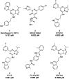
Fig. 3. The most potent AHR virtual screening hits. MolPort and vendor IDs, and measured AHR activity (in % with respect to untreated AHR) are given under the compound molecular structure. AHR activity was measured in the presence of 25 μM compound, and in the presence of 25 μM compound in combination with 10 nM AHR agonist TCDD.
Fig. 4
Fig. 4. A Pyrazolo[1,5-a]pyrimidine core and substituent numbering of the hit compound MolPort-002-650-228 (7). B Docked pose of compound 7 in complex with AHR PAS-B domain homology model. The antagonist (teal) and key AHR residues (salmon) are shown as sticks. Yellow and cyan dashed lines indicate hydrogen bonds and aromatic stacking, respectively. Hydrogens are omitted for clarity.
Fig. 5
Fig. 5. A Docked pose of compound 7 showing the vicinity of the AHR hydrophobic sub-pocket that accommodates the R1 substituent. B SAR or R1 substituent. The hit compound R1 substituent is shown in teal. Compound concentrations at which TCDD-upregulated AHR activity is reduced by 50% (IC50; in μM) are shown under the molecular structures. Compounds are arranged by activity. C Inhibition dose–response curves of BAY2416964 (BAY), GNF351 (GNF), comp. 7a and comp. 7c. HuH7 cells were transfected as described in materials and methods and then treated with 10 nM TCDD in combination with increasing doses of BAY2416964 (BAY), GNF351 (GNF), comp. 7a and comp. 7c. D Western blot for AHR protein in extracts from HuH7 cells after 24 h treatment with DMSO and 1 μM of compounds shown. Beta-actin (ACTB) was used as a loading control.
Fig. 6
Fig. 6. A Docked pose of compound 7 showing the AHR sub-pocket accommodating the R2 substituent. B and C show SAR of R2 substituent (B – commercially available; and C – synthesised compound 7 analogues). Hit compound 7 is shown in teal. Compound concentrations at which TCDD-upregulated AHR activity is reduced by 50% (IC50; in μM) are shown under the molecular structures. Compounds are arranged by activity.
Fig. 7
Fig. 7. A Docked pose of compound 7 showing the AHR sub-pocket around the R3 substituent. B SAR of R3 substituent. Hit compound R3 substituent is shown in teal. Compound concentrations at which TCDD-upregulated AHR activity is reduced by 50% (IC50; in μM) are shown under the molecular structures. Compounds are arranged by activity.
Fig. 8
Fig. 8. Compounds containing individually optimised R1, R2 and R3 substituents. Hit compound 7 is shown in teal. Compound concentrations at which TCDD-upregulated AHR activity is reduced by 50% (IC50; in μM) are shown under the molecular structures. Compounds are arranged by activity.

References

    1. Stockinger B. Di Meglio P. Gialitakis M. Duarte J. H. Annu. Rev. Immunol. 2014;32:403–432. doi: 10.1146/annurev-immunol-032713-120245. - DOI - PubMed
    1. Wen Z. Zhang Y. Zhang B. Hang Y. Xu L. Chen Y. Xie Q. Zhao Q. Zhang L. Li G. Zhao B. Sun F. Zhai Y. Zhu Y. Structure. 2023;31:295–308.e4. doi: 10.1016/j.str.2022.12.013. - DOI - PubMed
    1. Dai S. Qu L. Li J. Zhang Y. Jiang L. Wei H. Guo M. Chen X. Chen Y. Nat. Commun. 2022;13:6234. doi: 10.1038/s41467-022-33858-w. - DOI - PMC - PubMed
    1. Seok S. H. Lee W. Jiang L. Molugu K. Zheng A. Li Y. Park S. Bradfield C. A. Xing Y. Proc. Natl. Acad. Sci. U. S. A. 2017;114:5431–5436. doi: 10.1073/pnas.1617035114. - DOI - PMC - PubMed
    1. Schulte K. W. Green E. Wilz A. Platten M. Daumke O. Structure. 2017;25:1025–1033.e3. doi: 10.1016/j.str.2017.05.008. - DOI - PubMed

LinkOut - more resources