Case Reports
. 2024 Aug 17;19(11):5000-5006.
doi: 10.1016/j.radcr.2024.07.158. eCollection 2024 Nov.

Dual manifestations: spinal and cerebellar hemangioblastomas indicative of von Hippel-Lindau syndrome

Affiliations
Case Reports

Dual manifestations: spinal and cerebellar hemangioblastomas indicative of von Hippel-Lindau syndrome

Nurhuda Hendra Setyawan et al. Radiol Case Rep. .

Abstract

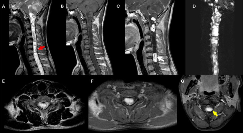
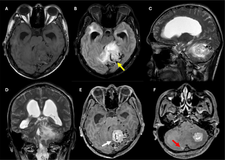
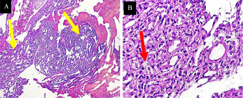
Hemangioblastomas are rare, benign, and highly vascular tumors of the central nervous system, often associated with von Hippel-Lindau (VHL) syndrome, an autosomal dominant disorder characterized by multiple tumors. We present a case of a 32-year-old male with progressive headaches, visual disturbances, and motor deficits, who was diagnosed with multiple hemangioblastomas in the cervical-thoracic spinal cord and bilateral cerebellum through MRI. Surgical resection and histopathological biopsy confirmed the diagnosis. This case highlights the critical role of MRI in diagnosing and managing VHL-associated hemangioblastomas and underscores the importance of regular imaging for early detection and intervention of new or recurring tumors, optimizing patient outcomes.

Keywords: Central nervous system tumors; Hemangioblastoma; MRI; Von Hippel-Lindau syndrome.


Figures

Fig 1
Fig. 1
MRI of the cervical and thoracic spine demonstrating multiple spinal lesions highly suggestive of hemangioblastomas. (A) Sagittal T2-weighted image highlighting the hyperintense cystic component of the suspected lesion. The surrounding spinal cord appears enlarged, resembling a syrinx. The red arrow indicates the focal flow void within the lesion. (B) Sagittal T1-weighted image showing a well-circumscribed, intramedullary lesion extending from the C2 level to the upper thoracic segment. (C) Sagittal postcontrast T1-weighted image illustrating the vivid enhancement of the nodular component of the suspected spinal lesions (white arrow). (D) Coronal STIR (short tau inversion recovery) myelogram showing multiple hyperintense lesions throughout the cervical and upper thoracic spine, consistent with lesions that enlarge the spinal cord. (E) Axial T2-weighted image at the C6-C7 level demonstrating the hyperintense intramedullary cystic component. (F) Axial postcontrast T1-weighted image at the C6-C7 level showing the nodular vivid enhancement of the suspected lesion. (G) Axial postcontrast T1-weighted image at the C2-C3 level showing the eccentric enhancement pattern typically seen in hemangioblastomas (yellow arrow).
Fig 2
Fig. 2
MRI of the brain demonstrating multiple cerebellar lesions highly suggestive of hemangioblastomas. (A) Axial T1-weighted image showing an intra-axial hypointense lesion in the left cerebellar hemisphere. (B) Axial T2-FLAIR image highlighting the hyperintense components of the cerebellar lesion with enlarged vessels appearing as flow voids (yellow arrow), and surrounding edema. (C) Sagittal T2-weighted image showing the location and extent of the lesion in the cerebellum, which is compressing the fourth ventricle. (D) Coronal T2-weighted image revealing hydrocephalus with periventricular edema, secondary to the mass effect from the cerebellar lesions. (E) Axial T1-weighted postcontrast image showing vivid enhancement of a large lesion in the left cerebellum (white arrow), indicating its highly vascular nature. (F) Axial T1-weighted postcontrast image at a lower level than (E), showing a smaller enhancing lesion in the right cerebellar hemisphere (red arrow).
Fig 3
Fig. 3
Intraoperative images of the suboccipital craniotomy for tumor removal. (A) The patient positioned in the prone position, with markings indicating the planned surgical site. (B) Postdurotomy, revealing the underlying structures. (C) Visualization of the cerebellar cortex following dual opening. (D) Post-transvermian corticotomy, white arrow shows the hypervascular, encapsulated tumor at a depth of 1 cm. (E) Surgical field post-tumor biopsy and hemostasis control. (F) Tumor samples obtained postbiopsy.
Fig 4
Fig. 4
Morphological features of hemangioblastoma using hematoxylin-eosin staining are depicted. (A) The hemangioblastoma demonstrates noninfiltrative growth with variable lobularity. Yellow arrows show neoplastic stromal cells arranged amidst numerous small vessels and shows evidence of red blood cell leakage. (B) The stromal component exhibits mild nuclear pleomorphism, degenerative atypia, clear foamy cytoplasm with lipid-filled vacuoles, and a few hyaline globules (red arrow).

References

    1. Klingler J-H, Gläsker S, Bausch B, Urbach H, Krauss T, Jilg CA, et al. Hemangioblastoma and von Hippel-Lindau disease: genetic background, spectrum of disease, and neurosurgical treatment. Child’s Nerv Syst. 2020;36:2537–2552. doi: 10.1007/s00381-020-04712-5. - DOI - PMC - PubMed
    1. Yin X, Duan H, Yi Z, Li C, Lu R, Li L. Incidence, Prognostic factors and survival for hemangioblastoma of the central nervous system: Analysis based on the surveillance, Epidemiology, and end results database. Front Oncol. 2020;10:1–10. doi: 10.3389/fonc.2020.570103. - DOI - PMC - PubMed
    1. Gaillard F: Hemangioblastoma (central nervous system) | radiology reference article | radiopaedia.org. Radiopaedia. Accessed June 25, 2024. https://radiopaedia.org/articles/haemangioblastoma-central-nervous-system-2. 10.53347/rID-1412.
    1. dos Santos AL, S Trevas, Rosado ML, Santos A, Trevas S, Rosado ML. A challenge in diagnosis of cerebellar hemangioblastoma. Cureus. 2022;14:1–5. doi: 10.7759/cureus.21713. - DOI - PMC - PubMed
    1. Yoda RA, Cimino PJ. Neuropathologic features of central nervous system hemangioblastoma. J Pathol Transl Med. 2022;56:115–125. doi: 10.4132/jptm.2022.04.13. - DOI - PMC - PubMed

Publication types

LinkOut - more resources