. 2024 Oct 14;52(18):e88.
doi: 10.1093/nar/gkae760.

DFF-ChIP: a method to detect and quantify complex interactions between RNA polymerase II, transcription factors, and chromatin

Affiliations

DFF-ChIP: a method to detect and quantify complex interactions between RNA polymerase II, transcription factors, and chromatin

Benjamin M Spector et al. Nucleic Acids Res. .

Abstract

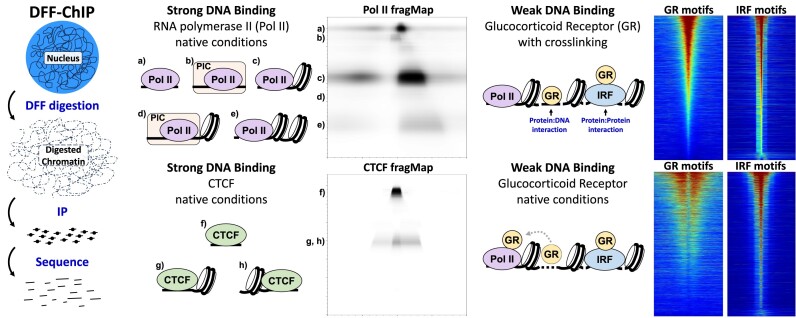
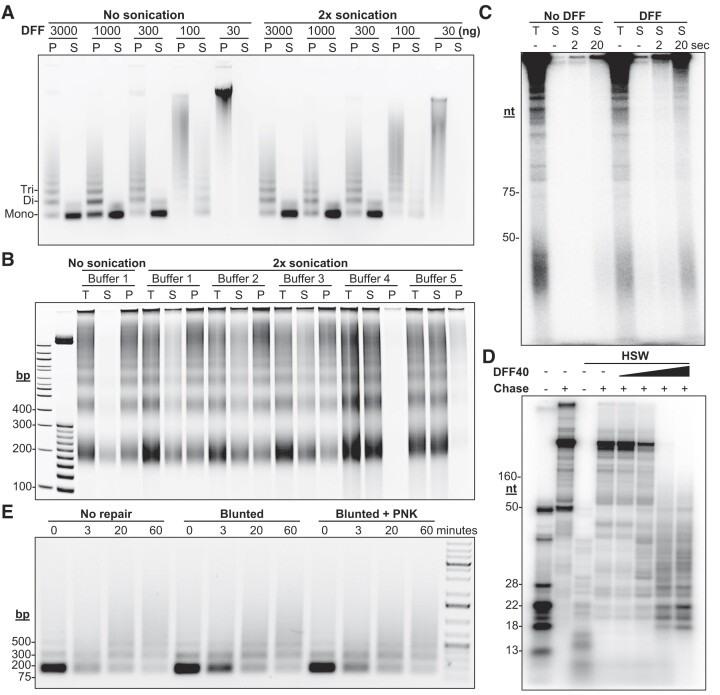
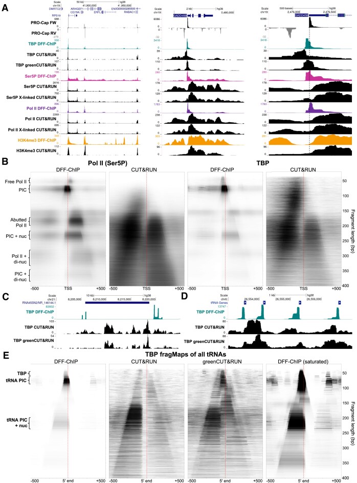
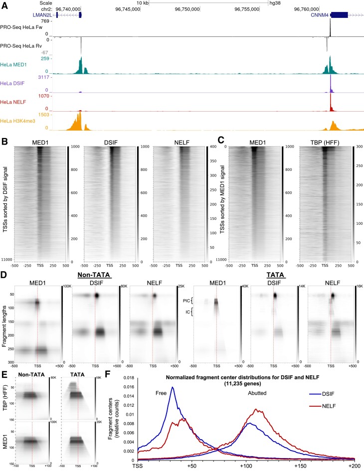
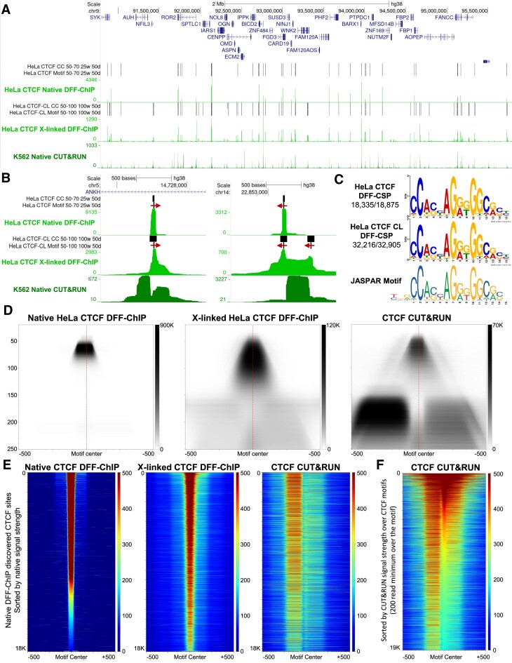
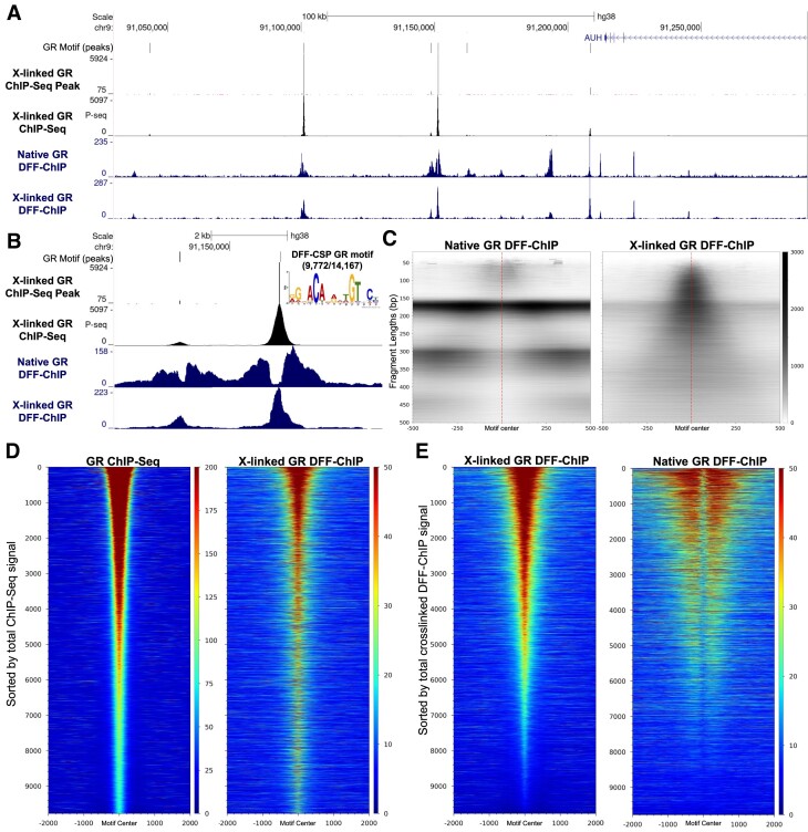
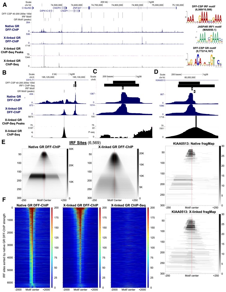
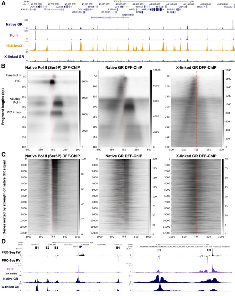
Recently, we introduced a chromatin immunoprecipitation (ChIP) technique utilizing the human DNA Fragmentation Factor (DFF) to digest the DNA prior to immunoprecipitation (DFF-ChIP) that provides the precise location of transcription complexes and their interactions with neighboring nucleosomes. Here we expand the technique to new targets and provide useful information concerning purification of DFF, digestion conditions, and the impact of crosslinking. DFF-ChIP analysis was performed individually for subunits of Mediator, DSIF, and NELF that that do not interact with DNA directly, but rather interact with RNA polymerase II (Pol II). We found that Mediator was associated almost exclusively with preinitiation complexes (PICs). DSIF and NELF were associated with engaged Pol II and, in addition, potential intermediates between PICs and early initiation complexes. DFF-ChIP was then used to analyze the occupancy of a tight binding transcription factor, CTCF, and a much weaker binding factor, glucocorticoid receptor (GR), with and without crosslinking. These results were compared to those from standard ChIP-Seq that employs sonication and to CUT&RUN which utilizes MNase to fragment the genomic DNA. Our findings indicate that DFF-ChIP reveals details of occupancy that are not available using other methods including information revealing pertinent protein:protein interactions.


Figures

Graphical Abstract
Graphical Abstract
Figure 1.
Figure 1.
Applicability of DFF to ChIP protocols. (A) Digestion of 2 million native HAP1 nuclei with the indicated amounts of DFF prior to separation of soluble (S) and insoluble pellet (P) material via centrifugation or prior to sonication and separation. Resulting products were run on an agarose gel and stained with ethidium bromide. (B) 40 million crosslinked HAP1 cell nuclei were digested with 10 μg of DFF for 2 h. The reaction was split into 6 tubes which were then supplemented with buffer to generate the differing conditions to alter solubility of DNA. All buffers had 25 mM Tris (pH 7.8) and 1 mM EDTA and the following additions: 100 mM sodium chloride (1), 100 mM sodium chloride and 0.1% TritonX (2), 150 mM sodium chloride, 1% TritonX, and 0.25% sodium deoxycholate (3), 150 mM sodium chloride and 0.2% Sarkosyl (4), or 150 mM sodium chloride and 0.1% SDS (5). After buffer supplementation samples were lightly sonicated prior to separation via centrifugation. The resulting products were run on a native polyacrylamide gel and stained with ethidium bromide. Samples are total (T), soluble (S) and insoluble pellet (P). (C) HAP1 nuclei were incubated alone or digested with DFF for 30 min at 37°C. Following digestion, transcripts were labeled via incubation with α-32P-CTP. Next, reactions were either sonicated lightly for 2 or 20 s or immediately spun to isolate soluble materials (S) for comparison against the total reaction (T) using a denaturing polyacrylamide gel. (D) An immobilized DNA template was incubated with HNE for 30 min and pulsed for 30 s with limiting α-32P-CTP. The resultant EECs were then washed and treated with a titration of DFF for 10 min. Next, EECs were subsequently chased for 1 minute. The resulting products were run on a denaturing polyacrylamide gel and radioactivity imaged. As a note, the presence of magnesium in the mock buffer allowed for slight backtracking of the paused transcripts in the mock buffer incubated sample. (E) HeLa nuclei were digested with DFF to primarily mono-nucleosomes and DNA isolated. The DNA was then subjected to blunting by T4 DNA polymerase, blunting and an additional end repair utilizing T4 Polynucleotide Kinase (PNK), or left unrepaired. The resulting DNA was then incubated with T4 DNA ligase for differing times and run on an agarose gel and stained.
Figure 2.
Figure 2.
Comparison of DFF-ChIP to CUT&RUN. (A) UCSC genome browser tracks showing HFF PRO-Cap dataset (GSE113394), CUT&RUN for TBP (GSE163049), Ser5P (GSE155666), Pol II (GSE155666) and H3K4me3 (GSE156787) compared to DFF-ChIP datasets (GSE185763). DFF-ChIP and CUT&RUN were not performed utilizing the same antibodies. The first region approximately depicts chr19:41850000–42000000, the second region chr19:2474000- 2481000, and the final region chr19:2475500–2476750. In the right-most region, the primary TSS for GADD45B is demarcated by a dotted line for ease of alignment. (B) FragMaps comparing DFF-ChIP targeting Ser5P and TBP to CUT&RUN targeting the same factors. FragMaps are centered on the MaxTSS of 11,229 genes expressed in HFF cells discovered utlizing the truQuant on the PRO-Cap dataset. (C) UCSC genome browser tracks of DFF-ChIP, CUT&RUN, and greenCUT&RUN targeting TBP showing a 45s preRNA region. (D) UCSC genome browser tracks of DFF-ChIP, CUT&RUN, and greenCUT&RUN targeting TBP showing four tRNA genes. (E) FragMaps comparing DFF-ChIP targeting TBP to CUT&RUN (GSE163049) and greenCUT&RUN (SRP278136) targeting TBP. FragMaps are centered on the mature 5′ ends of 534 tRNA genes.
Figure 3.
Figure 3.
DFF-ChIP allows targeting of non-DNA bound factors. (A) UCSC genome browser tracks showing HeLa PRO-Seq (GSE100742) and DFF-ChIP targeng MED1, DSIF, NELF, and H3K4me3 (GSE185763) on two promoters. (B) MED1, DSIF, and NELF DFF-ChIP heatmaps of 11235 HeLa expressed genes sorted by DSIF signal from -50 to + 250. (C) MED1 DFF-ChIP and TBP DFFChIP (GSE185763) heatmaps of 11,235 HeLa expressed genes sorted by MED1 signal from -50 to + 250. (D) FragMaps of Non-TATA (10380) and TATA (500) genes from the MED1, DSIF, and NELF datasets. (E) Narrow range fragMaps of TBP from HFF cells and MED1 over Non-TATA and TATA genes. (F) Fragment center distribution of either paused (40–60 bp) or abutted (170–210 bp) sized fragments from DSIF (blue) or NELF (red) DFF-ChIP datasets relative to 11235 HeLa TSSs (see Supplementary Data File).
Figure 4.
Figure 4.
DFF-ChIP targeting CTCF. (A) UCSC genome browser tracks showing native CTCF DFF-ChIP, crosslinked CTCF DFF-ChIP, and CTCF CUT&RUN (GSE84474). Every called center cluster (CC) and every CC with a CTCF motif are shown for two different runs of DFF-CSP on either the native or crosslinked CTCF DFF-ChIP datasets. (B) UCSC genome browser tracks around two CTCF sites showing native and crosslinked CTCF DFF-ChIP and CUT&RUN. The direction of the motif is indicated by a red arrow. (C) CTCF motifs discovered by utilizing the MEME motif discovery tool on DFF-CSP outputs for native and crosslinked CTCF DFF-ChIP. Below is the CTCF motif from the JASPAR database (MA0139.1). (D) FragMaps comparing CTCF DFF-ChIP to CTCF CUT&RUN. FragMaps are centered on the 15 bp motif oriented in the direction of the motif in (C) of the 18335 sites identified in the native CTCF DFF-ChIP dataset. (E) Heatmaps centered on the 15 bp motif of 18335 sites identified in the native CTCF dataset sorted by over the CTCF motif from the native dataset. (F) Heatmap centered on the 15 bp motif from the CTCF CUT&RUN dataset. Sites were discovered utilizing motifs identified with the MEME suite FIMO tool to identify every occurrence of the CTCF motif across the genome. Of these motifs, those with 200 reads covering the 15 bp motif were selected and sorted by coverage over the CTCF motif in the CUT&RUN dataset (n = 19 184).
Figure 5.
Figure 5.
Analysis of GR DFF-ChIP over GR DNA binding sites. (A) UCSC genome browser track showing native and crosslinked GR DFF-ChIP, GR ChIP-Seq and the ChIP-Seq-Peak program called peaks (refer to Materials and Methods regarding input for peak calling). (B) Zoomed in view of the same tracks as in (A) focusing on two of the 9,772 peaks with a GR motif identified by the ChIP-Seq-Peak program. Additionally, the MEME motif found for 9,772 of the 14,167 identified peaks is shown. (C) FragMaps centered on the center of 9,772 GR motifs utilizing the native and crosslinked GR DFF-ChIP datasets. (D) Heatmaps centered on the 15 bp GR motif utilizing ChIP-Seq and crosslinked DFF-ChIP datasets sorted by coverage in the ChIP-Seq dataset ±500 bp at the 9,772 GR peaks with a GR motif. (E) Same as (D) except that the heatmaps utilized the crosslinked and native GR DFF-ChIP datasets sorted based on the crosslinked GR DFF-ChIP dataset ±500 bp of the 9,772 GR peaks with a GR motif.
Figure 6.
Figure 6.
DFF-ChIP reveals GR tethered to IRF binding sites. (A) UCSC genome browser track showing GR DFF-ChIP, GR ChIP-Seq and the ChIP-Seq-Peak program called peaks. Sites unique to DFF-ChIP were identified utilizing DFF-CSP run on fragments of 40–200 bp in length from the native GR DFF-ChIP dataset with windows of 200 bp requiring at least 100 centers. The 10,506 resulting regions were used as input into the MEME motif discovery tool to identify 6569 center clusters containing the shown DFF-CSP IRF motif. Comparisons to the JASPAR IRF1 motif (MA0050.1) and the discovered DFF-CSP GR motif are shown. (B) Same as (A) highlighting a GR and IRF binding site. Also, regions enriched from IRF1 ChIP-Seq are also shown (85). (C) Same as (B) highlighting an IRF binding site with no detectable GR ChIP-Seq signal. (D) Same as (B) highlighting proximal IRF and GR binding sites. Below are native and crosslinked DFF-ChIP fragMaps depicting only this region highlighting two distinct footprints aligned with the marked IRF and GR motifs. (E) FragMaps centered on the 15 bp DFF-CSP IRF motif shown in (A) comparing native GR DFF-ChIP to crosslinked GR DFF-ChIP at the 6569 IRF Sites. (F) Heatmaps centered on the 6,569 15 bp DFF-CSP IRF motifs shown in (A) utilizing data from the native and crosslinked GR DFF-ChIP and the GR ChIP-Seq datasets. Heatmaps were sorted based on coverage of the 15 bp IRF motif in the native GR DFF-ChIP dataset.
Figure 7.
Figure 7.
GR associates with engaged Pol II. (A) UCSC genome browser tracks showing DFF-ChIP targeting GR under both native and crosslinked conditions (SUP-B15 cells) with Pol II and H3K4me3 DFF-ChIP (HFFs). (B) FragMaps comparing DFF-ChIP targeting Pol II (Ser5P in HFFs) to native and crosslinked DFF-ChIP targeng GR (SUP-B15 cells). FragMaps are centered on the TSSs of 11,229 genes expressed in HFF cells discovered utilizing the truQuant program on a PRO-Cap dataset. (C) Heatmaps utilizing Pol II (Ser5P) DFF-ChIP and both crosslinked and native GR DFF-ChIP datasets sorted by abutted Pol II signal (+1 to +250) from the Ser5P dataset at the 12,229 genes expressed in HFF cells. (D) UCSC genome browser tracks showing four enhancers (E1–4) surrounding DDIT4. Tracks shown are PRO-Seq and DSIF (HeLa), native and crosslinked GR DFF-ChIP (SUP-B15 cells), as well as the GR ChIP-Seq peaks containing GR motifs.

Similar articles

Cited by

References

    1. Luse D.S., Parida M., Spector B.M., Nilson K.A., Price D.H. A unified view of the sequence and functional organization of the human RNA polymerase II promoter. Nucleic Acids Res. 2020; 48:7767–7785. - PMC - PubMed
    1. Yamaguchi Y., Shibata H., Handa H. Transcription elongation factors DSIF and NELF: promoter-proximal pausing and beyond. Biochim. Biophys. Acta. 2013; 1829:98–104. - PubMed
    1. Core L., Adelman K. Promoter-proximal pausing of RNA polymerase II: a nexus of gene regulation. Genes Dev. 2019; 33:960–982. - PMC - PubMed
    1. Dollinger R., Gilmour D.S. Regulation of promoter proximal pausing of RNA polymerase II in metazoans. J. Mol. Biol. 2021; 433:166897. - PMC - PubMed
    1. Peterlin B.M., Price D.H. Controlling the elongation phase of transcription with P-TEFb. Mol. Cell. 2006; 23:297–305. - PubMed

MeSH terms