. 2024 Nov 1;30(21):4987-4994.
doi: 10.1158/1078-0432.CCR-23-3775.

Tumor-Infiltrating Lymphocytes in Necrotic Tumors after Melanoma Neoadjuvant Anti-PD-1 Therapy Correlate with Pathologic Response and Recurrence-Free Survival

Affiliations

Tumor-Infiltrating Lymphocytes in Necrotic Tumors after Melanoma Neoadjuvant Anti-PD-1 Therapy Correlate with Pathologic Response and Recurrence-Free Survival

Kevin L Ma et al. Clin Cancer Res. .

Abstract

Purpose: Neoadjuvant anti-PD-1 therapy in melanoma may increase tumor-infiltrating lymphocytes (TIL), and more TIL are associated with better treatment response. A major pathologic response (MPR) in melanoma after neoadjuvant anti-PD-1 therapy usually comprises tumor necrosis and fibrosis. The role of TIL in necrotic tumor necrosis (nTIL) has not been explored.

Experimental design: We performed CD3 and CD8 IHC stains on 41 melanomas with geographic necrosis. Of the 41, 14 were immunotherapy-naïve, and 27 had been treated with one dose of neoadjuvant anti-PD-1 in two clinical trials. CD3+ and CD8+ nTIL were graded as absent/minimal or moderate/brisk. The percentage of necrotic areas in the tumor bed before and after treatment was quantified. The endpoints were MPR and 5-year recurrence-free survival (RFS).

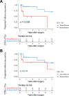
Results: In the immunotherapy-naïve cohort, 3/14 (21%) specimens had moderate/brisk CD3+, and 2/14 (14%) had moderate/brisk CD8+ nTIL. In the treated cohort, 16/27 (59%) specimens had moderate/brisk CD3+, and 15/27 (56%) had moderate/brisk CD8+ nTIL, higher than those of the naïve cohort (CD3, P = 0.046; CD8, P = 0.018). Tumor necrosis was significantly increased after anti-PD-1 therapy (P = 0.007). In the treated cohort, moderate/brisk CD3+ and CD8+ nTIL correlated with MPR (P = 0.042; P = 0.019, respectively). Treated patients with moderate/brisk CD3+ nTIL had higher 5-year RFS than those with absent/minimal nTIL (69% vs. 0%; P = 0.006). This persisted on multivariate analysis (HR, 0.16; 95% confidence interval, 0.03-0.84; P = 0.03), adjusted for pathologic response, which was borderline significant (HR, 0.26; 95% confidence interval, 0.07-1.01; P = 0.051).

Conclusions: CD3+ and CD8+ nTIL are associated with pathologic response and 5-year RFS in patients with melanoma after neoadjuvant anti-PD-1 therapy.


Conflict of interest statement

Conflicts of Interest: Dr. Karakousis serves on the advisory board of Merck. The remaining authors have no conflicts of interest to declare.

Figures

Figure 1.
Figure 1.. Immunohistochemistry of CD3 and CD8 in areas with melanoma tumor necrosis.
A) Grading schema of density of immunohistochemistry positive lymphocytes. Representative images of brisk, moderate, and minimal immunohistochemistry positive lymphocytes in the area of melanoma necrosis. B) Representative histology of a necrotic melanoma from an immunotherapy-naïve patient, with absent/minimal immunohistochemistry positive CD3+ and CD8+ T cells. C) Representative histology of a necrotic melanoma from a neoadjuvant anti-PD-1 treated patient with moderate/brisk immunohistochemistry positive CD3+ and CD8+ T cells. This patient had a major pathological response to a single neoadjuvant dose of pembrolizumab. Images taken at 40x magnification, and horizontal bars represent 50 μm. Abbreviations: H&E, hematoxylin & eosin.
Figure 2.
Figure 2.. More CD3+ and CD8+ nTILs are present in specimens treated with neoadjuvant anti-PD-1 therapy than immunotherapy-naïve specimens.
A) CD3+ nTILs by immunotherapy status. B) CD8+ nTILs by immunotherapy status. * indicates p<0.05. Error bars indicate 95% confidence intervals (Wilson-Brown method). Abbreviations: nTILs, tumor-infiltrating lymphocytes in necrotic tumor.
Figure 3.
Figure 3.. MPR to neoadjuvant anti-PD-1 therapy is associated with increased necrosis in the tumor bed.
A) Necrosis in all patients. B) Necrosis in patients with pNR or pPR. C) Necrosis in patients with MPR. Necrosis in the tumor bed was measured in the pre- and post-treatment tissue specimens. * indicates p<0.05; ** indicates p<0.01; ns indicates p>0.05. Abbreviations: pNR, pathological non-response; pPR, pathological partial response; MPR, major pathological response.
Figure 4.
Figure 4.. CD3+ and CD8+ nTILs are correlated with major pathological response.
A) CD3+ nTILs by pathological response. B) CD8+ nTILs by pathological response. * indicates p<0.05. Error bars indicate 95% confidence intervals (Wilson-Brown method). Abbreviations: nTILs, tumor-infiltrating lymphocytes in necrotic tumor; pNR, pathological non-response; pPR, pathological partial response; MPR, major pathological response.
Figure 5.
Figure 5.. Five-year recurrence-free survival of melanoma patients treated with neoadjuvant anti-PD-1 is significantly associated with CD3+ nTILs.
A) Recurrence-free survival by CD3+ nTILs. B) Recurrence-free survival by CD8+ nTILs. p-values correspond to log-rank tests. Tick marks indicate censored events. Abbreviations: nTILs, tumor-infiltrating lymphocytes in necrotic tumor.

References

    1. Pichler M, Hutterer GC, Chromecki TF, et al. Histologic tumor necrosis is an independent prognostic indicator for clear cell and papillary renal cell carcinoma. Am J Clin Pathol. Feb 2012;137(2):283–9. doi:10.1309/ajcplbk9l9kdyqzp - DOI - PubMed
    1. Liu ZG, Jiao D. Necroptosis, tumor necrosis and tumorigenesis. Cell Stress. Dec 19 2019;4(1):1–8. doi:10.15698/cst2020.01.208 - DOI - PMC - PubMed
    1. Jimenez RE, Wallis T, Visscher DW. Centrally necrotizing carcinomas of the breast: a distinct histologic subtype with aggressive clinical behavior. Am J Surg Pathol. Mar 2001;25(3):331–7. doi:10.1097/00000478-200103000-00007 - DOI - PubMed
    1. Ladstein RG, Bachmann IM, Straume O, Akslen LA. Tumor necrosis is a prognostic factor in thick cutaneous melanoma. Am J Surg Pathol. Oct 2012;36(10):1477–82. doi:10.1097/PAS.0b013e31825a5b45 - DOI - PubMed
    1. Bachmann IM, Ladstein RG, Straume O, Naumov GN, Akslen LA. Tumor necrosis is associated with increased alphavbeta3integrin expression and poor prognosis in nodular cutaneous melanomas. BMC Cancer. 2008/12/05 2008;8(1):362. doi:10.1186/1471-2407-8-362 - DOI - PMC - PubMed

MeSH terms

Substances