Review
. 2024 Oct 14;45(39):4156-4169.
doi: 10.1093/eurheartj/ehae526.

Management of aortic disease in children with FBN1-related Marfan syndrome

Affiliations
Review

Management of aortic disease in children with FBN1-related Marfan syndrome

Laura Muiño-Mosquera et al. Eur Heart J. .

Abstract

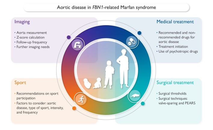
Marfan syndrome (MFS) is a hereditary connective tissue disorder with an estimated prevalence of 1:5000-1:10 000 individuals. It is a pleiotropic disease characterized by specific ocular, cardiovascular, and skeletal features. The most common cardiovascular complication is aortic root dilatation which untreated can lead to life-threatening aortic root dissection, mainly occurring in adult patients. Prompt diagnosis, appropriate follow-up, and timely treatment can prevent aortic events. Currently there are no specific recommendations for treatment of children with MFS, and management is greatly based on adult guidelines. Furthermore, due to the scarcity of studies including children, there is a lack of uniform treatment across different centres. This consensus document aims at bridging these gaps of knowledge. This work is a joint collaboration between the paediatric subgroup of the European Network of Vascular Diseases (VASCERN, Heritable Thoracic Aortic Disease Working Group) and the Association for European Paediatric and Congenital Cardiology (AEPC). A group of experts from 12 different centres and 8 different countries participated in this effort. This document reviews four main subjects, namely, (i) imaging of the aorta at diagnosis and follow-up, (ii) recommendations on medical treatment, (iii) recommendations on surgical treatment, and (iv) recommendations on sport participation.

Keywords: Aortic disease treatment; Aortic imaging; Children; Exercise recommendation; Marfan syndrome.


Figures

Graphical Abstract
Graphical Abstract
Overview of the four main topics addressed in this joint statement and the most important questions addressed within each topic. This document focuses on follow-up and treatment of aortic disease in children with Marfan syndrome and carrying a pathogenic variant in the fibrillin-1 (FBN1) gene. PEARS, personalized external aortic root support.
Figure 1
Figure 1
Stepwise process of the creation of the consensus document. The four main topics treated were (i) imaging of the aorta, (ii) medical treatment, (iii) surgical treatment, and (iv) sport participation. Literature search was mainly performed in PubMed and Web of Science. The online discussions were followed by dedicated surveys to evaluate the level of agreement to each statement. If a statement had <75% agreement, a new round of online questions was performed based on the former feedback of all group members. This process was repeated until statements were formulated with a minimum of 75% agreement. The document was validated in a final step by several independent expert centres as indicated in Acknowledgements
Figure 2
Figure 2
Echocardiographic imaging of the aorta at different levels. A Parasternal long-axis view of the aortic annulus, sinus of Valsalva, and sinotubular junction measured in systole using the inner-to-inner method. Measurements should be taken perpendicular to blood flow. B Parasternal long-axis view of the sinus of Valsalva and sinotubular junction measured in diastole using the leading edge-to-leading edge method. C Parasternal short-axis view of the aortic valve measured in diastole and using the largest aortic diameter. D Parasternal long-axis of the ascending aorta, measured in systole at the level of the right pulmonary artery, using the inner-to-inner method. E Suprasternal view of the aortic arch measured in systole between the truncus brachiocephalicus and the left carotid artery, using the inner-to-inner diameter. F Modified apical two-chamber view of the descending aorta measured at the level of the left atrium in systole using the inner-to-inner method. G Subcostal view of the abdominal aorta at the level of the liver, measured in systole using the inner-to-inner diameter. Ao, aorta; AP2CH, apical two-chamber view; LA, left atrium; LV, left ventricle; PSLAX, parasternal long-axis view; PSSAX, parastenal short-axis view
Figure 3
Figure 3
Differentiation of sports in relation to the predominant isotonic and isometric component, intensity, and frequency (adapted from the 2020 ESC guidelines on sport cardiology and exercise in patients with cardiovascular disease)

References

    1. Magenis RE, Maslen CL, Smith L, Allen L, Sakai LY. Localization of the fibrillin (FBN) gene to chromosome 15, band q21.1. Genomics 1991;11:346–51. 10.1016/0888-7543(91)90142-2 - DOI - PubMed
    1. Loeys BL, Dietz HC, Braverman AC, Callewaert BL, De Backer J, Devereux RB et al. The revised Ghent nosology for the Marfan syndrome. J Med Genet 2010;47:476–85. 10.1136/jmg.2009.072785 - DOI - PubMed
    1. Stheneur C, Tubach F, Jouneaux M, Roy C, Benoist G, Chevallier B et al. Study of phenotype evolution during childhood in Marfan syndrome to improve clinical recognition. Genet Med 2014;16:246–50. 10.1038/gim.2013.123 - DOI - PubMed
    1. Zarate YA, Morris SA, Blackshare A, Algaze CA, Connor BS, Kim AJ et al. A clinical scoring system for early onset (neonatal) Marfan syndrome. Genet Med 2022;24:1503–11. 10.1016/j.gim.2022.03.016 - DOI - PubMed
    1. Hennekam RCM. Severe infantile Marfan syndrome versus neonatal Marfan syndrome. Am J Med Genet A 2005;139A:1. 10.1002/ajmg.a.30979 - DOI - PubMed