

Neuroinflammation, cerebrovascular dysfunction and diurnal cortisol biomarkers in a memory clinic cohort: Findings from the Co-STAR study
- PMID: 39251589
- PMCID: PMC11385239
- DOI: 10.1038/s41398-024-03072-x
Neuroinflammation, cerebrovascular dysfunction and diurnal cortisol biomarkers in a memory clinic cohort: Findings from the Co-STAR study
Abstract
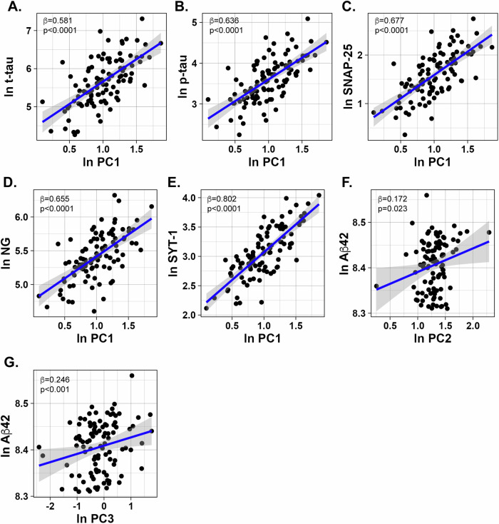
Cortisol dysregulation, neuroinflammation, and cerebrovascular dysfunction are biological processes that have been separately shown to be affected in Alzheimer's disease (AD). Here, we aimed to identify biomarker signatures reflecting these pathways in 108 memory clinic patients with subjective cognitive decline (SCD, N = 40), mild cognitive impairment (MCI, N = 39), and AD (N = 29). Participants were from the well-characterized Cortisol and Stress in Alzheimer's Disease (Co-STAR) cohort, recruited at Karolinska University Hospital. Salivary diurnal cortisol measures and 41 CSF proteins were analyzed. Principal component analysis was applied to identify combined biosignatures related to AD pathology, synaptic loss, and neuropsychological assessments, in linear regressions adjusted for confounders, such as age, sex, education and diagnosis. We found increased CSF levels of C-reactive protein (CRP), interferon γ-inducible protein (IP-10), thymus and activation-regulated chemokine (TARC), intercellular adhesion molecule-1 (ICAM-1), and vascular cell adhesion molecule-1 (VCAM-1) in MCI patients. Further, markers of cortisol dysregulation (flattened salivary cortisol awakening response and flattened cortisol slope) correlated with increased levels of placental growth factor (PlGF), IP-10, and chitinase 3-like 1 (YKL-40) in the total cohort. A biosignature composed of cortisol awakening response, cortisol slope, and CSF IL-6 was downregulated in AD patients. Moreover, biomarker signatures reflecting overlapping pathophysiological processes of neuroinflammation and vascular injury were associated with AD pathology, synaptic loss, and worsened processing speed. Our findings suggest an early dysregulation of immune and cerebrovascular processes during the MCI stage and provide insights into the interrelationship of chronic stress and neuroinflammation in AD.
© 2024. The Author(s).
Conflict of interest statement
HZ has served at scientific advisory boards and/or as a consultant for AbbVie, Alector, Annexon, Artery Therapeutics, AZTherapies, CogRx, Denali, Eisai, Nervgen, Novo Nordisk, Pinteon Therapeutics, Red Abbey Labs, Passage Bio, Roche, Samumed, Siemens Healthineers, Triplet Therapeutics, and Wave, has given lectures in symposia sponsored by Cellectricon, Fujirebio, Alzecure, Biogen, and Roche, and is a co-founder of Brain Biomarker Solutions in Gothenburg AB (BBS), which is a part of the GU Ventures Incubator Program (outside submitted work). MK has served at scientific advisory boards at Biogen, Roche, Combinostics and Swedish Care International and given lectures in symposia sponsored by Biogen, Roche, Nutricia, Lundbeck, and Nestlé. All other authors reported no biomedical financial interests or potential conflicts of interest.
Figures


References
MeSH terms
Substances
Grants and funding
LinkOut - more resources
Full Text Sources
Medical
Research Materials
Miscellaneous
