. 2024 Sep 9;15(1):7873.
doi: 10.1038/s41467-024-52284-8.

MerlinS13 phosphorylation regulates meningioma Wnt signaling and magnetic resonance imaging features

Affiliations

MerlinS13 phosphorylation regulates meningioma Wnt signaling and magnetic resonance imaging features

Charlotte D Eaton et al. Nat Commun. .

Abstract

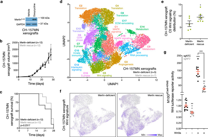
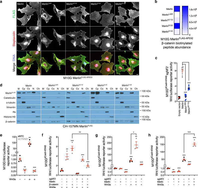
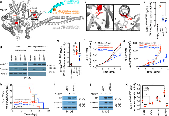
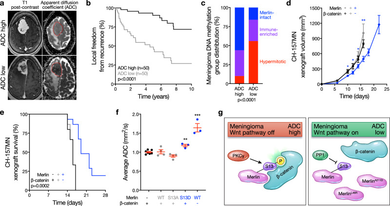
Meningiomas are associated with inactivation of NF2/Merlin, but approximately one-third of meningiomas with favorable clinical outcomes retain Merlin expression. Biochemical mechanisms underlying Merlin-intact meningioma growth are incompletely understood, and non-invasive biomarkers that may be used to guide treatment de-escalation or imaging surveillance are lacking. Here, we use single-cell RNA sequencing, proximity-labeling proteomic mass spectrometry, mechanistic and functional approaches, and magnetic resonance imaging (MRI) across meningioma xenografts and patients to define biochemical mechanisms and an imaging biomarker that underlie Merlin-intact meningiomas. We find Merlin serine 13 (S13) dephosphorylation drives meningioma Wnt signaling and tumor growth by attenuating inhibitory interactions with β-catenin and activating the Wnt pathway. MRI analyses show Merlin-intact meningiomas with S13 phosphorylation and favorable clinical outcomes are associated with high apparent diffusion coefficient (ADC). These results define mechanisms underlying a potential imaging biomarker that could be used to guide treatment de-escalation or imaging surveillance for patients with Merlin-intact meningiomas.


Conflict of interest statement

The authors declare no competing interests.

Figures

Fig. 1
Fig. 1. Merlin drives meningioma Wnt signaling.
a Immunoblots for FLAG (Merlin) or GAPDH in CH-157MN meningioma xenografts with or without 24 h of doxycycline-inducible Merlin rescue (200 μg/ml). Representative of four experimental repeats. b CH-157MN xenograft measurements in NU/NU mice with (n = 12) or without (n = 12) doxycycline-inducible Merlin rescue as in (a). c Kaplan-Meier survival curve for CH-157MN xenograft overall survival in NU/NU mice as in (b) (log-rank test). d Uniform manifold approximation and projection (UMAP) of single-cell RNA sequencing transcriptomes of 40,765 CH-157 cells from 12 xenografts (n = 5 Merlin-deficient CH-157MN xenografts, n = 7 CH-157MN xenografts with Merlin rescue) as in (ac) colored by assignments from Louvain clustering. e Quantification of single-cell types from (d) that were differentially enriched in Merlin-deficient compared to Merlin rescue xenografts. f Feature plots for the MSigDB Hallmark Wnt target gene expression signature in single-cell transcriptomes from Merlin-deficient (n = 5) compared to Merlin rescue xenografts (n = 7). g TOP-Flash Tcf/Lef luciferase reporter assay in M10GdCas9-KRAB meningioma cells expressing non-targeted control sgRNAs (sgNTC) or sgRNAs suppressing NF2 (sgNF2) with or without 24 h of Wnt3a treatment (100 ng/μl). Representative of three experimental repeats. Lines represent means, and error bars represent standard error of the means. *P ≤ 0.05, **P ≤ 0.01, ***P ≤ 0.0001 (Student’s t test, one sided). Source data are provided as a Source Data file.
Fig. 2
Fig. 2. Merlin regulates β-catenin localization in meningioma cells.
a Immunofluorescence for FLAG (Merlin) or streptavidin in M10G meningioma cells expressing MerlinFLAG-APEX2 constructs after proximity-labelling. Representative of five biological replicates. Scale bar, 10 μm. b Heatmap of β-catenin biotinylation peptide intensity from proximity-labeling proteomic mass spectrometry as in (a) (n = 3/condition). c TOP-Flash Tcf/Lef luciferase reporter assay in M10GdCas9-KRAB meningioma cells expressing sgRNAs suppressing NF2 (sgNF2) with or without rescue of MerlinFLAG-APEX2 wildtype or cancer-associated missense constructs, n = 3. d Immunoblots for FLAG (Merlin) or β-catenin after biochemical fractionation of CH-157MN meningioma cells expressing MerlinFLAG-APEX2 rescue constructs. Immunoblots for calreticulin, α-tubulin, vimentin, Rb, or histone H3 mark membrane (M), cytoplasmic (Cp), cytoskeletal (Cs), nuclear (N), or chromatin fractions (Ch), respectively. Representative of six biological replicates. e TOP-Flash Tcf/Lef luciferase reporter assay in M10G meningioma cells expressing non-targeted control siRNAs (siNTC) or siRNAs suppressing β-catenin (siCTNNB1) with or without 24 h of Wnt3a treatment (100 ng/μl). Data are representative of four biological replicates. fh TOP-Flash Tcf/Lef luciferase reporter assays in M10GdCas9-KRAB meningioma cells expressing non-targeted control sgRNAs (sgNTC) or sgNF2 with or without 24 h of Wnt3a treatment (100 ng/μl) in the presence or absence of β-catenin or Merlin overexpression or rescue. Data are representative of four biological replicates. f shows β-catenin overexpression fails to hyperactivate the Wnt pathway in the absence of Merlin. Data are representative of four biological replicates. g, h show Wnt stimulation is necessary for Merlin overexpression to hyperactivate the Wnt pathway. Data are representative of four biological replicates. Lines represent means, and error bars represent standard error of the means. **P ≤ 0.01, ***P ≤ 0.0001 (Student’s t test, one sided). Source data are provided as a Source Data file.
Fig. 3
Fig. 3. Merlin dephosphorylation at Serine 13 activates meningioma Wnt signaling.
a Full-length structural model of Merlin, based on the crystal template 4RZJ. Red shows cancer-associated missense mutations. Cyan shows the NTD, which is not conserved in other FERM family members, containing a consensus PP1A/PKC phosphorylation motif and phosphorylation site (S13) in orange. b the 4RZJ X-ray structure of Merlin shows L46 and A211 in hydrophobic pockets that may be destabilized by charged cancer-associated missense mutations. c TOP-Flash Tcf/Lef luciferase reporter assay in M10GdCas9-KRAB meningioma cells expressing sgRNAs suppressing NF2 (sgNF2) with or without rescue of Merlin constructs or overexpression of the FERM family member Moesin. Data are representative of four biological replicates. d Immunoblots for FLAG (Merlin), β-catenin, or GAPDH before versus after FLAG immunoprecipitation from M10G meningioma cells with or without doxycycline-inducible MerlinFLAG overexpression (1 μg/ml). e TOP-Flash Tcf/Lef luciferase reporter assay in M10GdCas9-KRAB meningioma cells expressing sgNF2 with or without rescue of Merlin wildtype or S13 unphosphorylatable (S13A) or phospho-mimetic (S13D) constructs. Representative of 3 experimental repeats. f CH-157MN meningioma cell MTT assays for cell proliferation with or without doxycycline-inducible Merlin rescue (100 ng/ml). Representative of four experimental repeats. g CH-157MN xenograft measurements in NU/NU mice with or without doxycycline-inducible Merlin rescue (20 μg/ml) as in (f). h Kaplan-Meier survival curve for CH-157MN xenograft overall survival in NU/NU mice as in g (log-rank test). i, j Immunoblots for HA (Merlin) or Merlin with phosphorylated S13 (MerlinpS13) after Merlin immunoprecipitation from M10G cells with or without Merlin overexpression and concurrent expression of non-targeted control siRNAs (siNTC) or siRNAs suppressing PP1A or PKC isoforms. GAPDH immunoblots are shown from immunoprecipitation inputs as a loading control. Representative of four experimental repeats. k TOP-Flash Tcf/Lef luciferase reporter assay in M10GdCas9-KRAB meningioma cells expressing non-targeted control sgRNAs (sgNTC) or sgNF2 with concurrent siNTC or siRNA suppression of PP1A or PKC isoforms. Data are representative of four biological replicates. Lines represent means, and error bars represent standard error of the means. *P ≤ 0.05, **P ≤ 0.01, ***P ≤ 0.0001 (Student’s t test, one sided). Source data are provided as a Source Data file.
Fig. 4
Fig. 4. High MRI apparent diffusion coefficient distinguishes Merlin-intact meningiomas with favorable clinical outcomes.
a Example brain magnetic resonance imaging (MRI) T1 post-contrast and apparent diffusion coefficient (ADC) maps. Red dotted lines show meningiomas on ADC maps. Representative of n = 100 meningiomas. b Kaplan-Meier survival curve for local freedom from recurrence in 100 human meningioma patients with pre-operative MRI analysis and post-operative DNA methylation profiling dichotomized at the mean normalized ADC (1.21, log-rank test). c Meningioma DNA methylation groups across ADC high versus ADC low strata from the 100 patients in (b) (Chi-squared test). d CH-157MN xenograft measurements in NU/NU mice with or without doxycycline-inducible Merlin rescue or shRNA suppression of β-catenin (n = 15/condition). e Kaplan-Meier survival curve for CH-157MN xenograft overall survival in NU/NU mice as in (d) (n = 15/condition, log-rank test). f Normalized ADC from MRI of CH-157MN xenografts as in (d, e). g Model of meningioma Wnt signaling in the context of Merlin post-translational modifications, PP1A and PKC activity, cancer-associated missense mutants, and meningioma ADC. Lines represent means, and error bars represent standard error of the means. *P ≤ 0.05, **P ≤ 0.01, ***P ≤ 0.0001 (Student’s t test, one sided). Source data are provided as a Source Data file.

Update of

References

    1. Ostrom, Q. T. et al. CBTRUS statistical report: primary brain and other central nervous system tumors diagnosed in the United States in 2015–2019. Neuro-Oncology24, v1–v95 (2022). 10.1093/neuonc/noac202 - DOI - PMC - PubMed
    1. Goldbrunner, R. et al. EANO guideline on the diagnosis and management of meningiomas. Neuro-Oncology23, 1821–1834 (2021). 10.1093/neuonc/noab150 - DOI - PMC - PubMed
    1. Lin, D. et al. Trends in intracranial meningioma incidence in the United States, 2004–2015. Cancer Med.8, 6458–6467 (2019). 10.1002/cam4.2516 - DOI - PMC - PubMed
    1. Brastianos, P. K. et al. Advances in multidisciplinary therapy for meningiomas. Neuro-Oncology.21, i18–i31 (2019). 10.1093/neuonc/noy136 - DOI - PMC - PubMed
    1. Sahm, F. et al. DNA methylation-based classification and grading system for meningioma: a multicentre, retrospective analysis. Lancet Oncol. 10.1016/s1470-2045(17)30155-9 (2017). - PubMed

Publication types

MeSH terms