. 2024 Sep 9;9(1):164.
doi: 10.1038/s41541-024-00950-9.

Computationally designed Spike antigens induce neutralising responses against the breadth of SARS-COV-2 variants

Affiliations

Computationally designed Spike antigens induce neutralising responses against the breadth of SARS-COV-2 variants

Sneha Vishwanath et al. NPJ Vaccines. .

Abstract

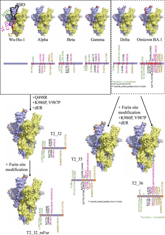
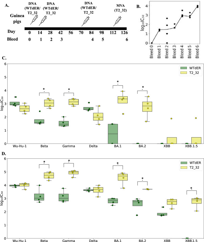
Updates of SARS-CoV-2 vaccines are required to generate immunity in the population against constantly evolving SARS-CoV-2 variants of concerns (VOCs). Here we describe three novel in-silico designed spike-based antigens capable of inducing neutralising antibodies across a spectrum of SARS-CoV-2 VOCs. Three sets of antigens utilising pre-Delta (T2_32), and post-Gamma sequence data (T2_35 and T2_36) were designed. T2_32 elicited superior neutralising responses against VOCs compared to the Wuhan-1 spike antigen in DNA prime-boost immunisation regime in guinea pigs. Heterologous boosting with the attenuated poxvirus - Modified vaccinia Ankara expressing T2_32 induced broader neutralising immune responses in all primed animals. T2_32, T2_35 and T2_36 elicited broader neutralising capacity compared to the Omicron BA.1 spike antigen administered by mRNA immunisation in mice. These findings demonstrate the utility of structure-informed computationally derived modifications of spike-based antigens for inducing broad immune responses covering more than 2 years of evolved SARS-CoV-2 variants.


Conflict of interest statement

R.K., R.W., and J.L.H. hold shares of DIOSynVax; R.K. is an employee of DIOSynVax. I.J and V.S are employees of ProBioGenAG, Berlin, Germany. J.G, C.D, V.M., A.R.S., C.P are employee of Ethris GmbH; Semmelweisstraße 3, 82152 Planegg, Germany. All other authors declare that the research was conducted in the absence of any commercial or financial relationships that could be construed as a potential conflict of interest.

Figures

Fig. 1
Fig. 1. In-silico design of Spike antigens.
Surface representation of the extra-virion region of the Spike protein of SARS-CoV-2. The three subunits are coloured in pale yellow, pale blue, and grey. The structural domains – N-terminal domain (NTD), receptor binding domain (RBD), C-terminal domain of the S1 region (S1-CTD) and the stalk region (S2) are highlighted by green, black, magenta, and yellow-brown outlines respectively. The mutations reported in different variants are coloured as red sphere in the surface representation and indicated by red lines in the linear representation. The mutations introduced in the spike vaccine antigens are coloured as orange spheres in the surface representation and indicated by orange lines in the linear representation for T2_32, T2_35, T2_36 and T2_32_mFur. The surface representation was generated and rendered using PyMol using PDB id. 7ZR9.
Fig. 2
Fig. 2. Immunogenicity of T2_32 in Guinea pigs.
A Immunisation and bleeding schedule in Guinea pigs. B Distribution of the neutralisation titres against Wu-Hu-1 pseudotype on immunisation with WTdER. The x-axis represents the bleed number, and the y-axis represents the log10(IC50) values. C Distribution of the neutralisation titres at bleed 4 against Wu-Hu-1 and the VOCs; Beta, Gamma, Delta, BA.1, BA.2, XBB, XBB.1.5. The x-axis represents the pseudoviruses tested for neutralisation, and the y-axis represents the log10(IC50) values. D Distribution of the neutralisation titre of bleed 6 against Wu-Hu-1 and VOCs – Beta, Gamma, Delta, BA.1, BA.2, XBB, XBB.1.5. The x-axis represents the pseudoviruses tested for neutralisation, and the y-axis represents the log10(IC50) values. The boxplots are colour coded according to vaccines. The boxes represent the quartiles (25th, 50th and 75th percentiles) of the distribution, and the whiskers represent the minimum and maximum of the distribution (excluding outliers) and the fliers represented as filled circle represent the outliers. Mann–Whitney U test is used as statistical significance test in all the plots (p value: * ≤0.05, **<0.01, *** ≤ 0.001). The distributions that are not statistically significant are not labelled in the plot. n= 4 for C and D.
Fig. 3
Fig. 3. Comparison of breadth of neutralisation of T2_35, T2_36, and T2_32_mFur in mRNA immunised mice.
A Immunisation and bleed schedule in mice. B, C Distribution of the neutralisation titres of terminal bleed against Wu-Hu-1 and VOCs. The x-axis represents the pseudoviruses tested for neutralisation, and the y-axis represents the log10(IC50) values. The boxplots are colour coded according to vaccines. The boxes represent the quartiles (25th, 50th and 75th percentiles) of the distribution, and the whiskers represent the minimum and maximum of the distribution (excluding outliers) with outliers are represented as filled circles. Mann–Whitney U test is used as statistical significance test in all the plots (p value: * ≤0.05, **<0.01, *** ≤ 0.001). The distributions that are not statistically significant are not labelled in the plot. n= 6 for B and C.

References

    1. Carabelli, A. M. et al. SARS-CoV-2 variant biology: immune escape, transmission and fitness. Nat. Rev. Microbiol21, 162–177 (2023). - PMC - PubMed
    1. Tan, C. C. S. et al. Transmission of SARS-CoV-2 from humans to animals and potential host adaptation. Nat. Commun.13, 2988 (2022). 10.1038/s41467-022-30698-6 - DOI - PMC - PubMed
    1. Divergent SARS-CoV-2 variant emerges in white-tailed deer with deer-to-human transmission - PMC. https://www.ncbi.nlm.nih.gov/pmc/articles/PMC9712111/. - PMC - PubMed
    1. Porter, A. F., Purcell, D. F. J., Howden, B. P. & Duchene, S. Evolutionary rate of SARS-CoV-2 increases during zoonotic infection of farmed mink. Virus Evol.9, vead002 (2023). 10.1093/ve/vead002 - DOI - PMC - PubMed
    1. Starr, T. N. et al. Deep Mutational Scanning of SARS-CoV-2 Receptor Binding Domain Reveals Constraints on Folding and ACE2 Binding. Cell182, 1295–1310.e20 (2020). 10.1016/j.cell.2020.08.012 - DOI - PMC - PubMed

LinkOut - more resources