. 2024 Sep 9;10(1):170.
doi: 10.1038/s41531-024-00785-0.

Uric acid and alterations of purine recycling disorders in Parkinson's disease: a cross-sectional study

Affiliations

Uric acid and alterations of purine recycling disorders in Parkinson's disease: a cross-sectional study

Sayuri Shima et al. NPJ Parkinsons Dis. .

Abstract

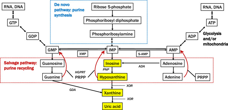
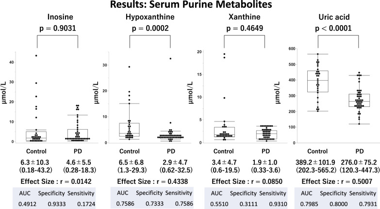
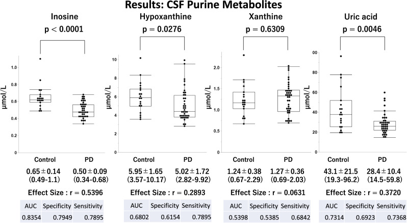
The relationship between reduced serum uric acid (UA) levels and Parkinson's disease (PD), particularly purine metabolic pathways, is not fully understood. Our study compared serum and cerebrospinal fluid (CSF) levels of inosine, hypoxanthine, xanthine, and UA in PD patients and healthy controls. We analyzed 132 samples (serum, 45 PD, and 29 age- and sex-matched healthy controls; CSF, 39 PD, and 19 age- and sex-matched healthy controls) using liquid chromatography-tandem mass spectrometry. Results showed significantly lower serum and CSF UA levels in PD patients than in controls (p < 0.0001; effect size r = 0.5007 in serum, p = 0.0046; r = 0.3720 in CSF). Decreased serum hypoxanthine levels were observed (p = 0.0002; r = 0.4338) in PD patients compared to controls with decreased CSF inosine and hypoxanthine levels (p < 0.0001, r = 0.5396: p = 0.0276, r = 0.2893). A general linear model analysis indicated that the reduced UA levels were mainly due to external factors such as sex and weight in serum and age and weight in CSF unrelated to the purine metabolic pathway. Our findings highlight that decreased UA levels in PD are influenced by factors beyond purine metabolism, including external factors such as sex, weight, and age, emphasizing the need for further research into the underlying mechanisms and potential therapeutic approaches.


Conflict of interest statement

The authors declare no competing interests.

Figures

Fig. 1
Fig. 1. Purine metabolite pathway.
UA is produced through the following process: inosine monophosphate (IMP) → inosine → hypoxanthine → xanthine → UA. We evaluated metabolites colored with yellow in this study. GTP guanosine triphosphate, GDP guanosine diphosphate, GMP guanosine monophosphate, XMP 5’-xanthylic acid, IMP inosine monophosphate, S-AMP succinyl adenosine monophosphate, AMP adenosine monophosphate, ADP adenosine diphosphate, ATP adenosine triphosphate, PPRP phosphoribosyl pyrophosphate, PNP purine nucleoside phosphorylase, HGPRT hypoxanthine-guanine phosphoribosyltransferase, XOR xanthine oxidoreductase, GDA guanine deaminase, ADA adenine deaminase.
Fig. 2
Fig. 2. Comparison of serum purine metabolites between patients with Parkinson’s disease (PD) and healthy controls.
This figure underscores the altered metabolism of serum purine compounds in PD. Serum hypoxanthine levels are significantly reduced in PD patients compared to controls (p = 0.0002). PD patients also exhibit significantly lower serum uric acid levels than controls (p < 0.0001). Analysis shows no significant variation in serum inosine and xanthine levels between the control and PD groups. Each panel presents data as mean ± standard deviation with the range in parentheses. Additionally, the effect size (r), representing the magnitude of difference between groups, along with the Area Under the Curve (AUC), specificity, and sensitivity values from Receiver Operating Characteristic (ROC) analysis, are provided to assess the diagnostic performance of these metabolites in distinguishing PD from control subjects.
Fig. 3
Fig. 3. Comparison of CSF purine metabolites between patients with Parkinson’s disease (PD) and healthy controls.
This figure underscores the altered metabolism of CSF purine compounds in PD. CSF inosine, hypoxanthine, and UA levels are significantly reduced in PD patients compared to controls. Analysis shows no significant variation in CSF xanthine levels between the control and PD groups. Each panel presents data as mean ± standard deviation with the range in parentheses. Additionally, the effect size (r), representing the magnitude of difference between groups, along with the Area Under the Curve (AUC), specificity, and sensitivity values from Receiver Operating Characteristic (ROC) analysis, are provided to assess the diagnostic performance of these metabolites in distinguishing PD from control subjects.

References

    1. Schwarzschild, M. A. Parkinson Study Group PRECEPT Investigators. et al. Serum urate as a predictor of clinical and radiographic progression in Parkinson disease. Arch. Neurol.65, 716–723 (2008). 10.1001/archneur.2008.65.6.nct70003 - DOI - PMC - PubMed
    1. Alonso, A., Rodríguez, L. A. G., Logroscino, G. & Hernán, M. A. Gout and risk of Parkinson disease: a prospective study. Neurology69, 1696–1700 (2007). 10.1212/01.wnl.0000279518.10072.df - DOI - PubMed
    1. Chang, H., Wang, B., Shi, Y. & Zhu, R. Dose-response meta-analysis on urate, gout, and the risk for Parkinson’s disease. NPJ Parkinsons Dis.8, 160 (2022). 10.1038/s41531-022-00433-5 - DOI - PMC - PubMed
    1. Ascherio, A. et al. Urate as a predictor of the rate of clinical decline in Parkinson disease. Arch. Neurol.66, 1460–1468 (2009). 10.1001/archneurol.2009.247 - DOI - PMC - PubMed
    1. Grażyńska, A. et al. The influence of serum uric acid level on non-motor symptoms occurrence and severity in patients with idiopathic Parkinson’s disease and atypical parkinsonisms-a systematic review. Medicina (Kaunas)57, 972 (2021). 10.3390/medicina57090972 - DOI - PMC - PubMed

LinkOut - more resources