Review
. 2024 Sep 9;23(1):193.
doi: 10.1186/s12943-024-02104-w.

Phenotypic and spatial heterogeneity of CD8+ tumour infiltrating lymphocytes

Affiliations
Review

Phenotypic and spatial heterogeneity of CD8+ tumour infiltrating lymphocytes

Yikan Sun et al. Mol Cancer. .

Abstract

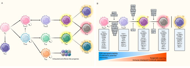
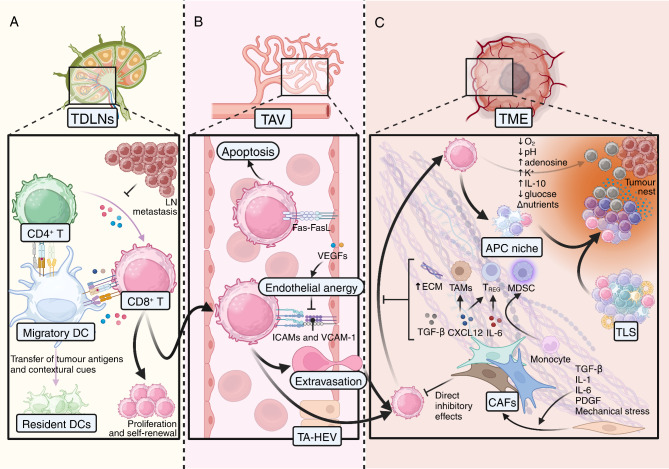
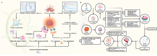
CD8+ T cells are the workhorses executing adaptive anti-tumour response, and targets of various cancer immunotherapies. Latest advances have unearthed the sheer heterogeneity of CD8+ tumour infiltrating lymphocytes, and made it increasingly clear that the bulk of the endogenous and therapeutically induced tumour-suppressive momentum hinges on a particular selection of CD8+ T cells with advantageous attributes, namely the memory and stem-like exhausted subsets. A scrutiny of the contemporary perception of CD8+ T cells in cancer and the subgroups of interest along with the factors arbitrating their infiltration contextures, presented herein, may serve as the groundwork for future endeavours to probe further into the regulatory networks underlying their differentiation and migration, and optimise T cell-based immunotherapies accordingly.

Keywords: CD8+ T cells; Cancer immunotherapy.; Caner immunotypes; T cell exclusion; T cell nomenclature; Tumour-infiltrating lymphocytes.


Conflict of interest statement

The authors declare no competing interests.

Figures

Fig. 1
Fig. 1
Outlining CD8+ T cell evolution in cancer. (A) On the way to terminally differentiated states, naïve CD8+ T cells in cancer move through a meshwork of transcriptomically distinct phenotypes. The cells may stroll down the exhausted (top) or memory (bottom) thoroughfare, though memory cells can give rise to exhausted cells after acquiring the epigenetic imprints and the TRM branch has clear transcriptional idiosyncrasies. In general terms, progression through either lane sees the waning of stem-like features and reprogrammability by immunotherapy and eventual fate convergence at the TTEX terminus. TEXEFF and TEMRA are lineage-wise juxtaposed with TPEX but infused with heightened effector capacities. (B) Subsets of TEX are identifiable by the expression of an assortment of TFs and surface proteins. TPEX are vital to maintaining the exhausted T cell populations due to their stem-like features and capabilities to self-renew, as indicated by TCF1 expression, whereas TOX marks the inability of exhausted cells to fully reconstitute the memory or effector functions. The quiescent TPEX1 subset is distinguished from the mobilised ones by SLAMF6 and CD69 expression profiles. The activated state of TPEX2 is illustrated by the acquisition of the proliferative marker MKI67 and effector molecules IFN-γ and TNF-α, whose expression plateaus in TINT before returning to low levels in TTEX. In addition to escalation of immune checkpoint molecules (e.g., PD-1), TTEX also express terminal exhaustion makers (e.g., CD39 and TIM-3). TEXEFF may form in place of TTEX only under certain circumstances such as TH-derived IL-21 induction. TEX differentiation is under the oversight of an extensive, tightly regulated TF network, as shown in the figure. Notably, many of the TFs maintaining the TPEX population are implicated in memory formation, and those propelling their differentiation support the effector cells during acute infection.
Fig. 2
Fig. 2
Critical anatomical and microenvironmental waypoints dictating success of CD8+ T cell infiltration. (A) TDLNs are the ideal venue for tumour antigen-bearing migratory DCs from the TME to condition the TH and CD8+ T cells. The migratory DCs also pass on the antigens and TME cues to their resident counterparts. With provision of the primary, costimulatory, and cytokine signals by the DCs and TH sans the inhibitory TME elements, CD8+ T cells have the privilege of remaining in the stem-like states and serving as a reservoir of tumour-repressive cells. Establishment of metastases in the LNs demolishes the supportive context for these population-sustaining CD8+ T cells. (B) Tumour-associated vascular endothelium is a major barrier against T cell infiltration. Endothelial expression of FasL can precipitate direct CD8+ T cell death. Aberrant adhesion molecule expression secondary to VEGF-induced endothelial cell anergy renders it impregnable to CD8+ T cells. Tumour-associated high endothelial venules are important sites for CD8+ T cell extravasation and aggregation. (C) CAFs orchestrate the tumour-associated stroma and mediate T cell exclusion in a multitude of ways, as illustrated. The APC niches and TLSs are on-site powerhouses that harbour stem-like CD8+ T cells and, by safeguarding their survival, greatly amplifies anti-tumour immunity. Conversion to the more terminally differentiated phenotypes ensues the longer the dwell time and the closer the distance to the tumour cells, which are known to foment an immunosuppressive aura.
Fig. 3
Fig. 3
Modifying CD8+ T response in cancer. (A) Immune checkpoint blockade radically changes the CD8+ T landscape via mobilisation of the stem-like populations, which primarily occurs in the company of supportive cells in the TDLNs and microenvironmental niches. Cancer cell death as a result of amplified response fosters a self-amplifying cycle owing to tumour antigen release and subsequent increased uptake and priming by APCs. ICBs can simultaneously stimulate TRM, which contributes to increased tumour killing and long-lasting immunosurveillance that deters recurrence and metastasis. (B) In addition to the conventional steps implicated in adoptive cell therapies (lymphocyte procurement, isolation, activation and expansion, pre-infusion lymphodepletion and post-infusion IL-2 administration), there have been multiple strategies in the phases of ex vivo manipulation and in vivo augmentation (marked with plus signs) to maximise the utility of transferred cells. A deeper understanding of the transcriptional, epigenetic, and metabolic controls of CD8+ T phenotypes will shed light on novel ways to engineer cells for the desirable attributes.

Similar articles

Cited by

References

    1. Sun L, Su Y, Jiao A, Wang X, Zhang B. T cells in health and disease. Signal Transduct Target Ther. 2023;8:235. 10.1038/s41392-023-01471-y - DOI - PMC - PubMed
    1. Bruni D, Angell HK, Galon J. The immune contexture and immunoscore in cancer prognosis and therapeutic efficacy. Nat Rev Cancer. 2020;20:662–80. 10.1038/s41568-020-0285-7 - DOI - PubMed
    1. Brummel K, Eerkens AL, de Bruyn M, Nijman HW. Tumour-infiltrating lymphocytes: from prognosis to treatment selection. Br J Cancer. 2023;128:451–8. 10.1038/s41416-022-02119-4 - DOI - PMC - PubMed
    1. Zheng L, Qin S, Si W, Wang A, Xing B, Gao R, et al. Pan-cancer single-cell landscape of tumor-infiltrating T cells. Science. 2021;374:abe6474. 10.1126/science.abe6474 - DOI - PubMed
    1. Rao A, Barkley D, França GS, Yanai I. Exploring tissue architecture using spatial transcriptomics. Nature. 2021;596:211–20. 10.1038/s41586-021-03634-9 - DOI - PMC - PubMed

MeSH terms

Grants and funding