. 2024 Sep 9;23(1):331.
doi: 10.1186/s12933-024-02404-x.

MiRNA-132/212 encapsulated by adipose tissue-derived exosomes worsen atherosclerosis progression

Affiliations

MiRNA-132/212 encapsulated by adipose tissue-derived exosomes worsen atherosclerosis progression

Bei Guo et al. Cardiovasc Diabetol. .

Abstract

Background: Visceral adipose tissue in individuals with obesity is an independent cardiovascular risk indicator. However, it remains unclear whether adipose tissue influences common cardiovascular diseases, such as atherosclerosis, through its secreted exosomes.

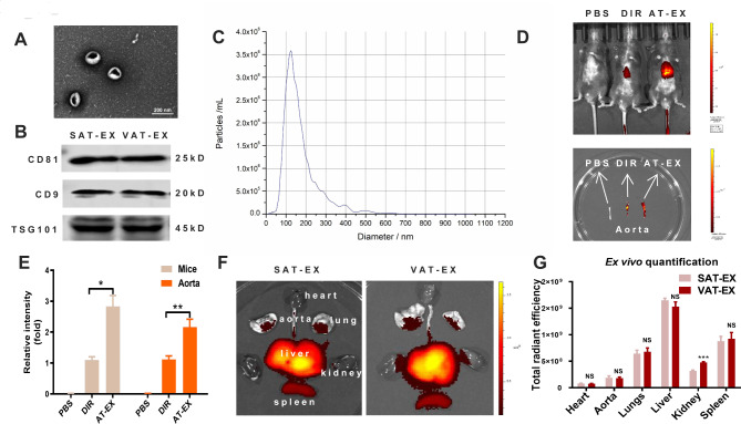
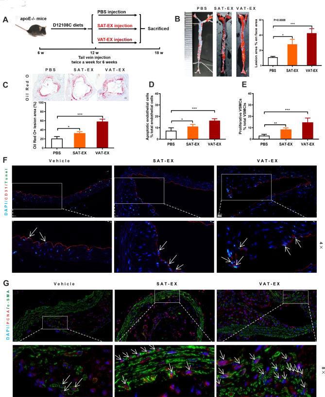
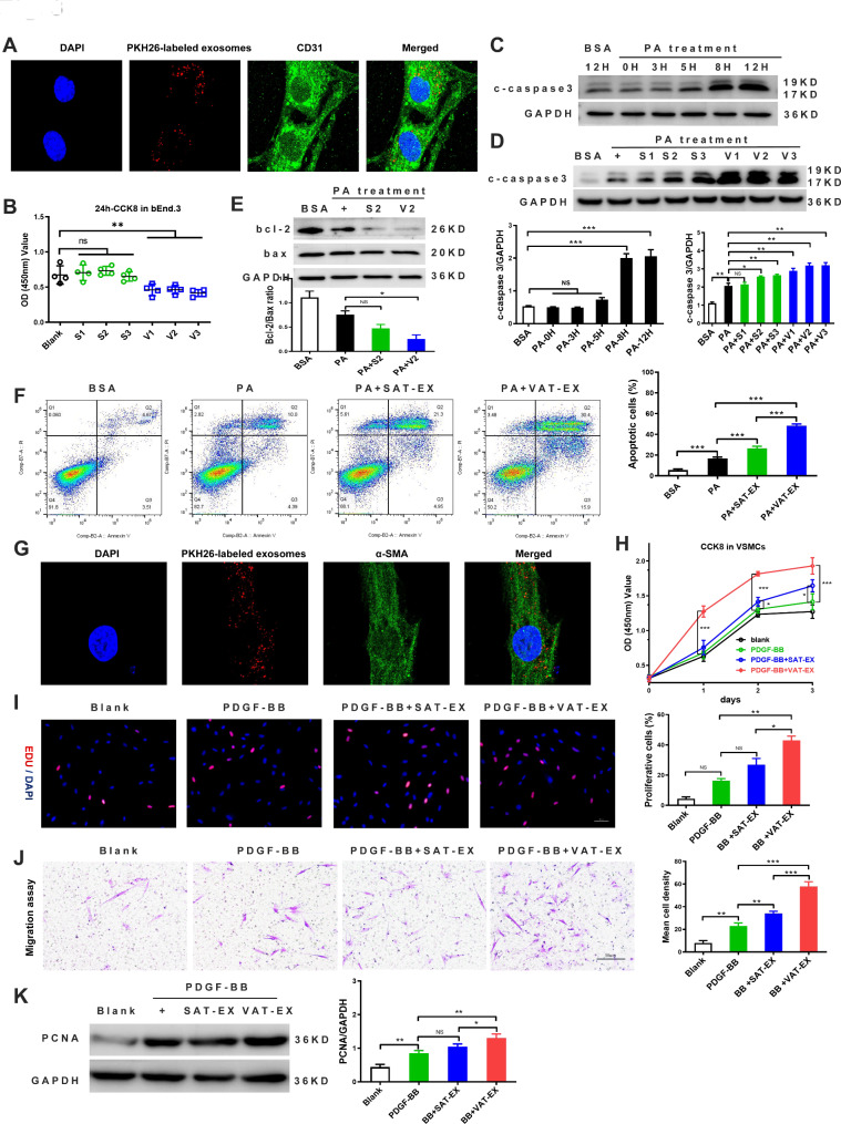
Methods: The exosomes secreted by adipose tissue from diet-induced obesity mice were isolated to examine their impact on the progression of atherosclerosis and the associated mechanism. Endothelial apoptosis and the proliferation and migration of vascular smooth muscle cells (VSMCs) within the atherosclerotic plaque were evaluated. Statistical significance was analyzed using GraphPad Prism 9.0 with appropriate statistical tests.

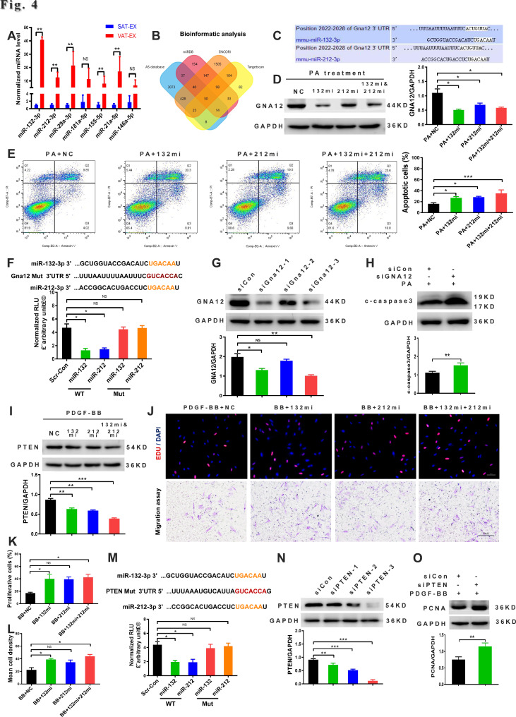
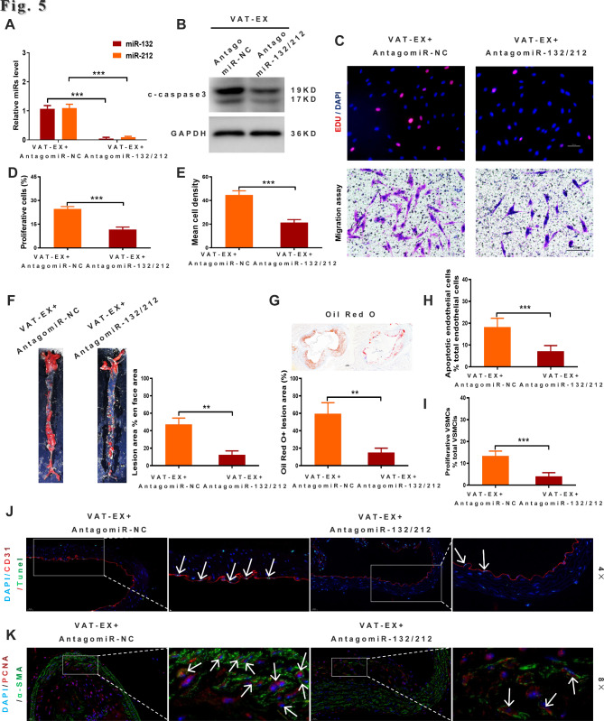
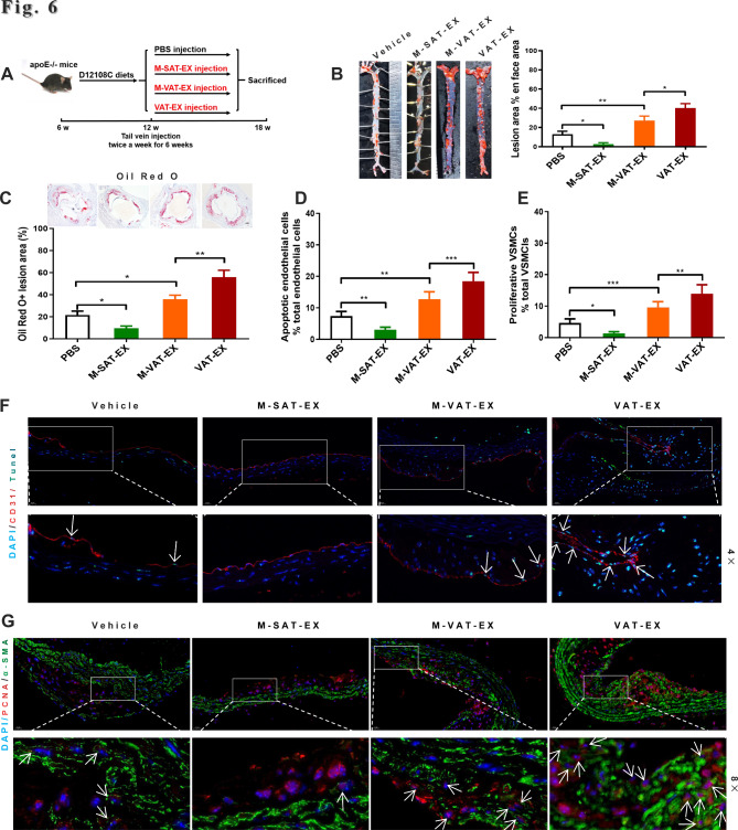
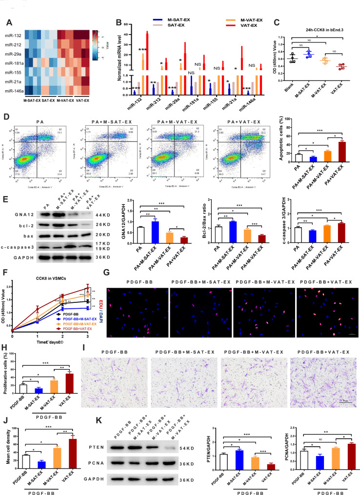
Results: We demonstrate that adipose tissue-derived exosomes (AT-EX) exacerbate atherosclerosis progression by promoting endothelial apoptosis, proliferation, and migration of VSMCs within the plaque in vivo. MicroRNA-132/212 (miR-132/212) was detected within AT-EX cargo. Mechanistically, miR-132/212-enriched AT-EX exacerbates palmitate acid-induced endothelial apoptosis via targeting G protein subunit alpha 12 and enhances platelet-derived growth factor type BB-induced VSMC proliferation and migration by targeting phosphatase and tensin homolog in vitro. Importantly, melatonin decreases exosomal miR-132/212 levels, thereby mitigating the pro-atherosclerotic impact of AT-EX.

Conclusion: These data uncover the pathological mechanism by which adipose tissue-derived exosomes regulate the progression of atherosclerosis and identify miR-132/212 as potential diagnostic and therapeutic targets for atherosclerosis.

Keywords: Adipose tissue; Atherosclerosis; Melatonin; MiR-132/212; Obesity.


Conflict of interest statement

The authors declare no competing interests.

Figures

Fig. 1
Fig. 1
Identification of AT-EX and uptake of DiR-labelled AT-EX in the aortas of apoE−/− mice. A Representative image of the ultrastructure of adipose tissue exosomes (AT-EX) observed by transmission electron microscopy, scale bar = 200 nm. B Expression of exosome markers (CD81, CD9 and TSG 101) in exosomes derived from subcutaneous adipose tissue (SAT) and visceral adipose tissue (VAT) in diet-induced obese mice (DIO). C The average particle size distribution of AT-EX using nanoparticle tracking analysis. D Approximately 100 µg (at protein level) in 100 µL of AT-EX labelled with DiR was injected via tail vein. Fluorescence images of the mice in vivo and the aorta ex vivo were obtained 12 h after intravenous injection. E Quantification of D. F Distribution of other organs ex vivo after DiR-labeled AT-EX injection. G Quantification of F. ***P < 0.001. Data are represented as mean ± SD
Fig. 2
Fig. 2
AT-EX from obese mice aggravate atherosclerosis by augmenting endothelial apoptosis and the proliferation and migration of VSMCs in apoE−/− mice. A The animal experimental procedure for detecting the effects of SAT exosomes or VAT exosomes treatments on atherosclerosis (AS). B Representative images showing Oil Red O staining (red) of neutral lipids in en face atherosclerotic lesions and the quantification, N = 6. C Representative micrographs showing Oil Red O staining of neutral lipids in the aortic root 300 μm from the aortic sinus and the quantification (scale bar = 200 μm, N = 6). D The percentage of apoptotic ECs in the thoracic aorta. E The percentage of proliferative VSMCs in the thoracic aorta. F Representative images of TUNEL (apoptotic cells, green) and CD31 (red) co-staining in sections of thoracic aortas, arrowheads show TUNEL/CD31 co-localization, scale bar = 20 μm, N = 6. G Representative images of α-SMA (green) and PCNA (red) co-staining in sections of thoracic aortas, arrowheads and dotted line show α-SMA/PCNA co-localization, scale bar = 20 μm, N = 5. NS no significance, *P < 0.05, **P < 0.01, ***P < 0.001. Data are represented as mean ± SD
Fig. 3
Fig. 3
AT-EX from obese mice exacerbates endothelial apoptosis and the proliferation and migration of VSMC in vitro. A Representative confocal micrograph of PKH-26-labelled exosomes (red) internalized by bEnd.3 (CD31 positive, green). B The effect of SAT-EX and VAT-EX on the viability of bEnd.3 at 24 h (S1, S2 and S3 represent 50 µg/mL, 100 µg/mL and 200 µg/mL SAT-EX, respectively; V1, V2 and V3 show 50 µg/mL, 100 µg/mL and 200 µg/mL VAT-EX, respectively). C Expression of c-caspase 3 at different time after palmitic acid (PA, 0.4 mmol/L) treatment. D Expression of c-caspase3 in PA-induced bEnd.3 after 12 h of AT-EX (SAT-EX or VAT-EX) treatment. E Protein levels of BCL-2 and Bax in PA-induced bEnd.3 after AT-EX treatment. F Apoptotic bEnd.3 were stained with Annexin V-FITC and propidium iodide (PI). G Representative confocal micrograph of PKH-26-labelled exosomes (red) internalized by primary VSMCs. H Effect of AT-EX on the viability of primary VSMCs. I Representative proliferation images of primary VSMCs, scale bar = 50 μm. J Representative migration images of primary VSMCs, scale bar = 50 μm. K PCNA expression in PDGF-BB-induced primary VSMCs after AT treatment. NS no significance, *P < 0.05, **P < 0.01, ***P < 0.001, N = 6 independent experiments. Data are represented as mean ± SD
Fig. 4
Fig. 4
The pro-atherosclerotic effect of AT-EX is associated with the MiR-132/212-GNA12/PTEN signaling in vitro. A Normalized atherosclerosis-related miRNA levels in SAT-EX and VAT-EX. B The potential target genes of the miR-132/212 cluster were predicted by bioinformatic analysis. C G protein subunit alpha 12 (Gna12) 3′UTR includes a potential binding site for miR-132-3p and miR-212-3p. D GNA12 expression in bEnd.3 after PA treatment for 24 h post-transfection of miR-132 or/and miR-212 mimics, NC negative control. E Apoptotic bEnd.3 were stained with Annexin V-FITC and PI. F The luciferase activity in target cells co-transfected with WT or mutant Gna12 3′UTR luciferase constructs and miR-132/212 mimics; Scr-Con, scramble control. G Silencing efficiency of small interfering RNA (siRNA) on Gna12. H The expression of c-caspase3 in Gna12-silenced bEnd.3. I PTEN expression in VSMCs after PDGF-BB treatment for 24 h post-transfection of miR-132 or/and miR-212 mimics. J Representative proliferation and migration images of VSMCs. K, L quantitative analysis of J. (M) The luciferase activity in target cells co-transfected with WT or mutant PTEN 3′UTR luciferase constructs and miR-132/212 mimics; Scr-Con, scramble control. N Silencing efficiency of siRNA on PTEN. O The expression of PCNA in PTEN-silenced VSMCs. NS no significance, *P < 0.05, **P < 0.01, ***P < 0.001, N = 6 independent experiments. Data are represented as mean ± SD
Fig. 5
Fig. 5
AntagomiR-132/212 abolish the pro-atherosclerotic effect of AT-EX in vitro and in vivo. A qRT-PCR analysis of miR-132/212 expression in VAT-EX + AntagomiR-132/212. ***P < 0.001, N = 6 independent experiments. B Expression of c-caspase3 in PA-induced bEnd.3 after 12 h of VAT-EX (AntagomiR-NC or AntagomiR-132/212) treatment. C Representative proliferation and migration images of primary VSMCs, scale bar = 50 μm. D Quantification of proliferation images. E Quantification of migration images. F Representative images showing Oil Red O staining (red) of neutral lipids in en face atherosclerotic lesions and the quantification, N = 5. G Representative micrographs showing Oil Red O staining of neutral lipids in the aortic root 300 μm from the aortic sinus and the quantification (scale bar = 200 μm, N = 5). H The percentage of apoptotic ECs in the thoracic aorta. I The percentage of proliferative VSMCs in the thoracic aorta. J Representative images of TUNEL (apoptotic cells, green) and CD31 (red) co-staining in sections of thoracic aortas, arrowheads show TUNEL/CD31 co-localization, scale bar = 20 μm, N = 5. K Representative images of α-SMA (green) and PCNA (red) co-staining in sections of thoracic aortas, arrowheads and dotted line show α-SMA/PCNA co-localization, scale bar = 20 μm, N = 5. NS no significance, *P < 0.05, **P < 0.01, ***P < 0.001. Data are represented as mean ± SD
Fig. 6
Fig. 6
Melatonin alleviates the pro-atherosclerotic effect of at exosomes derived from obese mice and modulates miR-132/212-GNA12/PTEN signaling in apoE−/− mice. A The animal experimental procedure for detecting the effects of SAT-EX and VAT-EX after melatonin intervention (M-SAT-EX, M-VAT-EX) for one month on AS. B Representative images showing Oil Red O staining (red) of neutral lipids in en face atherosclerotic lesions and the quantitative analysis, N = 6. C Representative micrographs showing Oil Red O staining in the aortic root 300 μm from the aortic sinus and the quantitative analysis, scale bar = 200 μm, N = 6. D the percentage of apoptotic ECs in the thoracic aorta. E The percentage of proliferative VSMCs in the thoracic aorta. F Representative images of TUNEL (green) and CD31 (red) co-staining in sections of thoracic aortas, arrowheads show TUNEL/CD31 co-localization, scale bar = 20 μm, N = 6. G Representative images of α-SMA (green) and PCNA (red) co-staining in sections of thoracic aortas, arrowheads and dotted line show α-SMA/PCNA co-localization, scale bar = 20 μm, N = 5. NS no significance, *P < 0.05, **P < 0.01, ***P < 0.001. Data are represented as mean ± SD
Fig. 7
Fig. 7
The endothelial miR-132/212-Gna12 axis and medial miR-132/212-PTEN axis are essential for the effects of AT-EX on atherosclerosis. A Heat map depicting changes in atherosclerosis-relevant microRNAs in four kinds of exosomes. B The miRNAs level in four kinds of exosomes examined by qRT-PCR assay, taking SAT-EX data as reference. M-SAT-EX vs. SAT-EX, M-VAT-EX vs. VAT-EX. C Effect of M-SAT-EX and M-VAT-EX on the viability of bEnd.3 at 24 h. D Apoptotic bEnd.3 were stained with Annexin V-FITC and PI. E Expression of GNA12, BCL-2, Bax and c-caspase3 in PA-induced bEnd.3 after SAT-EX or VAT-EX treatment. F Effect of M-SAT-EX and M-VAT-EX on the viability of primary VSMCs. G Representative proliferation images of primary VSMCs, scale bar = 50 μm. H uantitative analysis of G. I Representative migration images of primary VSMCs, scale bar = 50 μm. J Quantitative analysis of I. K Expression of PTEN and PCNA in PDGF-BB-induced primary VSMCs after M-SAT-EX or M-VAT-EX treatment. NS no significance, *P < 0.05, **P < 0.01, ***P < 0.001, N = 6 independent experiments. Data are represented as mean ± SD

Similar articles

Cited by

References

    1. Libby P. The changing landscape of atherosclerosis. Nature. 2021;592:524–33. 10.1038/s41586-021-03392-8 - DOI - PubMed
    1. Fuster JJ, Ouchi N, Gokce N, Walsh K. Obesity-induced changes in adipose tissue microenvironment and their impact on cardiovascular disease. Circ Res. 2016;118:1786–807. 10.1161/CIRCRESAHA.115.306885 - DOI - PMC - PubMed
    1. Francisco V, Pino J, Gonzalez-Gay MA, Lago F, Karppinen J, Tervonen O, Mobasheri A, Gualillo O. A new immunometabolic perspective of intervertebral disc degeneration. Nat Rev Rheumatol. 2022;18:47–60. 10.1038/s41584-021-00713-z - DOI - PubMed
    1. Hiuge-Shimizu A, Kishida K, Funahashi T, Ishizaka Y, Oka R, Okada M, Suzuki S, Takaya N, Nakagawa T, Fukui T, Fukuda H, Watanabe N, Yoshizumi T, Nakamura T, Matsuzawa Y, Yamakado M, Shimomura I. Absolute value of visceral fat area measured on computed tomography scans and obesity-related cardiovascular risk factors in large-scale Japanese general population (the VACATION-J study). Ann Med. 2012;44:82–92. 10.3109/07853890.2010.526138 - DOI - PubMed
    1. Neeland IJ, Ross R, Despres JP, Matsuzawa Y, Yamashita S, Shai I, Seidell J, Magni P, Santos RD, Arsenault B, Cuevas A, Hu FB, Griffin B, Zambon A, Barter P, Fruchart JC, Eckel RH. Visceral and ectopic fat, atherosclerosis, and cardiometabolic disease: a position statement. Lancet Diabetes Endocrinol. 2019;7:715–25. 10.1016/S2213-8587(19)30084-1 - DOI - PubMed

Publication types

MeSH terms