

Commentary on "Structural characterization of SLYM - a 4th meningeal membrane"
- PMID: 39252039
- PMCID: PMC11385822
- DOI: 10.1186/s12987-024-00568-y
Commentary on "Structural characterization of SLYM - a 4th meningeal membrane"
Abstract
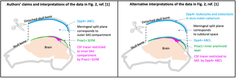
For centuries, the meninges have been described as three membranes: the inner pia, middle arachnoid and outer dura. It was therefore sensational when in early 2023 Science magazine published a report of a previously unrecognized - 4th - meningeal membrane located between the pia and arachnoid. Multiple features were claimed for this new membrane: a single cell layer marked by the transcription factor Prox1 that formed a barrier to low molecular weight substances and separated the subarachnoid space (SAS) into two fluid-filled compartments, not one as previously described. These features were further claimed to facilitate unidirectional glymphatic cerebrospinal fluid transport. These claims were immediately questioned by several researchers as misinterpretations of the authors' own data. The critics argued that (i) the 4th meningeal membrane as claimed did not exist as a separate structure but was part of the arachnoid, (ii) the "outer SAS" compartment was likely an artifactual subdural space created by the experimental procedures, and (iii) the 4th membrane barrier property was confused with the arachnoid barrier. Subsequent publications in late 2023 indeed showed that Prox1 + cells are embedded within the arachnoid and located immediately inside of and firmly attached to the arachnoid barrier cells by adherens junctions and gap junctions. In a follow-up study, published in this journal, the lead authors of the Science paper Kjeld Møllgård and Maiken Nedergaard reported additional observations they claim support the existence of a 4th meningeal membrane and the compartmentalization of the SAS into two non-communicating spaces. Their minor modification to the original paper was the 4th meningeal membrane was better observable at the ventral side of the brain than at the dorsal side where it was originally reported. The authors also claimed support for the existence of a 4th meningeal membrane in classical literature. Here, we outline multiple concerns over the new data and interpretation and argue against the claim there is prior support in the literature for a 4th meningeal membrane.
Keywords: Arachnoid barrier; Cerebrospinal fluid; Claudin-11; Dpp4; E-cadherin; Inner arachnoid; Meninges; Prox1.
© 2024. The Author(s).
Conflict of interest statement
The authors declare no competing interests.
Figures


Comment on
-
Structural characterization of SLYM-a 4th meningeal membrane.Fluids Barriers CNS. 2023 Dec 14;20(1):93. doi: 10.1186/s12987-023-00500-w. Fluids Barriers CNS. 2023. PMID: 38098084 Free PMC article.
References
Publication types
MeSH terms
Grants and funding
LinkOut - more resources
Full Text Sources
Miscellaneous
