. 2024 Sep 9;57(1):64.
doi: 10.1186/s40659-024-00544-8.

Mouse testicular macrophages can independently produce testosterone and are regulated by Cebpb

Affiliations

Mouse testicular macrophages can independently produce testosterone and are regulated by Cebpb

Nengliang Duan et al. Biol Res. .

Abstract

Background: Testicular macrophages (TM) have long been recognized for their role in immune response within the testicular environment. However, their involvement in steroid hormone synthesis, particularly testosterone, has not been fully elucidated. This study aims to explore the capability of TM to synthesize and secrete testosterone de novo and to investigate the regulatory mechanisms involved.

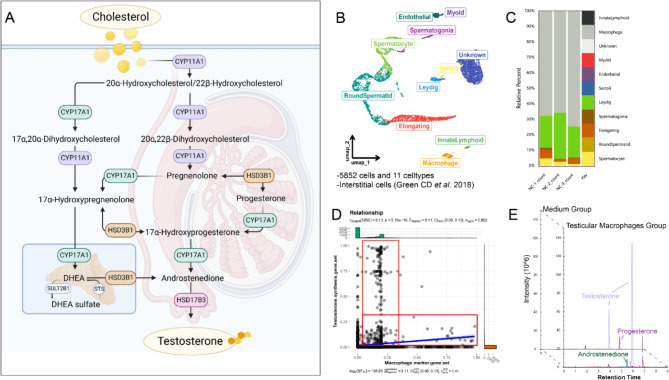
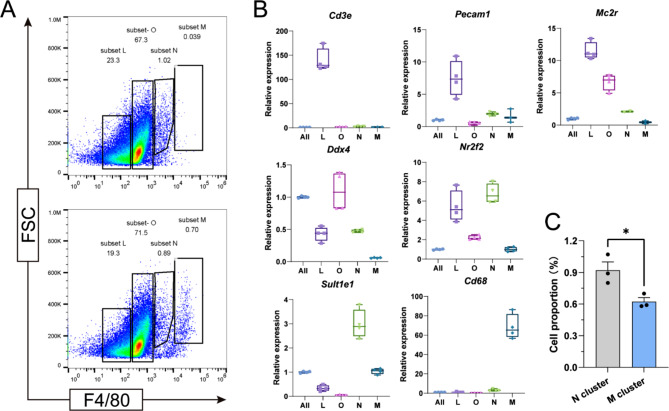
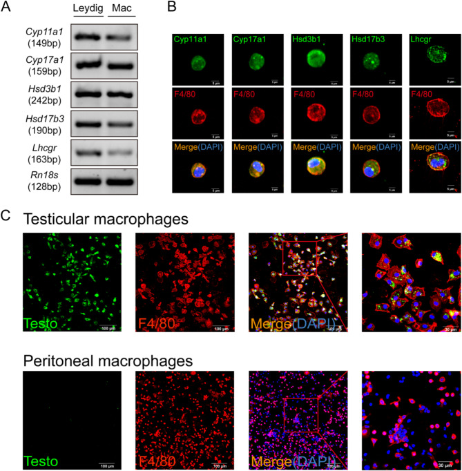
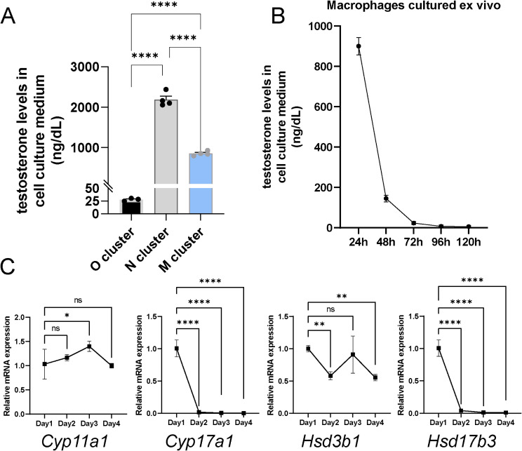
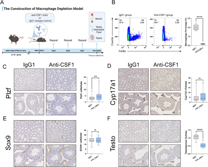
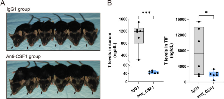
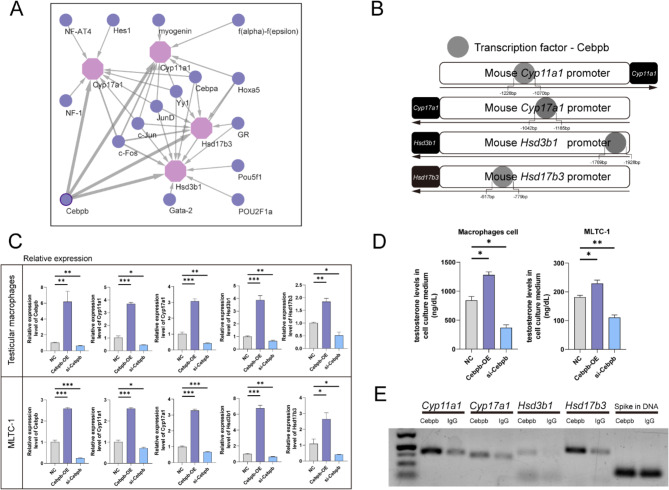
Results: Transcriptomic analysis revealed significant expression of Cyp11a1, Cyp17a1, Hsd3b1, and Hsd17b3 in TM, which are key enzymes in the testosterone synthesis pathway. qPCR analysis and immunofluorescence validation confirmed the autonomous capability of TM to synthesize testosterone. Ablation of TM in mice resulted in decreased physiological testosterone levels, underscoring the significance of TM in maintaining testicular testosterone levels. Additionally, the study also demonstrated that Cebpb regulates the expression of these crucial genes, thereby modulating testosterone synthesis.

Conclusions: This research establishes that TM possess the autonomous capacity to synthesize and secrete testosterone, contributing significantly to testicular testosterone levels. The transcription factor Cebpb plays a crucial role in this process by regulating the expression of key genes involved in testosterone synthesis.

Keywords: Cebpb; De novo synthesis; Testicular macrophage; Testosterone.


Conflict of interest statement

The authors declare that they have no conflict of interest.

Figures

Fig. 1
Fig. 1
TM and Testosterone Synthesis Genes. A: A schematic representation of the complete pathway for de novo testosterone synthesis; B: A UMAP plot showing the distribution of different cell clusters in the testis, with each colored dot representing a cell; C: The estimated proportions of various cell types following deconvolution analysis of transcriptomic sequencing from three groups of TM; D: A scatter plot illustrating the correlation analysis results after scoring each cell for two gene sets; E: The results from targeted steroid hormone metabolomics revealed the levels of various steroid hormones in macrophage culture medium and in control blank culture medium
Fig. 2
Fig. 2
Sorting and Identification of TM and Adult Leydig Cells. A: Flow cytometry results of testicular interstitial cells following F4/80–488 staining; B: Quantitative PCR results for various marker genes in each cell group
Fig. 3
Fig. 3
Expression of Testosterone Synthesis Genes in TM. A: Comparison of mRNA expression levels of key genes and Lhcgr involved in testosterone synthesis in Adult Leydig cells and TM; B: Immunofluorescence images of the four key enzymes, Lhcgr, and the macrophage marker protein F4/80 in mouse TM. Scale bars, 5 μm; C: Immunofluorescence images of testosterone and F4/80 in mouse TM and peritoneal macrophages. Scale bars, 100 μm
Fig. 4
Fig. 4
Quantification and Tracking of Testicular Macrophage Secretory Capability. A: Testosterone secretion levels in equal O, N, and M cell groups; B: Time-dependent changes in testosterone secretion by TM in vitro culture; C: Changes in mRNA levels of four key genes in macrophages at different culture times
Fig. 5
Fig. 5
Construction and Validation of Macrophage Depletion Mouse Model. A: Schematic diagram of the construction of the testicular macrophage depletion model in mice; B: Flow cytometry validation of the proportion of F4/80 positive cells in the total testis of macrophage-depleted mice compared to control mice; C, D, E, F: Immunohistochemical analysis showing changes in spermatogonial stem cells (Plzf), Interstitial cells (Cyp17a1), Sertoli cells (Sox9), and Testosterone after macrophage depletion, with bar graphs summarizing the results of the immunohistochemical analysis
Fig. 6
Fig. 6
Whiskers and Steroid Hormone Levels in Macrophage Depletion Mice. A: Whiskers of control group and macrophage depletion mice, indicated by arrows; B: Changes in testosterone levels in serum and testicular interstitial fluid
Fig. 7
Fig. 7
Cebpb is an upstream regulator of testosterone secretion in TM. A: Network diagram showing all transcription factors predicted based on the four target genes, with the thickness of the arrows corresponding to the RE values; B: A schematic illustrating the binding sites of Cebpb with various gene promoter regions; C: Bar graphs showing the changes in relative mRNA expression levels of different genes in cells with overexpression and knockdown of Cebpb; D: Bar graphs depicting the variations in testosterone hormone secretion in two groups of cells following overexpression and knockdown of Cebpb; E: Agarose gel electrophoresis displaying the content of different gene 5` regulatory region fragments, with Spike in DNA serving as an internal control to ensure equal total DNA content in both groups

References

    1. Lawrence BM, O’Donnell L, Smith LB, Rebourcet D. New insights into Testosterone Biosynthesis: novel observations from HSD17B3 deficient mice. Int J Mol Sci 2022, 23(24). - PMC - PubMed
    1. Storer TW, Basaria S, Traustadottir T, Harman SM, Pencina K, Li Z, Travison TG, Miciek R, Tsitouras P, Hally K, et al. Effects of Testosterone Supplementation for 3 years on muscle performance and physical function in older men. J Clin Endocrinol Metab. 2017;102(2):583–93. - PMC - PubMed
    1. Buendia-Gonzalez FO, Legorreta-Herrera M. The Similarities and Differences between the Effects of Testosterone and DHEA on the Innate and Adaptive Immune Response. Biomolecules 2022, 12(12). - PMC - PubMed
    1. Guo W, Schmidt PJ, Fleming MD, Bhasin S. Hepcidin is not essential for mediating testosterone’s effects on erythropoiesis. Andrology. 2020;8(1):82–90. 10.1111/andr.12622 - DOI - PubMed
    1. Tuck S, Francis R. Testosterone, bone and osteoporosis. Front Horm Res. 2009;37:123–32. 10.1159/000176049 - DOI - PubMed

Substances

LinkOut - more resources