. 2024 Aug 26:11:1454762.
doi: 10.3389/fvets.2024.1454762. eCollection 2024.

Impact of swine influenza A virus on porcine reproductive and respiratory syndrome virus infection in alveolar macrophages

Affiliations

Impact of swine influenza A virus on porcine reproductive and respiratory syndrome virus infection in alveolar macrophages

Janaïna Grevelinger et al. Front Vet Sci. .

Abstract

Porcine respiratory disease complex represents a major challenge for the swine industry, with swine influenza A virus (swIAV) and porcine reproductive and respiratory syndrome virus (PRRSV) being major contributors. Epidemiological studies have confirmed the co-circulation of these viruses in pig herds, making swIAV-PRRSV co-infections expected. A couple of in vivo co-infection studies have reported replication interferences between these two viruses. Herein, using a reductionist in vitro model, we investigated the potential mechanisms of these in vivo interferences. We first examined the impact of swIAV on porcine alveolar macrophages (AMs) and its effects on AMs co-infection by PRRSV. This was done either in monoculture or in co-culture with respiratory tracheal epithelial cells to represent the complexity of the interactions between the viruses and their respective target cells (epithelial cells for swIAV and AMs for PRRSV). AMs were obtained either from conventional or specific pathogen-free (SPF) pigs. SwIAV replication was abortive in AMs, inducing cell death at high multiplicity of infections. In AMs from three out of four conventional animals, swIAV showed no impact on PRRSV replication. However, inhibition of PRRSV multiplication was observed in AMs from one animal, accompanied by an early increase in the expression of interferon (IFN)-I and IFN-stimulated genes. In AMs from six SPF pigs, swIAV inhibited PRRSV replication in all animals, with an early induction of antiviral genes. Co-culture experiments involving tracheal epithelial cells and AMs from either SPF or conventional pigs all showed swIAV-induced inhibition of PRRSV replication, together with early induction of antiviral genes. These findings highlight the complex interactions between swIAV and PRRSV in porcine AMs, and would suggest a role of host factors, such as sanitary status, in modulating viral propagation. Our co-culture experiments demonstrated that swIAV inhibits PRRSV replication more effectively in the presence of respiratory tracheal epithelial cells, suggesting a synergistic antiviral response between AMs and epithelial cells, consistent with in vivo experiments.

Keywords: co-infection; co-inoculation; interferon; lung; porcine respiratory disease complex.


Conflict of interest statement

The authors declare that the research was conducted in the absence of any commercial or financial relationships that could be construed as a potential conflict of interest. The author(s) declared that they were an editorial board member of Frontiers, at the time of submission. This had no impact on the peer review process and the final decision.

Figures
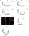
Figure 1
Figure 1
Inoculation of swine influenza A virus on conventional porcine alveolar macrophages. (A,B) Conventional porcine AMs and NPTr were inoculated with swIAV at MOI 0.1. Adherent cells and supernatants were sampled at different times post-inoculation (1, 12, 24, 36, 48 h). Expression of the swIAV viral genome was quantified by RT-qPCR (A), and the virus was titrated in supernatants (B) (mean ± SD; for AMs n = 2, and for NPTr n = 3). (C,D) Conventional porcine AMs were inoculated with swIAV at different MOI 0.1, 0.5, and 2. Adherent cells and supernatants were sampled at different times post-inoculation (1, 12, 24, 36, 48 h). Expression of the swIAV viral genome was quantified by RT-qPCR (C), and the virus was titrated in supernatants (D) (mean ± SD; n = 2). (E) AMs and NPTr cells were inoculated with swIAV at a MOI of 0.5. Cells were fixed after 6 h and 12 h post-inoculation and stained with an antibody against the nucleoprotein to detect infected cells (in red). Cell nuclei were stained (in white) using 4′,6′-diamidino-2-phenylindole (DAPI). (E1) Confocal imaging of NPTr and AMs cells, 12 h post inoculation with swIAV. Upper panel NPTr cells. Lower panel AMs cells. Nuclear staining with DAPI, swIAV immunodetection and merge (from left to right). Scale bars = 20 μm. (E2) The number of infected cells was calculated in relation to the total number of cells (n = 3, technical replicate). (F) AMs were inoculated with either medium alone or swIAV at a MOI of 0.5 or 2; or PRRSV at a MOI of 2. After 48 h, cells were stained with propidium iodide/Hoechst. Each symbol corresponds to AMs from a single animal. The number of dead cells was calculated in relation to the total number of cells. Statistics Mann–Whitney unpaired, non-parametric test, (*) p < 0.05 or (**) p < 0.01 (mean ± SD; n = 6).
Figure 2
Figure 2
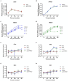
Simultaneous co-inoculation of swIAV and PRRSV viruses on AMs from conventional pigs. AMs were inoculated with either culture medium, swIAV at a MOI of 0.5, PRRSV at a MOI of 1, or both viruses simultaneously. Samples were collected at 1, 12, 24, 36 and 48 h. (A,B) The expression of the swIAV (A) and PRRSV (B) viral genomes was quantified using RT-qPCR in cell lysates (mean ± SD; n = 4). (C,D) For the PRRSV inoculated and swIAV/PRRSV co-inoculated conditions, the expression of the PRRSV genome was individually evaluated for AMs from each animal. (E–H) The expression of the antiviral genes IFNα, IFNβ, MX2, and PKR were analyzed by RT-qPCR. Statistics Kruskal-Walis, non-parametric test (mean ± SD; n = 4). Data are combined from 2 independent experiments.
Figure 3
Figure 3
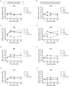
Simultaneous co-inoculation of swIAV and PRRSV viruses on AMs from specific pathogen-free piglets. AMs were inoculated with either culture medium, swIAV at a MOI of 0.5, PRRSV at a MOI of 1, or both viruses simultaneously. Samples were collected at 1, 12, 24, 36 and 48 h. (A–D) The expression of swIAV viral genome was quantified in the cell lysates using RT-qPCR (A), and the virus was titrated in supernatants (B). The expression of PRRSV viral genome was quantified in the cell lysates using RT-qPCR (C), and the virus was titrated in supernatants (D). Statistics Mann–Whitney unpaired, non-parametric test, (*) p < 0.05 or (**) p < 0.01 (mean ± SD; n = 5–6). (E–H) The expression of the antiviral genes IFNα, IFNβ, MX2 and PKR were analyzed by RT-qPCR. Statistics Kruskal-Walis, non-parametric test. Different letters (a–d) indicate that the considered group (specified by its color) was significantly different from the Mock group (a), from the swIAV group (b), from the PRRSV group (c) or from the swIAV/PRRSV group (d) with p < 0.05 (mean ± SD; n = 6). Data are combined from 2 independent experiments.
Figure 4
Figure 4
Simultaneous co-inoculation of swIAV and PRRSV viruses on epithelial cell line/AMs co-cultures derived from both specific pathogen-free and conventional swine. NPTr epithelial cells and AMs were inoculated with either culture medium, swIAV at a MOI of 0.5, PRRSV at a MOI of 1, or both viruses simultaneously. Samples were collected at 1, 12, 24, and 48 h. On the left side are presented the results for SPF pig AMs, and on the right are the results for conventional pig AMs. (A,B) The expression of the swIAV viral genome was quantified in the cell lysates using RT-qPCR. (C,D) The expression of the PRRSV viral genome was quantified in the cell lysates using RT-qPCR. Statistics Mann–Whitney unpaired, non-parametric test, (*) p < 0.05 or (**) p < 0.01 (mean ± SD; n = 6 for SPF swine AMs; n = 4 for conventional swine AMs). Data are combined from 2 independent experiments for AMs originating from SPF or conventional swine.
Figure 5
Figure 5
Simultaneous co-inoculation of swIAV and PRRSV viruses on epithelial cell line/AMs co-cultures derived from both specific pathogen-free and conventional swine. NPTr epithelial cells and AMs were inoculated with either culture medium, swIAV at a MOI of 0.5, PRRSV at a MOI of 1, or both viruses simultaneously. Samples were collected at 1, 12, 24, and 48 h. On the left side are presented the results for SPF pig AMs and on the right are the results for conventional pig AMs. (A–H) The expression of the antiviral genes IFNα, IFNβ, MX2, and PKR were analyzed by RT-qPCR. Statistics Kruskal-Walis, non-parametric test. Different letters (a–d) indicate that the considered group (specified by its color) was significantly different from the Mock group (a), from the swIAV group (b), from the PRRSV group (c) or from the swIAV/PRRSV group (d) with p < 0.05 (mean ± SD; n = 6 for SPF swine AMs; n = 4 for conventional swine AMs). Data are combined from 2 independent experiments for AMs originating from SPF or conventional swine.

References

    1. Opriessnig T, Giménez-Lirola LG, Halbur PG. Polymicrobial respiratory disease in pigs. Anim Health Res Rev. (2011) 12:133–48. doi: 10.1017/S1466252311000120, PMID: - DOI - PubMed
    1. Boeters M, Garcia-Morante B, van Schaik G, Segalés J, Rushton J, Steeneveld W. The economic impact of endemic respiratory disease in pigs and related interventions - a systematic review. Porcine Health Manag. (2023) 9:45. doi: 10.1186/s40813-023-00342-w, PMID: - DOI - PMC - PubMed
    1. Fablet C, Marois-Créhan C, Simon G, Grasland B, Jestin A, Kobisch M, et al. Infectious agents associated with respiratory diseases in 125 farrow-to-finish pig herds: a cross-sectional study. Vet Microbiol. (2012) 157:152–63. doi: 10.1016/j.vetmic.2011.12.015, PMID: - DOI - PubMed
    1. Fablet C, Marois C, Kuntz-Simon G, Rose N, Dorenlor V, Eono F, et al. Longitudinal study of respiratory infection patterns of breeding sows in five farrow-to-finish herds. Vet Microbiol. (2011) 147:329–39. doi: 10.1016/j.vetmic.2010.07.005, PMID: - DOI - PMC - PubMed
    1. Saade G, Deblanc C, Bougon J, Marois-Créhan C, Fablet C, Auray G, et al. Coinfections and their molecular consequences in the porcine respiratory tract. Vet Res. (2020) 51:80. doi: 10.1186/s13567-020-00807-8, PMID: - DOI - PMC - PubMed

LinkOut - more resources