

Non-Vascularized Fibular Cortical Strut Graft with Intramedullary Nailing for Fibrous Dysplasia of the Proximal Radius - A Case Report
- PMID: 39253674
- PMCID: PMC11381070
- DOI: 10.13107/jocr.2024.v14.i09.4766
Non-Vascularized Fibular Cortical Strut Graft with Intramedullary Nailing for Fibrous Dysplasia of the Proximal Radius - A Case Report
Abstract
Introduction: Fibrous dysplasia (FD) is a skeletal developmental abnormality commonly affecting the ribs, femur, tibia, skull, pelvis, spine, and shoulder. FD of the proximal radius is extremely rare and very few cases have been reported. In addition, monostotic lesions of FD in the upper extremity go unnoticed as they are usually asymptomatic. Symptomatic lesions warrant surgical intervention. Here, we present a rare case of FD of the proximal radius treated with curettage and non-vascularized fibular cortical strut graft with intramedullary elastic nailing. We believe that this is the first report in the literature wherein this treatment modality has been undertaken.
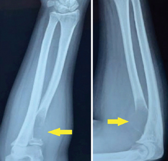
Case report: A 27-year-old woman presented with excruciating pain and swelling in her right elbow for 4 weeks, with no inciting event or trauma leading to the pain. Plain radiographs revealed a well-circumscribed radiolucent lesion in the proximal radius with cortical thinning at the metaphysis and a rim of epiphyseal bone. Clinically, the patient had restricted supination (50°) and limited elbow range of motion (ROM) (20-130°), mostly because of the pain but had full pronation. With these radiographic and clinical features, FD and giant cell tumor were kept as differential diagnoses and surgical treatment was planned. The lesion was excised leaving the normal epiphysis of the radius intact and samples were sent for histopathological examination. A non-vascularized fibular cortical strut graft was harvested from the same side and was fluted into the radial shaft. Final stabilization was done using a 2.5 mm intramedullary elastic nail. The arm was immobilized in an above-elbow slab. Histopathology confirmed our diagnosis of FD. The slab was removed after 6 weeks, and a gentle ROM was started in the form of active-assisted ROM. At the end of 1 year, complete union and almost full ROM were achieved and the patient was completely pain-free.
Conclusion: Non-vascularized fibular strut grafting with intramedullary nailing provides a comparatively quicker, cost-effective way of treating this lesion with a minimum insult of the bony cortex and quicker rehabilitation.
Keywords: Fibrous dysplasia; intramedullary nailing; proximal radius; strut graft.
Copyright: © Indian Orthopaedic Research Group.
Conflict of interest statement
Conflict of Interest: Nil
Figures


References
-
- Resnick D. Diagnosis of Bone and Joint Disorders. 4th ed. Philadelphia, PA: WB Saunders; 2002. Fibrous dysplasia; pp. 4825–43.
-
- Kumar R, Madewell JE, Lindell MM, Swischuk LE. Fibrous lesions of bones. Radiographics. 1990;10:237–56. - PubMed
-
- Parekh SG, Donthineni-Rao R, Ricchetti E, Lackman RD. Fibrous dysplasia. J Am Acad Orthop Surg. 2004;12:305–13. - PubMed
-
- Wilner D. In:Radiology of Bone Tumors and Allied Disorders. Philadelphia, PA: Saunders; 1982. Fibrous dysplasia of bone; pp. 1443–80.
-
- Kokkalis ZT, Jain S, Sotereanos DG. Fibrous dysplasia around the elbow. J Shoulder Elbow Surg. 2010;19:e6–11. - PubMed
Publication types
LinkOut - more resources
Full Text Sources