Review
. 2025 Jan;169(1):e16219.
doi: 10.1111/jnc.16219. Epub 2024 Sep 10.

From BBB to PPP: Bioenergetic requirements and challenges for oligodendrocytes in health and disease

Affiliations
Review

From BBB to PPP: Bioenergetic requirements and challenges for oligodendrocytes in health and disease

Milton Guilherme Forestieri Fernandes et al. J Neurochem. 2025 Jan.

Abstract

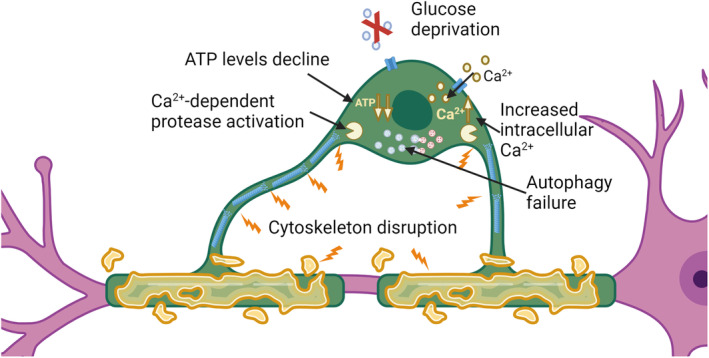
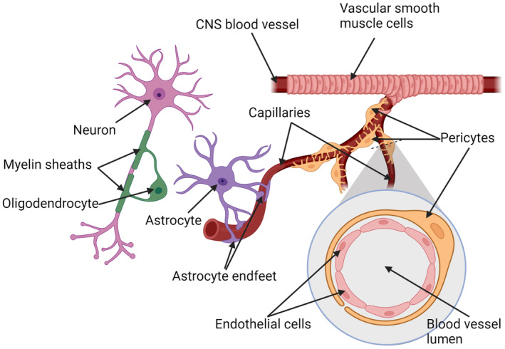
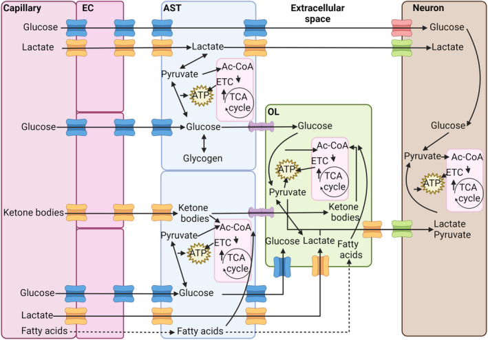
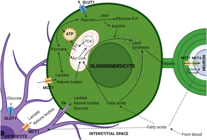
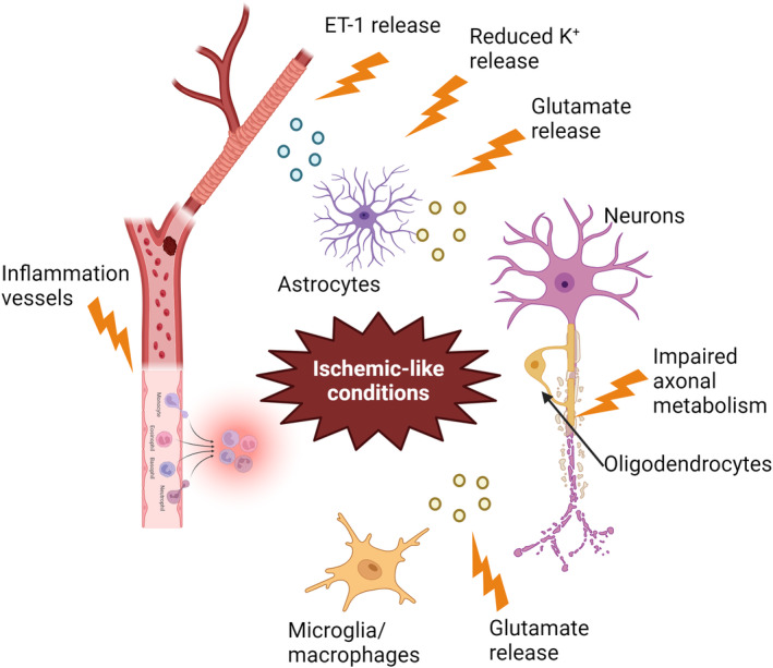
Mature myelinating oligodendrocytes, the cells that produce the myelin sheath that insulates axons in the central nervous system, have distinct energetic and metabolic requirements compared to neurons. Neurons require substantial energy to execute action potentials, while the energy needs of oligodendrocytes are directed toward building the lipid-rich components of myelin and supporting neuronal metabolism by transferring glycolytic products to axons as additional fuel. The utilization of energy metabolites in the brain parenchyma is tightly regulated to meet the needs of different cell types. Disruption of the supply of metabolites can lead to stress and oligodendrocyte injury, contributing to various neurological disorders, including some demyelinating diseases. Understanding the physiological properties, structures, and mechanisms involved in oligodendrocyte energy metabolism, as well as the relationship between oligodendrocytes and neighboring cells, is crucial to investigate the underlying pathophysiology caused by metabolic impairment in these disorders. In this review, we describe the particular physiological properties of oligodendrocyte energy metabolism and the response of oligodendrocytes to metabolic stress. We delineate the relationship between oligodendrocytes and other cells in the context of the neurovascular unit, and the regulation of metabolite supply according to energetic needs. We focus on the specific bioenergetic requirements of oligodendrocytes and address the disruption of metabolic energy in demyelinating diseases. We encourage further studies to increase understanding of the significance of metabolic stress on oligodendrocyte injury, to support the development of novel therapeutic approaches for the treatment of demyelinating diseases.

Keywords: hypoperfusion; metabolic stress; metabolism; multiple sclerosis; myelin; oligodendrocyte injury.


Figures

FIGURE 1
FIGURE 1
Oligodendrocyte injury mechanisms resulting from metabolic stress. Glucose deprivation causes failure of ATP production. As ATP decreases, the capacity to execute autophagy is lost and autophagosomes accumulate. ATP‐dependent plasma membrane pumps are compromised and the cell becomes incapable of pumping Ca2+ out of the cytosol. The increased intracellular Ca2+ concentration activates Ca2+‐dependent proteases leading to degradation. Failure of autophagy, which is also an ATP‐dependent process, further aggravates the process as cellular components can no longer be recycled as substrates for energy production. Created with BioRender.
FIGURE 2
FIGURE 2
Components of the neurovascular unit. Central nervous system (CNS) blood vessels are surrounded by contractile vascular smooth muscle cells. Substance exchange occurs across the capillaries which are surrounded by pericytes. Astrocyte endfeet embrace the capillaries, forming a key element of the blood–brain barrier that regulates substance exchange with the blood. Astrocytes also reach out to oligodendrocytes and neurons, passing along trophic support. Oligodendrocytes form myelin sheaths and provide metabolic support to neurons. Created with BioRender.
FIGURE 3
FIGURE 3
Flux of energetic metabolites in the central nervous system (CNS). Glucose, lactate, ketone bodies, and fatty acids pass into the CNS via endothelial cells and are then transferred to astrocytes, oligodendrocytes, and neurons. Glucose trafficking is mediated by glucose transporter (GLUT) 1 (blue channels) in endothelial cells, astrocytes, and oligodendrocytes and by GLUT3 (red channels) in neurons. Lactate and ketone bodies pass through MCT1 in endothelial cells, astrocytes, and oligodendrocytes (orange channels) and MCT4 in neurons. Fatty acids can diffuse directly through plasma membranes. Metabolites can be shared between astrocytes and oligodendrocytes through gap junctions formed by connexins (purple). Glucose can be converted and stored in astrocytes as glycogen. Glucose and lactate can be converted into pyruvate by glycolysis to yield ATP. Pyruvate, ketone bodies, and fatty acids can be converted into acetyl‐CoA (Ac‐CoA) and used to produce considerable amounts of ATP by oxidative phosphorylation in mitochondria via the TCA cycle and the electron transport chain (ETC). This source of ATP is particularly important for neurons because of their higher energetic demands. Fatty acids are mainly used by oligodendrocytes to produce myelin, as is a substantial portion of the acetyl‐CoA produced from other metabolites. Created with BioRender.
FIGURE 4
FIGURE 4
Oligodendrocyte energy metabolism and its relationship with neighboring cells. Oligodendrocytes import glucose from the central nervous system interstitial space via glucose transporter (GLUT) 1, and lactate and ketone bodies by monocarboxylate transporter (MCT) 1. These metabolites are transferred to the interstitial space by astrocytes largely using the same channels, or they can be transferred to oligodendrocytes via gap junctions formed by connexins (Cx). Once taken up, glucose can be converted to pyruvate through glycolysis, generating ATP, or feed the pentose phosphate pathway (PPP), generating NADPH, an important metabolite for lipid synthesis and the cellular anti‐oxidant system. Lactate can also be converted to pyruvate, which in turn can be converted to acetyl‐CoA (Ac‐CoA) in mitochondria. Ketone bodies may also be converted to Ac‐CoA, which can be used to produce ATP by oxidative phosphorylation involving the TCA cycle and electron transport chain (ETC). Alternatively, in oligodendrocytes a substantial portion of cellular Ac‐CoA is used for lipid synthesis to produce myelin. Fatty acids can diffuse through the cell plasma membrane and generate Ac‐CoA by β‐oxidation, or be used for lipid synthesis. Lactate, shuttled to the periaxonal space by oligodendrocytes can be taken up by neurons to support their energetic needs.
FIGURE 5
FIGURE 5
Potential causes of ischemic‐like conditions in MS. Inflammation of the vessel wall may trigger capillary thrombosis and stimulate leukocyte invasion, thereby reducing blood perfusion. Glutamate excitotoxicity may also induce ischemic‐like conditions because of metabolic stimulation. Impaired axonal metabolism may lead to mitochondrial dysfunction and oxidative stress. Abnormal ET‐1 released by astrocytes may cause vasoconstriction. Additionally, reduced K+ release by astrocytes, possibly due to decreased expression of β2‐adrenergic receptors may also induce vasoconstriction. Created with BioRender.

References

    1. Abbott, N. J. , Rönnbäck, L. , & Hansson, E. (2006). Astrocyte‐endothelial interactions at the blood‐brain barrier. Nature Reviews. Neuroscience, 7, 41–53. - PubMed
    1. Aboul‐Enein, F. , Krssák, M. , Höftberger, R. , Prayer, D. , & Kristoferitsch, W. (2010). Reduced NAA‐levels in the NAWM of patients with MS is a feature of progression. A study with quantitative magnetic resonance spectroscopy at 3 Tesla. PLoS One, 5, e11625. - PMC - PubMed
    1. Aboul‐Enein, F. , Rauschka, H. , Kornek, B. , Stadelmann, C. , Stefferl, A. , Brück, W. , Lucchinetti, C. , Schmidbauer, M. , Jellinger, K. , & Lassmann, H. (2003). Preferential loss of myelin‐associated glycoprotein reflects hypoxia‐like white matter damage in stroke and inflammatory brain diseases. Journal of Neuropathology and Experimental Neurology, 62, 25–33. - PubMed
    1. Adhya, S. , Johnson, G. , Herbert, J. , Jaggi, H. , Babb, J. S. , Grossman, R. I. , & Inglese, M. (2006). Pattern of hemodynamic impairment in multiple sclerosis: Dynamic susceptibility contrast perfusion MR imaging at 3.0 T. NeuroImage, 33, 1029–1035. - PMC - PubMed
    1. Alarcon‐Martinez, L. , Villafranca‐Baughman, D. , Quintero, H. , Kacerovsky, J. B. , Dotigny, F. , Murai, K. K. , Prat, A. , Drapeau, P. , & Di Polo, A. (2020). Interpericyte tunnelling nanotubes regulate neurovascular coupling. Nature, 585, 91–95. - PubMed

LinkOut - more resources