. 2024 Sep 10;9(17):e179291.
doi: 10.1172/jci.insight.179291.

Paternal hypercholesterolemia elicits sex-specific exacerbation of atherosclerosis in offspring

Affiliations

Paternal hypercholesterolemia elicits sex-specific exacerbation of atherosclerosis in offspring

Rebecca Hernandez et al. JCI Insight. .

Abstract

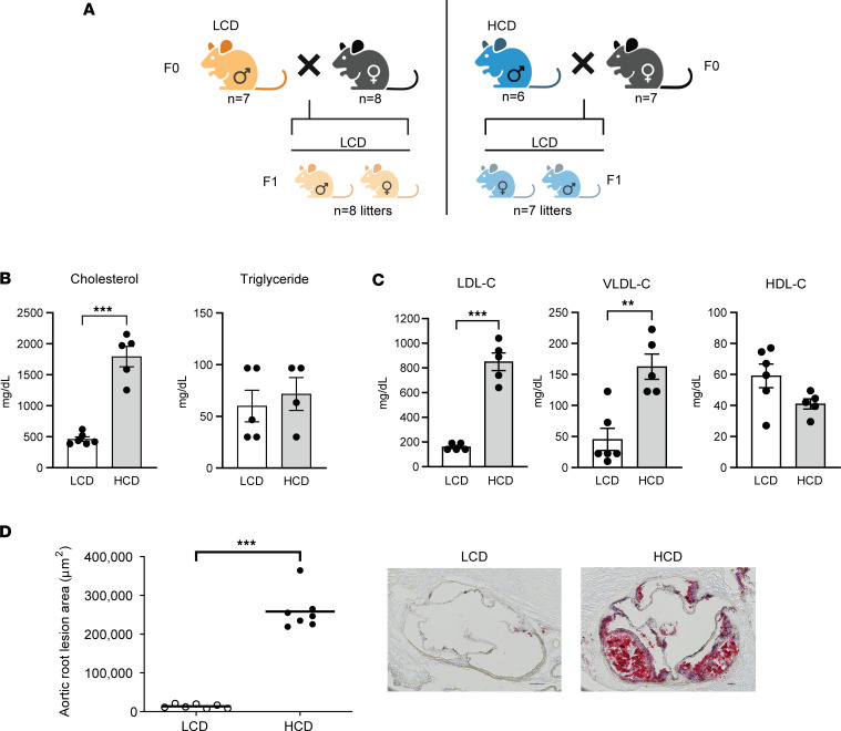
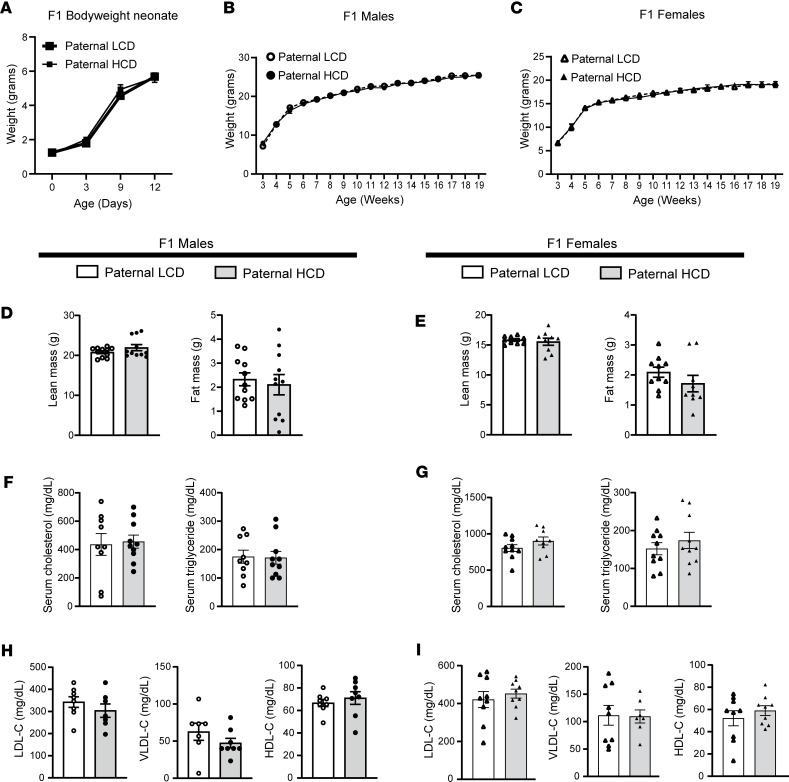
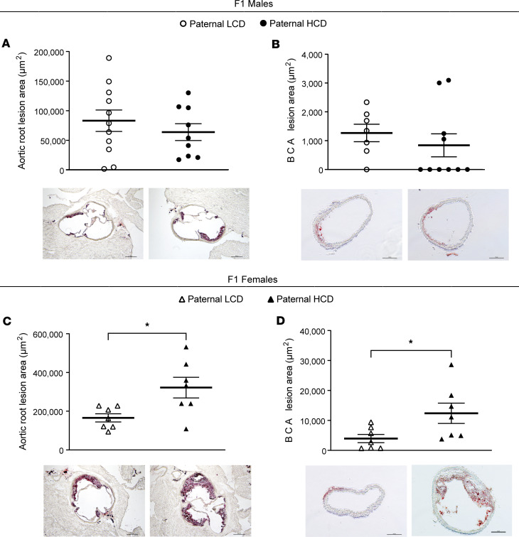
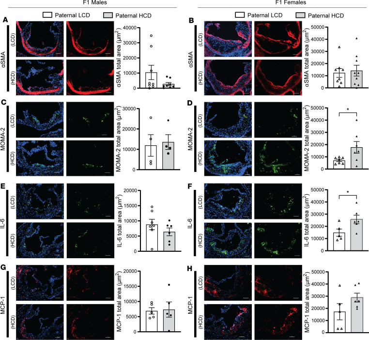
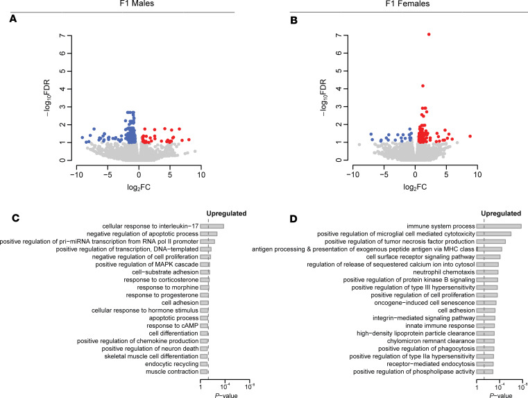
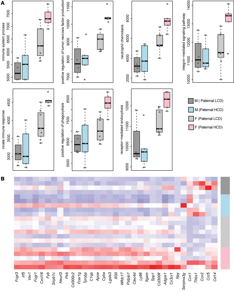
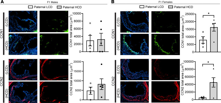
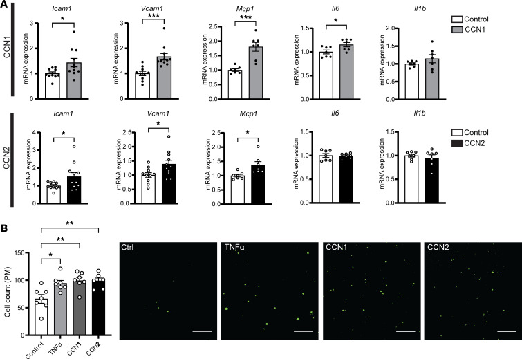
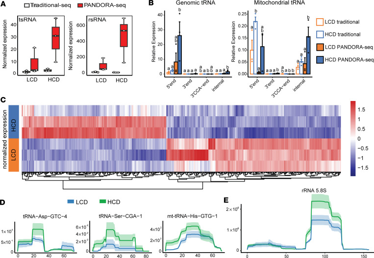
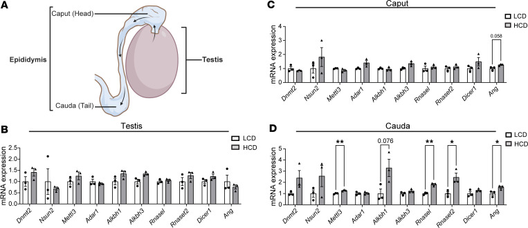
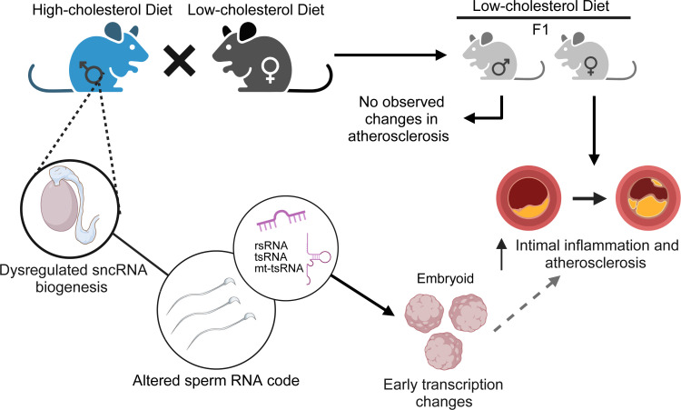
Emerging studies suggest that various parental exposures affect offspring cardiovascular health, yet the specific mechanisms, particularly the influence of paternal cardiovascular disease (CVD) risk factors on offspring cardiovascular health, remain elusive. The present study explores how paternal hypercholesterolemia affects offspring atherosclerosis development using the LDL receptor-deficient (LDLR-/-) mouse model. We found that paternal high-cholesterol diet feeding led to significantly increased atherosclerosis in F1 female, but not male, LDLR-/- offspring. Transcriptomic analysis highlighted that paternal hypercholesterolemia stimulated proatherogenic genes, including Ccn1 and Ccn2, in the intima of female offspring. Sperm small noncoding RNAs (sncRNAs), particularly transfer RNA-derived (tRNA-derived) small RNAs (tsRNAs) and rRNA-derived small RNAs (rsRNAs), contribute to the intergenerational transmission of paternally acquired metabolic phenotypes. Using a newly developed PANDORA-Seq method, we identified that high-cholesterol feeding elicited changes in sperm tsRNA/rsRNA profiles that were undetectable by traditional RNA-Seq, and these altered sperm sncRNAs were potentially key factors mediating paternal hypercholesterolemia-elicited atherogenesis in offspring. Interestingly, high-cholesterol feeding altered sncRNA biogenesis-related gene expression in the epididymis but not testis of LDLR-/- sires; this may have led to the modified sperm sncRNA landscape. Our results underscore the sex-specific intergenerational effect of paternal hypercholesterolemia on offspring cardiovascular health and contribute to the understanding of chronic disease etiology originating from parental exposures.

Keywords: Atherosclerosis; Cardiovascular disease; Cell biology; Epigenetics; Vascular biology.


Conflict of interest statement

Conflict of interest: The authors have declared that no conflict of interest exists.

Figures

Figure 1
Figure 1. Male LDL receptor–deficient mice fed a low-fat, high-cholesterol diet develop severe hypercholesterolemia-mediated atherosclerosis.
Three-week-old male LDLR–/– mice were fed a low-cholesterol diet (LCD, 0.02% cholesterol) or high-cholesterol diet (HCD, 0.5% cholesterol) for 8 weeks before mating with female LDLR–/– mice. The F1 offspring were weaned at 3 weeks old and were fed an LCD for 16 weeks. (A) Schematic representation of experimental design and generation of F1 offspring. (B) Serum total cholesterol and triglyceride levels were measured (n = 4–6, ***P < 0.001, 2-sample, 2-tailed Student’s t test). (C) Lipoprotein fractions (VLDL-C, LDL-C, and HDL-C) were isolated from serum, and the cholesterol levels of each fraction were measured (n = 5–6, **P < 0.01, ***P < 0.001; 2-sample, 2-tailed Student’s t test). (D) Quantitative analysis of the lesion area in the aortic root of LCD- and HCD-fed LDLR–/– mice (n = 7, ***P < 0.05, 2-sample, 2-tailed Student’s t test). Representative images are shown to the right. VLDL-C, very low-density lipoprotein cholesterol; LDL-C, low density lipoprotein cholesterol; HDL-C, high density lipoprotein cholesterol. All data are plotted as mean ± SEM. Scale bar: 100 μm.
Figure 2
Figure 2. Paternal high-cholesterol diet feeding does not affect body weight or serum lipid levels in F1 offspring.
Three-week-old male LDLR–/– mice were fed an LCD or HCD diet for 8 weeks before mating with control female LDLR–/– mice. Three-week-old F1 offspring were fed an LCD for 16 weeks and euthanized at 19 weeks of age. (A) Birth weight (day 0) and body weight of F1 pups before weaning (n = 5; 2-way ANOVA followed by Bonferroni’s multiple-comparison test). (B and C) Growth curves of male (B) and female (C) F1 offspring were measured (n = 9–14; 2-way ANOVA followed by Bonferroni’s multiple-comparison test). (D and E) Lean and fat mass were measure in male (D) and female (E) F1 offspring (n = 9–11, 2-sample, 2-tailed Student’s t test). (F and G) Serum cholesterol and triglyceride levels were measured in male and female offspring (n = 9–10, 2-sample, 2-tailed Student’s t test). (H and I) Serum lipoprotein fractions (VLDL-C, LDL-C, and HDL-C) were isolated from male (H) and female (I) F1 offspring and cholesterol levels from each fraction were measured (n = 7–9, 2-sample, 2-tailed Student’s t test). VLDL-C, very low-density lipoprotein cholesterol; LDL-C, low density lipoprotein cholesterol; HDL-C, high density lipoprotein cholesterol. All data are plotted as mean ± SEM.
Figure 3
Figure 3. Paternal hypercholesterolemia increases atherosclerosis development in F1 female LDL receptor–deficient offspring.
Three-week-old male LDLR–/– mice were fed an LCD or HCD diet for 8 weeks before mating with control female LDLR–/– mice. Three-week-old F1 descendants were fed an LCD for 16 weeks. (AD) Quantitative analysis of the lesion area at the aortic root (A and C) or brachiocephalic artery (B and D) of male (A and B) and female (C and D) offspring (n = 7–11, *P < 0.05, 2-sample, 2-tailed Student’s t test). Representative Oil Red O–stained sections displayed below the quantification data. Scale bar: 200 μm. All data are plotted as mean ± SEM.
Figure 4
Figure 4. Paternal high-cholesterol diet feeding elicits macrophage accumulation and inflammation in atherosclerotic plaques of F1 offspring.
Three-week-old male LDLR–/– mice were fed an LCD or HCD diet for 8 weeks before mating with control female LDLR–/– mice. Three-week-old F1 descendants were fed an LCD for 16 weeks. (AH) Representative images of immunofluorescence staining of α-SMA (A and B), MOMA-2 (C and D), IL-6 (E and F), and MCP-1 (G and H) at the aortic root of male and female offspring. Scale bar: 100 μm. The nuclei were stained with DAPI (blue). Quantification analysis of staining areas is displayed as indicated (n = 4–9, *P < 0.05, 2-sample, 2-tailed Student’s t test). All data are plotted as mean ± SEM.
Figure 5
Figure 5. Paternal hypercholesterolemia elicits transcriptomic changes in the intima of F1 LDL receptor–deficient mice.
Three-week-old male LDLR–/– mice were fed an LCD or HCD diet for 8 weeks before mating with control female LDLR–/– mice. Three-week-old F1 descendants were fed a LCD for 16 weeks. Total RNAs were isolated from the intima of F1 offspring and used for RNA-Seq analysis. (A and B) Volcano plots of differentially expressed genes (DEGs) in the intima of male offspring (A) and female offspring (B) from HCD-fed LDLR–/– sires. Colored dots represent the enriched (red dots) or depleted (blue dots) DEGs with a FDR of < 0.1 and a FC > 1.5 as a cut-off threshold. (C and D) GOBP terms significantly associated with upregulated DEGs in intima of male offspring (C) and female offspring (D) from HCD-fed sires. The P values were computed by the modified Fisher’s exact test using the DAVID bioinformatics tool. The vertical dash line indicates the significance level of α = 0.05. The y axis displays the GOBP terms, while the x axis displays the P values (n = 4–7 each group).
Figure 6
Figure 6. Paternal hypercholesterolemia alters atherosclerosis-related gene expression in the intima of female F1 offspring.
Three-week-old male LDLR–/– mice were fed an LCD or HCD diet for 8 weeks before mating with control female LDLR–/– mice. Three-week-old F1 descendants were fed a LCD for 16 weeks. Total RNAs were isolated from the intima of F1 offspring and used for RNA-Seq analysis. (A) Gene set scores of the prioritized GOBP terms of male and female offspring from LCD or HCD-fed sires. The gene set score was calculated using the FAIME algorithm. (B) Heatmap representation of DEGs involved in the indicated GOBP terms. Each column shows 1 individual gene, and each row shows a biological replicate of mouse. Red represents relatively increased gene expression, whereas blue denotes downregulation (n = 4–7 each group).
Figure 7
Figure 7. CCN1 and CCN2 proteins are elevated in the atherosclerotic lesions of F1 female LDL receptor–deficient descendants from high-cholesterol diet–fed sires.
Three-week-old male LDLR–/– mice were fed an LCD or HCD diet for 8 weeks before mating with control female LDLR–/– mice. Three-week-old F1 descendants were fed an LCD for 16 weeks. (A and B) Representative immunofluorescence images of CCN1 (green) and CCN2 (red) at the aortic root of F1 male (A) and female (B) offspring. The nuclei were stained with DAPI (blue). Scale bar: 100 μm. Quantification analysis of stating areas is displayed as indicated (n = 4–5, *P < 0.05, 2-sample, 2-tailed Student’s t test). All data are plotted as mean ± SEM.
Figure 8
Figure 8. CCN1 and CCN2 proteins promote proatherogenic gene expression in endothelial cells in vitro.
(A) Human endothelial cells, HMEC-1 cells, were treated with 1 μg/mL CCN1 or CCN2 for 4 hours followed by total RNA isolation. The expression levels of indicated genes were analyzed by qPCR (n = 7–11, *P < 0.05, ***P < 0.001, 2-sample, 2-tailed Student’s t test). (B) HMEC-1 endothelial cells were pretreated with 50 ng/mL CCN1 or CCN2 or 10 ng/mL TNF-α for 24 hours before incubating with calcein acetoxymethyl–stained peritoneal macrophages isolated from LDLR–/– mice for 4 hours. Adhered cells were counted under a fluorescence microscope. Quantitative analysis of the adhered cells is displayed to the left of representative images (n = 6–7, *P < 0.05, **P < 0.01, 1-way ANOVA followed by Bonferroni’s multiple-comparison test). All data are plotted as mean ± SEM.
Figure 9
Figure 9. PANDORA-Seq reveals significantly changed sperm tsRNAs and rsRNAs induced by high-cholesterol diet feeding in male LDL receptor–deficient mice.
Three-week-old male LDLR–/– mice were fed an LCD or HCD for 9 weeks. Total RNAs were isolated from the sperm and used for PANDORA-Seq and traditional small RNA sequencing. (A) Sperm tsRNA and rsRNA relative expression (normalized to miRNAs) under traditional sequencing and PANDORA-Seq protocols. (B) Sperm tsRNA responses to traditional sequencing and PANDORA-Seq in regard to different genomic or mitochondria tRNA origins (5′tsRNA, 3′tsRNA, 3′tsRNA-CCA end, and internal tsRNAs). The y axes represent the relative expression levels compared with total reads of miRNA. Different letters above the bars indicate statistically significant differences (P < 0.05). Same letters indicate P > 0.05. Statistical significance was determined by 2-sided 1-way ANOVA with uncorrected Fisher’s least significant difference test. All data are plotted as mean ± SEM. (C) Heatmap representation of differentially expressed sperm tsRNAs detected by PANDORA-Seq. Biological replicates are represented in each row. Red represents relatively increased expression, whereas blue represents decreased expression with adjusted P < 0.05 and FC > 2 as the cutoff threshold. (D and E) Dynamic responses to LCD or HCD of representative sperm tsRNAs (D) and rsRNAs (E) detected by PADNORA-Seq. Mapping plots are presented as mean ± SEM (n = 3 in each group).
Figure 10
Figure 10. Hypercholesterolemia alters the expression of sncRNA biogenesis–related genes in cauda epididymis.
(A) Schematic of the testis and epididymis. Sperm generated in the testis undergo maturational changes during transiting through the caput and cauda epididymis. (BD) Three-week-old male LDLR–/– mice were fed an LCD or HCD for 9 weeks. Total RNAs were isolated from the testis (B), and caput (C), and cauda (D) epididymis. The expression levels of indicated genes related to sncRNA biogenesis were analyzed by qPCR (n = 3, *P < 0.05, **P < 0.01, 2-sample, 2-tailed Student’s t test). All data are plotted as mean ± SEM.
Figure 11
Figure 11. Schematic of the effect of paternal exposure to the high-cholesterol diet on sperm sncRNAs and offspring atherosclerosis development.
Paternal high-cholesterol feeding led to significantly increased atherosclerosis and intimal inflammation in F1 female, but not male, LDLR–/– offspring. PANDORA-Seq identified an altered sncRNA landscape in the sperm of high-cholesterol diet–fed LDLR–/– sires. Overexpression of a pool of sperm tsRNAs/rsRNAs that were upregulated in the sperm of hypercholesterolemic sires induced transcription changes in embryoid bodies that may contribute the increased atherosclerosis in the adult offspring. The image was created with BioRender.com.

References

    1. Lusis AJ. Atherosclerosis. Nature. 2000;407(6801):233–241. doi: 10.1038/35025203. - DOI - PMC - PubMed
    1. Glass CK, Witztum JL. Atherosclerosis. The road ahead. Cell. 2001;104(4):503–516. doi: 10.1016/S0092-8674(01)00238-0. - DOI - PubMed
    1. Bjorkegren JLM, Lusis AJ. Atherosclerosis: Recent developments. Cell. 2022;185(10):1630–1645. doi: 10.1016/j.cell.2022.04.004. - DOI - PMC - PubMed
    1. Herrington W, et al. Epidemiology of atherosclerosis and the potential to reduce the global burden of atherothrombotic disease. Circ Res. 2016;118(4):535–546. doi: 10.1161/CIRCRESAHA.115.307611. - DOI - PubMed
    1. Roth GA, et al. Demographic and epidemiologic drivers of global cardiovascular mortality. N Engl J Med. 2015;372(14):1333–1341. doi: 10.1056/NEJMoa1406656. - DOI - PMC - PubMed