. 2024 Nov;20(11):7729-7744.
doi: 10.1002/alz.14233. Epub 2024 Sep 10.

Hypertension and cerebral blood flow in the development of Alzheimer's disease

Collaborators, Affiliations

Hypertension and cerebral blood flow in the development of Alzheimer's disease

Dario Bachmann et al. Alzheimers Dement. 2024 Nov.

Abstract

Introduction: We investigated the interactive associations between amyloid and hypertension on the entorhinal cortex (EC) tau and atrophy and the role of cerebral blood flow (CBF) as a shared mechanism by which amyloid and hypertension contribute to EC tau and regional white matter hyperintensities (WMHs).

Methods: We analyzed data from older adults without dementia participating in the Add-Tau study (NCT02958670, n = 138) or Alzheimer's Disease Neuroimaging Initiative (ADNI) (n = 523) who had available amyloid-positron emission tomography (PET), tau-PET, fluid-attenuated inversion recovery (FLAIR), and T1-weighted magnetic resonance imaging (MRI). A subsample in both cohorts had available arterial spin labeling (ASL) MRI (Add-Tau: n = 78; ADNI: n = 89).

Results: The detrimental effects of hypertension on AD pathology and EC thickness were more pronounced in the Add-Tau cohort. Increased amyloid burden was associated with decreased occipital gray matter CBF in the ADNI cohort. In both cohorts, lower regional gray matter CBF was associated with higher EC tau and posterior WMH burden.

Discussion: Reduced cerebral perfusion may be one common mechanism through which hypertension and amyloid are related to increased EC tau and WMH volume.

Highlights: Hypertension is associated with increased entorhinal cortex (EC) tau, particularly in the presence of amyloid. Decreased cortical cerebral blood flow (CBF) is associated with higher regional white matter hyperintensity volume. Increasing amyloid burden is associated with decreasing CBF in the occipital lobe. MTL CBF and amyloid are synergistically associated with EC tau.

Keywords: Alzheimer's disease; amyloid pathology; arterial spin labeling; cerebral perfusion; regional white matter hyperintensities; small vessel disease; tau pathology.


Conflict of interest statement

Christoph Hock and Roger M. Nitsch are employees and shareholders of Neurimmune AG, Switzerland. Dario Bachmann, Antje Saake, Sandro Studer, Andreas Buchmann, Katrin Rauen, Esmeralda Gruber, Lars Michels, Anton Gietl, and Valerie Treyer declare no relevant conflicts of interest. Author disclosures are available in the supporting information.

Figures
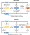
FIGURE 1
FIGURE 1
The deleterious effect of hypertension acts through several pathways, some of which differ depending on Aβ exposure. The left‐right‐headed arrow indicates the residual covariance between occipital rWMH burden and EC tau. The values in the boxes indicate path coefficients with the 95% confidence interval in brackets and a significance level of *p < .05, **p < .01, and ***p < .001. Path coefficients were estimated separately for low Aβ (Aβ−) and high Aβ (Aβ+) groups for paths where constraining the coefficients led to a significant reduction in model fit. EC, entorhinal cortex; rWMH, relative white matter hyperintensities.
FIGURE 2
FIGURE 2
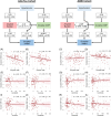
Relationships among hypertension, continuous Aβ burden, regional rCBF, and regional rWMH volume in Add‐Tau and ADNI cohorts. The models and scatterplots on the left‐hand side illustrate associations for the Add‐Tau cohort, while those on the right side illustrate associations for the ADNI cohort. The panels present scatterplots depicting the associations between regional rWMH and regional rCBF (A–D), Aβ burden and regional rWMH (E–H), and Aβ burden and regional rCBF (I–L). The values in the boxes in the structural equation models indicate path coefficients with the 95% confidence interval in brackets and significance levels of *p < .05 and ***p < .001. β coefficients and p‐value displayed in the scatterplots are extracted from the respective paths within the corresponding structural equation model. The values on the y‐axis in the plots represent residuals obtained after regressing the influences of age and sex. ADNI, Alzheimer's Disease Neuroimaging Initiative; rCBF, relative cerebral blood flow; rWMH, relative white matter hyperintensities.
FIGURE 3
FIGURE 3
Mediating role of MTL rCBF in association between hypertension and EC tau depending on Aβ status in Add‐Tau and ADNI cohorts. The models and plots on the left‐hand side illustrate associations for Add‐Tau cohort, while those on the right‐hand side illustrate associations for ADNI cohort. In the Add‐Tau cohort, one outlier was excluded from the plots but was included in the statistical analysis. The values in the boxes in the moderated‐mediation models indicate path coefficients with the 95% confidence interval in brackets and significance levels of *p < .05 and **p < .01. EC tau values in the plots represent residuals obtained after regressing the effects of age and sex. ADNI, Alzheimer's Disease Neuroimaging Initiative; EC, entorhinal cortex; MTL, medial temporal lobe; rCBF, relative cerebral blood flow.
FIGURE 4
FIGURE 4
MTL rCBF modifies the association between Aβ burden and EC tau in the ADNI cohort (A) and is lower in individuals with high SBP at sc (B, C). In A, tertiles of MTL rCBF divided ADNI participants into high, intermediate, and low groups. In B and C, β coefficients and p‐values displayed are extracted from the respective paths within the corresponding structural equation model. The values on the y‐axis in the plots represent residuals obtained after regressing the influences of age and sex. ADNI, Alzheimer's Disease Neuroimaging Initiative; MTL, medial temporal lobe; rCBF, relative cerebral blood flow; sc, screening visits.

References

    1. Ungvari Z, Toth P, Tarantini S, et al. Hypertension‐induced cognitive impairment: from pathophysiology to public health. Nat Rev Nephrol. 2021;17(10):639‐654. doi:10.1038/s41581-021-00430-6 - DOI - PMC - PubMed
    1. Yau W‐YW, Shirzadi Z, Yang H‐S, et al. Tau mediates synergistic influence of vascular risk and Aβ on cognitive decline. Ann Neurol. 2022;92(5):745‐755. doi:10.1002/ana.26460 - DOI - PMC - PubMed
    1. Braak H, Braak E. Frequency of stages of Alzheimer‐related lesions in different age categories. Neurobiol Aging. 1997;18(4):351‐357. - PubMed
    1. Vemuri P, Lesnick TG, Przybelski SA, et al. Age, vascular health, and Alzheimer disease biomarkers in an elderly sample. Ann Neurol. 2017;82(5):706‐718. doi:10.1002/ana.25071 - DOI - PMC - PubMed
    1. Kim HJ, Park S, Cho H, et al. Assessment of extent and role of tau in subcortical vascular cognitive impairment using 18F‐AV1451 positron emission tomography imaging. JAMA Neurol. 2018;75(8):999‐1007. doi:10.1001/jamaneurol.2018.0975 - DOI - PMC - PubMed

Publication types

MeSH terms