Review
. 2025 Jun;62(6):6785-6810.
doi: 10.1007/s12035-024-04468-y. Epub 2024 Sep 10.

Exploring the Mechanisms and Therapeutic Approaches of Mitochondrial Dysfunction in Alzheimer's Disease: An Educational Literature Review

Affiliations
Review

Exploring the Mechanisms and Therapeutic Approaches of Mitochondrial Dysfunction in Alzheimer's Disease: An Educational Literature Review

Mostafa Hossam El Din Moawad et al. Mol Neurobiol. 2025 Jun.

Abstract

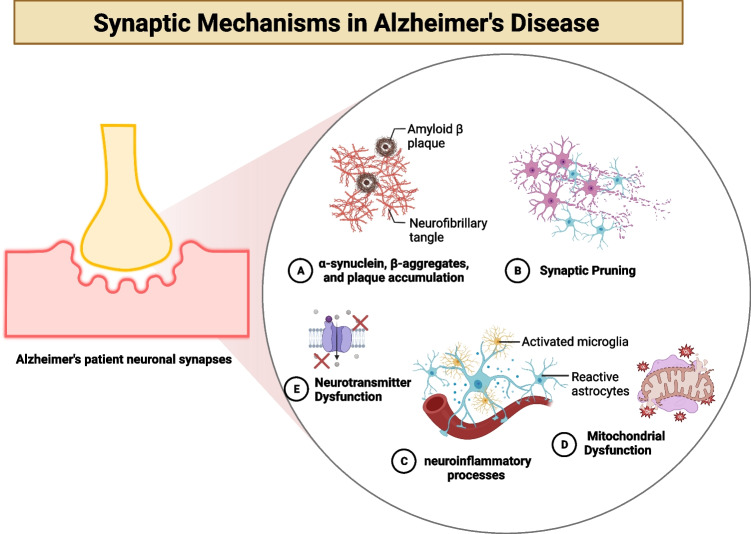
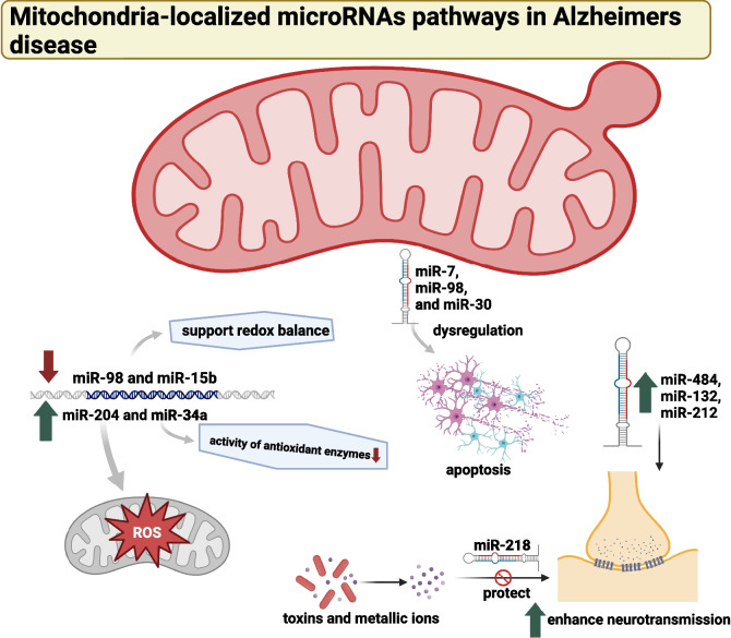
Alzheimer's disease (AD) presents a significant challenge to global health. It is characterized by progressive cognitive deterioration and increased rates of morbidity and mortality among older adults. Among the various pathophysiologies of AD, mitochondrial dysfunction, encompassing conditions such as increased reactive oxygen production, dysregulated calcium homeostasis, and impaired mitochondrial dynamics, plays a pivotal role. This review comprehensively investigates the mechanisms of mitochondrial dysfunction in AD, focusing on aspects such as glucose metabolism impairment, mitochondrial bioenergetics, calcium signaling, protein tau and amyloid-beta-associated synapse dysfunction, mitophagy, aging, inflammation, mitochondrial DNA, mitochondria-localized microRNAs, genetics, hormones, and the electron transport chain and Krebs cycle. While lecanemab is the only FDA-approved medication to treat AD, we explore various therapeutic modalities for mitigating mitochondrial dysfunction in AD, including antioxidant drugs, antidiabetic agents, acetylcholinesterase inhibitors (FDA-approved to manage symptoms), nutritional supplements, natural products, phenylpropanoids, vaccines, exercise, and other potential treatments.

Keywords: Alzheimer’s Disease; Mitochondrial Dysfunction; Therapeutic Modalities.


Conflict of interest statement

Declarations. Ethical Approval: Not applicable, and all data are available on the internet. Consent to Participate: Not applicable. Consent for Publication: Not applicable. Competing Interests: The authors declare no competing interests.

Figures

Fig. 1
Fig. 1
Synaptic mechanisms in Alzheimer’s disease (AD). A AD is characterized by the accumulation of tau protein tangles and amyloid beta (Aβ) plaques in the brain, disrupting synapses’ normal functioning. The regular operation of synapses is compromised due to the interference of oligomers with neurotransmitter action. Microtubules, which are essential for maintaining the structure and function of synapses, are adversely affected by tau protein tangles that disrupt their typical structure and function [14]; B synaptic pruning is a process through which the brain eliminates redundant or underused synapses. This process is essential for the normal functioning of the brain. However, it may be implicated in AD. In AD, synaptic pruning is excessively activated, leading to a reduction in functional synapses. Consequently, the brain ends up with fewer synapses, which could contribute to the cognitive decline associated with AD [15, 16]; C microglia, the brain’s immune cells, can become activated due to persistent inflammation. While microglia are necessary for removing Aβ plaques, they may also contribute to synaptic dysfunction. The pro-inflammatory cytokines secreted by activated microglia can impair synaptic function. Additionally, the activation of astrocytes, another type of brain cell, can release inflammatory substances that exacerbate synaptic dysfunction [17]; D the onset of AD has been linked to mitochondrial dysfunction. When mitochondria fail, they produce reactive oxygen compounds that can disrupt proteins, lipids, and DNA. This oxidative stress may play a role in the loss of functional connections, a characteristic feature of AD [13], and (E) neurotransmitters are chemical messengers essential for transmitting signals between neurons. AD is characterized by decreased neurotransmitters, such as acetylcholine, which plays a crucial role in memory and learning. The dysfunction of synapses caused by this neurotransmitter deficiency may contribute to the cognitive decline associated with AD [18]
Fig. 2
Fig. 2
Illustration of calcium ions (Ca2 +) signaling in mitochondrial dysfunction-associated neuronal apoptosis in Alzheimer’s disease (AD). The buildup of Aβ in cortical neurons is associated with releasing calcium from the endoplasmic reticulum, leading to increased cytosolic calcium ion levels and enhanced mitochondrial calcium absorption. Mitochondria and the endoplasmic reticulum play a crucial role in maintaining calcium homeostasis by transferring calcium ions out of the mitochondria and into the matrix via various channels and transporters [voltage-dependent anion-selective channel 1 (VDAC1); the Na + -dependent mitochondrial calcium efflux transporter (NCLX), and the mitochondrion calcium uniporter (MCU)]. Overloading mitochondria with calcium ions triggers the opening of mitochondrial transition pores (mPTPs), releasing cytochrome c, activating caspase activation, and initiating apoptosis [–54, 59]
Fig. 3
Fig. 3
The role of mitochondrial miRNAs in the pathogenesis of Alzheimer’s disease (AD). Dysregulation of specific miRNAs, often due to oxidative stress, can lead to increased production of ROS and neuronal death. Specific miRNAs, such as miR-98 and miR-15b, support redox balance, while others, like miR-204 and miR-34a, elevate ROS generation. The figure also highlights the role of miRNAs in synaptic information transmission and plasticity, with miR-484, miR-132, and miR-212 enhancing neurotransmission. Dysregulation of these miRNAs can contribute to synaptic dysfunction in AD. The figure further depicts the role of miRNAs in apoptosis, a mechanism regulating neuronal survival and death. Dysregulation of miRNAs implicated in apoptosis, such as miR-7, miR-98, and miR-30, can lead to increased apoptosis and neuronal death, disrupting learning and memory pathways [–125]
Fig. 4
Fig. 4
Summary of therapeutic modalities for mitigating mitochondrial dysfunction in Alzheimer’s disease. Preclinical models (catalase, N-acetylcysteine, Coenzyme Q10, melatonin, exenatide, metformin, carnosine, clove, berberine, ligstroside, and oleuroside, Egb761, quercetin, dihydroxyflavone, nilotinib, rapamycin, resveratrol, Aβ3-10-KLH vaccine and olesoxime), and clinical models (vitamin C and E, alfa-lipoic acid, thiazolidinediones, curcumin, lithium and small peptide SS-31)
Fig. 5
Fig. 5
Reactive oxygen species (ROS)-induced mitochondrial abnormalities in Alzheimer’s disease (AD). The overproduction of ROS or an impaired antioxidant system can shift the cellular redox balance towards oxidative imbalance. ROS, generated during cellular respiration, can harm mitochondria and neuronal function. An increase in ROS can lead to a reduction in mitochondrial membrane potential (ΔΨm) and ATP generation, negatively impacting mitochondrial energy stores, disrupting energy metabolism, and compromising dynamics and mitophagy. Furthermore, ROS can increase caspase activity, initiating apoptosis. Overproduction of ROS can also inhibit phosphatase 2A (PP2A), which activates glycogen synthase kinase (GSK) 3β, leading to tau hyperphosphorylation and the accumulation of neurofibrillary tangles [157]

Similar articles

Cited by

References

    1. Alkon D, Sun MK, Thompson R (2021) Evidence of significant cognitive improvement over baseline in advanced Alzheimer’s disease (AD) patients: a regenerative therapeutic strategy. Alzheimers Dement 17(S9):e050013
    1. Kashif M, Sivaprakasam P, Vijendra P, Waseem M, Pandurangan AK (2023) A recent update on pathophysiology and therapeutic interventions of Alzheimer’s disease. Curr Pharm Des. 10.2174/0113816128264355231121064704 - PubMed
    1. Varadharajan A, Davis AD, Ghosh A, Jagtap T, Xavier A, Menon AJ et al (2023) Guidelines for pharmacotherapy in Alzheimer’s disease – a primer on FDA-approved drugs. J Neurosci Rural Pract 14(4):566–573 - PMC - PubMed
    1. Gettman L (2024) Lecanemab-irmb (Leqembi™) for treatment of Alzheimer’s disease. Sr Care Pharm 39(2):75–77 - PubMed
    1. Huang M, Bargues-Carot A, Riaz Z, Wickham H, Zenitsky G, Jin H et al (2022) Impact of environmental risk factors on mitochondrial dysfunction, neuroinflammation, protein misfolding, and oxidative stress in the etiopathogenesis of Parkinson’s disease. Int J Mol Sci 23(18):10808 - PMC - PubMed

LinkOut - more resources